Lips of the mouth
(
labia oris
; Greek
chelos
). The upper lip (labium sup.) and the lower lip (labium inf.) in the area of the corners of the mouth (angulus oris), connecting by commissures (commissura labiorum), form the oral fissure (rima oris). The Upper Lip is limited by the base of the nose, the oral fissure and the nasolabial grooves (sulcus nasolabialis), the lower Lip - by the oral fissure and the labiomental groove (sulcus mentolabialis).
During ontogenesis, the lips are formed from the jaw processes. The lower Lip is formed at the end of the first month of uterine development as a result of the fusion of the mandibular processes, the upper - at the end of the second month as a result of the fusion of the right and left maxillary processes with the median nasal process (see Face). Musculature in G. is found only in mammals. In humans, bundles of facial muscles are embedded in the thickness of the G., thanks to which the Crimea G. have great mobility and participate not only in the act of grasping and processing food, but also in the act of speech and facial expressions.
Anatomy
Rice.
1. Lips are normal. The shape and size of the lips depend on the individual characteristics of the orbicularis oris muscle, the position or absence of the frontal teeth (see Bite), etc. In this regard, a distinction is made between protruded lips (procheilia) and straight lips (orthocheilia), sunken lips (opistocheilia), which usually observed in old and senile age with the loss of front teeth. Normally, the upper g. will stand somewhat in relation to the lower one. On the upper g., a groove (philtrum) runs in the vertical direction, dividing it into three parts: the middle and two lateral ones. In the area of the red border, the groove ends with a labial tubercle (tuberculum labii sup.). The dimensions of the labial tubercle vary significantly. The line that defines the border of the skin and the red border of the upper thigh is called Cupid's arc.
Lips consist of skin, subcutaneous tissue, muscle layer and mucous membrane. G.'s skin is thin, contains hair follicles and a large number of sebaceous glands. Near the oral fissure, the skin passes into the red border, or intermediate part (pars intermedia), where the structure of the skin changes, approaching the structure of the oral mucosa. In the red border, outer and inner zones are distinguished, especially sharply demarcated in newborns, in whom the inner zone is covered with papillae; During the first weeks of life, the papillae of the red border smooth out and become less noticeable. The epithelium covering the red border has a thin stratum corneum. In this part of the gland there are no hair follicles and sweat glands, but there are sebaceous glands, which are mainly concentrated in the area of the corners of the mouth, and there are more of them on the upper gland than on the lower one. The red border gradually passes into the mucous membrane of G.
Rice. 1. The vestibule of the oral cavity with the lips retracted, visible: a - frenulum of the upper lip; b — frenulum of the lower lip.
The mucous membrane of G., covered with stratified squamous non-keratinizing epithelium, has a pronounced submucosal layer where small salivary glands (glandulae labiales) are located. The mucous membrane of the mouth passes into the mucous membrane of the cheeks and gums, forming folds along the midline of the vestibule of the oral cavity - the frenulum (frenulum) of the upper and lower tongue (Fig. 1). The muscle layer is formed by the circular muscle of the mouth (m. orbicularis oris), into which fibers of some other facial muscles are woven.
Blood supply
G. occurs mainly from the facial artery, the edges at the level of the corners of the mouth are divided into the upper and lower labial arteries (a. labialis sup. et inf.). According to Yu. L. Zolotko, the upper blood supply from the facial artery occurs in 97.3% of cases, from the artery arising from the transverse artery of the face in 1.8%, and from both at the same time in 0.9%. The lower blood supply is supplied from the facial artery in 95.5% of cases, from the median artery of the chin in 0.8%, and from both in 3.6%. Typically, the arteries of the right and left sides merge along the midline and form a continuous ring. However, V. M. Kalinichenko (1970) found that in some cases the blood supply to the blood supply can be one-sided: to the lower blood supply in 19.6% of cases, to the upper blood supply in 16.1%; in this case, on one side the labial artery is absent or extends only to the corner of the mouth of the corresponding side.
The veins form a dense network and flow into the hl. arr. into the facial vein. M.A. Sreseli (1957) distinguishes two forms in the structure of the venous network of G.: with the first, a dense network of veins is observed with many anastomoses around the oral opening, spreading in depth; with the second, two veins of the upper and two veins of the lower vein are clearly visible, connected to each other by anastomoses.
Lymphatic vessels flow into the buccal, parotid, submandibular and cervical lymph nodes and into the deep cervical lymph nodes near the internal jugular vein (v. jugularis inf.). In addition, from the lower G. the outflow of lymph occurs into the submental lymph nodes.
Sensory innervation
the upper G. is carried out by the second branch, and the lower G. by the third branch of the trigeminal nerve; sympathetic nerve fibers arise from the superior cervical ganglion; motor nerve branches to the G. muscles come from the facial nerve.
Disturbance of oral microflora: causes
Oral dysbiosis can be caused by a variety of diseases and problems. Violation of opportunistic microflora of the oral cavity most often causes problems such as:
- Diseases of the gastrointestinal tract. Malfunctions of the digestive organs lead to a slowdown in metabolic processes in the body. The absorption of vitamins and nutrients deteriorates, the balance of the intestinal bacterial environment is disrupted, which provokes problems in other organs and systems.
- Decreased immunity. If the body's resistance deteriorates, the oral cavity automatically becomes more vulnerable to pathogenic microflora.
- Chronic diseases. Often, small caries or stomatitis, if left untreated, can spread from the source of inflammation to the entire oral cavity.
- Bad habits, such as systematic drinking of alcohol and smoking, inevitably affect the quality of the salivary glands. Prolonged drying out or too much moisture in the oral cavity has a detrimental effect on the composition of the microflora.
- Poor nutrition and lack of vitamins worsens the quality of saliva and makes the microflora of the oral cavity more vulnerable.
- Taking antibiotics and certain medications, such as hormones.
Pathology
Developmental defects
Congenital clefts occupy a significant place in the pathology of lip development
; according to most authors, they are found in one out of 1000 newborns. The occurrence of clefts is determined by Ch. arr. genetic factors, but may also be associated with impaired intrauterine development under the influence of endogenous and exogenous factors (complicated heredity, malnutrition, mental and physical trauma and illness of the mother at the beginning of pregnancy, etc.). Isolated cases of impaired fusion of the mandibular processes have been described, in which a median cleft occurs, as well as congenital fistulas of the lower gland in the form of blind canals of varying depths, lined with epithelium. Often there is a violation of the fusion of the maxillary and median nasal processes, which leads to the development of a congenital cleft of the upper nasal cavity (the so-called cleft lip). The shapes of the clefts are different - from a small notch at the red border to complete communication of the G. cleft with the opening of the nose. Sometimes the tissue cleft may be limited to only the muscle layer, which is called a hidden cleft; in this case, at the site of separation of the muscle layer, a sinking furrow of the skin or mucous membrane is visible. Clefts of the upper thigh can be one-sided or two-sided; in approximately 50% of cases they are combined with a cleft of the alveolar process and palate and are accompanied by deformation of the nose. A through bilateral cleft, as it were, separates the middle part of the upper nose along with the premaxillary bone, the edges stand forward, remaining connected to the vomer and the nasal septum. With a complete cleft of the upper gland, the child’s act of sucking becomes difficult, and in some cases impossible, breathing becomes shallow and frequent, and pneumonia often occurs as a complication.
Acheilia
(absence of lips) is rare with congenital atresia of the oral opening. Sometimes syncheilia is observed - fusion of the lateral parts of the mouth, leading to a decrease in the oral gap, as well as brachycheilia - a short middle part of the upper tongue.
Hypertrophy of the mucous glands and submucosal tissue manifests itself in the form of the so-called. double lips (labium duplex)—folds of the mucous membrane of G., the edges are especially evident when smiling.
Thickening and shortening of the frenulum
upper G.
Damage
Damage occurs as a result of falls, blows, bites, and gunshot wounds to the face. Wounds can be cut, torn, bruised, with or without tissue defects; by length - superficial, deep, through. Damage is accompanied by rapid development of swelling of the lips or significant bleeding. A feature of the wounds is the strong gaping of the wound, which creates the impression of a larger size than in reality, especially on the upper chin. Damage to the lower chin with a tissue defect leads to the leakage of saliva, which irritates and macerates the skin of the chin, making it difficult to eat.
Gunshot wounds of G. are usually not isolated: according to materials from the Great Patriotic War, isolated wounds of the lips accounted for 4% of facial wounds.
Diseases
The skin of the lips is often affected by eczema, characterized by a rash of blisters, weeping and a chronic recurrent course (see Eczema). In men, chronic inflammation of the hair follicles is more common (see Sycosis). The skin and mucous membrane of G. can be affected by herpes (see), lichen planus (see Lichen planus), lupus erythematosus (see), etc. Lesions of the mucous membrane of G. (without skin lesions) are observed with stomatitis (see .), sometimes with candidiasis (see); some forms of inflammation of the red border of G. are identified under the name cheilitis (see).
Macrocheilia (increase in the size of the lips) is a consequence of pathological processes in the tissues of the gland (persistent swelling, impaired lymph circulation after inflammatory and specific diseases), and can also be observed with acromegaly (see) and myxedema (see Hypothyroidism).
Boils and carbuncles
are difficult, especially when localized on the upper lip. M. A. Sreseli established that venous thrombosis, observed with purulent inflammation in the area of the upper G., sometimes spreads first along the facial vein, and then along the angular and superior ophthalmic veins, followed by a transition to the cavernous sinus; More often, thrombosis can spread through the venous anastomosis to the pterygoid plexus, then through the vein of the foramen ovale to the cavernous sinus. When purulent inflammation is localized on the lower lip, venous thrombosis can spread through the venous anastomoses of the face, the pterygoid plexus and the vein of the foramen ovale, less often - along the external jugular vein with subsequent transition to the sinuses of the dura mater.
For cutaneous anthrax
G.'s lesion resembles a banal boil or carbuncle, but the lesion is painless against the background of a sharp deterioration in the general condition and a rapid increase in intoxication of the body; When examining the discharge from the lesion, anthrax bacteria are detected (treatment - see Anthrax).
Tuberculous lesions of G. most often manifest themselves in the form of lupus (see Tuberculosis of the skin).
G.'s defeat during syphilis can occur in the primary period - the appearance of hard chancre on the lip, in the secondary - the appearance of papules, in the tertiary period - gumma may appear in the tissues of G.; characterized by painlessness (see Syphilis).
Tumors
Benign tumors include papilloma, keratoacanthoma, mixed tumors from minor salivary glands, tumor-like vascular neoplasms - hemangioma and lymphangioma (usually found in early childhood), retention cyst. The most common malignant tumor of G. is cancer; angiosarcoma, neurogenic sarcoma, melanoma, etc. are observed extremely rarely. Cancer of the lower gland often develops against the background of long-existing pretumor changes—dyskeratosis, less commonly papillomas, and keratoacanthoma. Dyskeratosis can be diffuse and focal: with diffuse, loss of shine, dryness, roughness, and peeling of the red border are observed; focal dyskeratosis is manifested by areas of leukoplakia (see) or hyperkeratosis (see) in the form of a flat or spiky horny protrusion. Erosion, ulcers, and slit-like fissures, characteristic of the malignant form of dyskeratosis, may be observed (see). The transition of dyskeratosis into cancer cannot always be detected clinically; if suspected, a histological examination should be performed (see Biopsy).
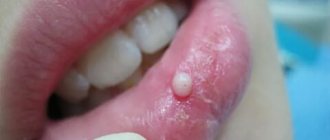
Rice. 2. Papillomas of the upper lip.
Papilloma
- a clearly demarcated papillary formation on the red border or on the mucous membrane of the lip.
The tumor is often single, less often in the form of several formations, usually small in size (up to 0.5-1 cm in diameter), pedunculated or broad based; acts as an exophyte above the surface of the red border or mucous membrane (color. Fig. 2). Its color is pink, its consistency is soft, covered with normal, sometimes slightly thinned epithelium (see Papilloma, papillomatosis). The appearance of ulceration, bleeding or infiltration of the base of the papilloma are signs that raise suspicion of the onset of cancer. Rice.
3. Keratoacanthoma of the lower lip: disease duration - 2 months Fig. 4. Keratoacanthoma of the lower lip: disease duration is about 1 year (beginning malignancy) Keratoacanthoma
occurs more often on the red border of the lower G. in the form of a towering spherical tumor measuring 1-2 units in diameter (tsvetn. Fig. 3 and 4).
The center of the tumor is crater-shaped, filled with horny masses, its edge is raised in the form of a clearly defined ridge. The tumor grows quite quickly in the first 3-4 weeks, then its growth stabilizes, and in some cases after 6-8 months. the tumor may disappear spontaneously, with the horny crust in the center falling off, the tumor flattening and a scar forming. Relapses are observed in 4-5% of cases (see Keratoacanthoma). The development of cancer from keratoacanthomas occurs in 20% of cases. Differential diagnosis with squamous cell carcinoma (clinically and even morphologically) is often difficult. Rice.
5. Mixed tumor of the lower lip. Mixed tumors from minor salivary glands
on G. are observed extremely rarely.
They are usually localized on the inner surface of the gland, covered with unchanged mucous membrane, clearly demarcated (tsvetn. Fig. 5). Their consistency is dense, the surface is smooth. These tumors rarely reach large sizes and grow slowly; according to gistol, the structure does not differ from similar tumors of the major salivary glands (see Mixed tumors). Rice.
7. Cavernous hemangioma of the lower lip. Hemangioma
, simple or cavernous, has the appearance of a node or a diffuse bluish-reddish tumor-like formation, causing G.’s deformation (tsvetn. Fig. 7).
Its consistency is usually soft, and when squeezed it decreases in size. The mucous membrane over the hemangioma is thinned, and sometimes there may be bleeding. Hemangioma grows slowly, but often spreads to adjacent areas of the face or oral cavity (see Hemangioma). Rice.
6. Lymphangioma of the upper lip. Lymphangioma manifests itself similarly (tsvetn. Fig. 6), but the red border or mucous membrane has a normal color, giving the impression of swelling of the lip (see Lymphangioma).
Rice. 8. Retention cyst of the lower lip.
Retention cyst of the mucous gland
quite often occurs on the inner surface of the lips, closer to the corner of the mouth (print. Fig. 8); has the appearance of a spherical bulge up to 0.5-1 cm in diameter. The mucous membrane over the cyst is thinned, translucent, less often whitish in the center. Upon palpation in the thickness of the G., a clearly demarcated node of soft-elastic consistency is determined. A retention cyst occurs due to retention of secretions or obstruction of the duct of the mucous gland and contains a light mucous fluid (see Cyst).
Cancer
in 90-95% of cases it is localized on the red border of the lower gland. In the upper gland, cancer often comes from the skin, spreading to the red border a second time. The majority of patients with lower gastrointestinal cancer are men aged 40–60 years. Predisposing factors are chronic mechanical, thermal and chemical. irritations, particularly smoking.
Lip cancer is most often keratinizing squamous cell (80-95% of all cases), less often non-keratinizing squamous cell and extremely rarely undifferentiated.
Rice. 9. Cancer of the lower lip: ulcerative - infiltrative form.
According to the wedge, the picture distinguishes papillary and ulcerative forms of cancer. The initial period of the papillary form is characterized by the appearance of a painless round-shaped compaction with fuzzy contours, covered with a crust; upon removal, a pink, easily bleeding area is revealed. As the process develops, a roller-like edge of the tumor becomes noticeable, and then an ulcer with uneven roller-like edges, with a necrotic bottom in the center, forms. In the ulcerative form, a fissure that does not heal for a long time is first discovered, turning into an ulcer with roller-like edges and infiltration in the underlying tissues; infiltration and destruction proceed faster than in the papillary form; the process involves not only the submucosal but also the muscular layer of the gland. In a later period, the differences in the manifestation of the papillary and ulcerative forms are erased, the ulcerative-infiltrative process predominates with the formation of an increasingly extensive defect of the gland (color fig. 9). Cancer of the lower gland is characterized by lymphogenous metastasis with damage to the submandibular and submental lymph nodes, and subsequently to the deep cervical lymph nodes. Distant metastases are rare.
It is customary to distinguish four stages of G cancer. Stage I is a limited tumor or ulcer with a diameter of 1-1.5 cm in the thickness of the mucous membrane and red border, without metastases. Stage II: a) tumor or ulcer with a diameter of more than 1.5 cm, limited to the mucous membrane and under the mucous layer, occupying no more than half of the lower g., without metastases; b) a tumor or ulcer of the same or smaller size, but in the presence of one or two mobile metastases in the regional lymph. nodes. Stage III: a) a tumor or ulcer that occupies most of the gastrointestinal tract with germination of its thickness or spread to the corner of the mouth, cheek and soft tissue of the chin; b) a tumor or ulcer of the same size or less widespread, but with the presence of limitedly mobile regional metastases. Stage IV - a disintegrating tumor that occupies most of the tumor with germination of its entire thickness and spread to the jaw bone, or a tumor with fixed metastases in the regional lymph. nodes, or a tumor of any size with distant metastases.
Thermal injuries of the oral mucosa in children.
They are rare in children, but are possible when eating hot food, especially milk and broth. The mucous membrane of the lips, the tip of the tongue, and the anterior part of the hard palate is mainly affected. It becomes swollen, hyperemic, and painful when touched. Less commonly, superficial intraepithelial vesicles form, which immediately burst. Upon examination in this case, fragments of white epithelium are visible on a hyperemic base. Prescribing antiseptics is unnecessary, since there is no deep defect in the epithelium, and therefore no conditions for secondary infection. For pain, painkillers are indicated: applications of a 0.5% solution of novocaine, lysozyme, 5–10% suspension of anesthesin in oil.
Treatment
For purulent processes on the Lip (furuncle, carbuncle), treatment is mainly conservative; you should not squeeze out the so-called. rods. Good results are obtained by using local novocaine blockade with antibiotics with simultaneous intramuscular administration of broad-spectrum antibiotics. In the first stage of inflammation, during the period of infiltration, radiotherapy at 120 kV, a 1-3 mm Al filter, a field that covers normal tissues surrounding the infiltrate by 1-1.5 cm, with a single dose of 15-25 r daily or every other day up to a total dose of 75-125 r. Under the influence of radiation, the infiltrate disappears, surgical intervention is not required. Surgical treatment is indicated for a formed abscess (see Carbuncle, Furuncle).
Treatment of malignant tumors can be divided into treatment of the primary tumor and regional metastases.
To treat the primary tumor, radiation therapy or a combined method is used (in the first stage - radiation therapy, in the second - wide excision with primary plastic surgery). Treatment of regional metastases is carried out mainly by surgery.
Radiation therapy for cancer of G. is carried out using the methods of interstitial gamma therapy (see), close-focus radiotherapy (see), electronic therapy (see), and, less often, application gamma therapy.
For the treatment of patients with stage I–II cancer, close-focus radiotherapy and interstitial gamma therapy are indicated. At stage III, interstitial gamma therapy and electron therapy have an advantage. For stage IV G. cancer, combined radiation therapy is indicated: remote gamma therapy or electron therapy, followed by the use of close-focus radiotherapy or interstitial gamma therapy. When the mucous membrane and skin of the mouth are affected, when the tumor is localized in the corners of the mouth, as well as when cancer recurs, the interstitial method has an advantage.
A contraindication for radiation therapy is the presence of a concomitant inflammatory process, upon elimination of which radiation therapy can be carried out. A contraindication for interstitial gamma therapy and close-focus radiotherapy is also the spread of the tumor to bone tissue and the inability to determine its boundaries, and in case of relapses, significant radiation changes in the surrounding normal tissues.
For close-focus radiotherapy, a single dose is 400-500 rad, the total dose to the lesion is 6000-6500 rad; irradiation field no more than 25 cm2.
In the interstitial method, needles with 226Ra, 60Co are used; The most convenient are nylon threads with 60Co granules. Radioactive drugs are administered after local anesthesia with 0.25% novocaine solution. Irradiation is continuous for 6-7 days. The total focal dose is 5000-7000 rad at a dose rate of 30-40 rad/hour.
For electronic therapy, Betatron type devices with radiation energy of 8-15 MeV are used. A single dose is 400 rad, the total dose is 5000-7000 rad, if; used as the only method. When combined with the interstitial method, the dose from electronic therapy is reduced.
The application method using 60Co preparations allows for fractional treatment with a daily dose of 500-600 rads and a total dose of 5000-6500 rads.
During radiation therapy, protection of the alveolar part of the lower jaw is required; the edges are provided with plexiglass or methyl methacrylate gaskets between the jaw and the jaw bone.
In stage I cancer of the lower cervix, a permanent cure is achieved in 95-96% of cases; Regional lymph nodes are not removed. Radiation therapy provides a high percentage of radical cure, better cosmetic and functional results compared to the surgical method, and fewer cases of relapses and metastases.
In stages II-IV of cancer, when curing the primary tumor, even in the absence of enlarged lymph nodes, an upper cervical excision operation should be performed, in which not only the submandibular and submental lymph nodes, but also the deep cervical lymph nodes located in the bifurcation area are removed carotid artery. In the presence of clinically significant regional metastases, preoperative external gamma or electron therapy with conventional dose fractionation and a total dose of 4000–4500 rad is indicated. The operation is performed after 2-3 weeks. after completion of radiation therapy.
Lip operations
taken to treat wounds, for purulent processes, for the treatment of tumors, etc.; A special place is occupied by operations on children and plastic surgeries.
Primary surgical treatment of wounds of G. should be performed taking into account functional and cosmetic requirements. Excision of tissue should be minimal and only tissue that is obviously non-viable and crushed. When applying layer-by-layer sutures, it is imperative to restore the continuity of the orbicularis oris muscle. The suture should be applied especially carefully to the skin and red border of the lips. In case of injury with a large defect in the tissue of the lips, when the edges of the wound cannot be sutured without tension, primary plastic surgery should be performed using tissue from areas of the face adjacent to the defect.
If the frenulum is thick and short, limiting G.’s mobility, it is excised ( frenectomy
) . To avoid scar formation, it is better to make a middle incision along the frenulum and use opposing triangular flaps.
With the so-called the double lip is surgically removed; excess submucosal tissue and mucous glands and fix the mucous membrane to the G muscle.
The retention cyst is removed with suturing of the mucous membrane. A mixed tumor should be removed along with the capsule and the mucous membrane covering it. The papilloma is excised with a small area of adjacent tissue. For small-sized hemangioma and lymphangioma, they are excised. With diffuse hemangioma, it can be reduced by introducing 70% alcohol into it to produce tissue sclerosis. For keratoacanthoma, they resort to either excision or close-focus radiotherapy.
Stages of dysbiosis
Depending on the degree of development of the disease, dentists distinguish four stages of dysbiosis:
- Latent. The first, latent stage is characterized by subtle changes in the number of microorganisms of one strain. The patient feels well and does not experience any symptoms of inflammation.
- Subcompensated. The number of lactobacilli decreases, the disease has a blurred picture. The patient may feel discomfort in the oral cavity, but does not always understand that this is dysbacteriosis.
- Pathogenic. Lactobacilli are observed in minimal quantities in the oral cavity. The oral cavity begins to be populated by a facultative pathogenic environment.
- Decompensated. In addition to severe inflammation in the mouth, uncontrolled growth of yeast-like fungi occurs. The functioning of the salivary glands is disrupted, and an unpleasant taste and burning sensation occurs in the mouth.
Advanced forms of dysbiosis are characterized by symptoms such as:
- inflammation of the gums and mucous membranes;
- plaque on the teeth and surface of the tongue;
- bleeding gums;
- ulcers and blisters on mucous membranes;
- increased body temperature;
- swelling and soreness of the tongue;
- dry skin, sticking in the corners of the mouth.