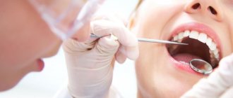
Treatment of the oral cavity is the area of expertise of the dentist. Therapy may include various techniques, as well as folk remedies, depending on what ailment has been diagnosed. Treatment of the oral mucosa and teeth is carried out in stages. At the first stage, the condition of the oral cavity is diagnosed through a visual examination and X-ray diagnostics. The second stage is preparing the oral cavity for treatment. It consists of professional hygiene - cleaning teeth from hard stones, as well as from soft plaque. Next, therapy is prescribed based on the stage of the pathology and the severity of the clinical picture.
Common pathologies
Treatment of the oral cavity in adults and children depends on what kind of pathological process is developing. The most common ailments include:
- inflammatory processes in periodontal tissues – gingivitis, stomatitis, periodontitis, periodontitis. Treatment for inflammation of the oral cavity involves only comprehensive treatment - local therapy, prescription of pharmaceuticals, physiotherapeutic procedures, etc.;
- carious lesion. Caries ranks first among all dental ailments. Its treatment involves removing the affected tissue, treating the tooth cavity and placing a filling;
- Oral candidiasis. This pathology is more typical for children, but there is also a possibility of its development in adults. Treatment of oral candidiasis is carried out both with pharmaceuticals and traditional medicine;
- pulpitis. Dangerous due to the formation of flux and the possibility of tooth loss (in the absence of adequate therapy;
- formation of ulcers in the mouth and tongue. This is most often observed after injury, burns, or stomatitis. But this symptom may be the first sign of the development of oral cancer, tuberculosis or syphilis.
The oral mucosa is constantly exposed to a variety of irritants - chemical, mechanical, thermal, numerous microbial agents and toxins. In addition, the oral cavity is a sensitive indicator, showing the state of the internal organs and promptly signaling the presence of problems in one or another body system. If at least one protective factor is weakened, there is a risk of developing inflammatory diseases of the oral mucosa. The most common of them are stomatitis and glossitis.
The cause of diseases of the oral mucosa can be: traumatic damage to the tissues of the oral cavity and other traumatic effects (chemical, thermal, etc.) with the development of traumatic erosion, ulcers, leukoplakia or leukokeratosis (keratinization of an area of the mucous membrane capable of malignant degeneration).
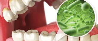
Infectious diseases that affect the oral mucosa due to the penetration of viruses, bacteria, and fungi. Quite often, the occurrence of pathological changes in the oral mucosa is associated with disruption of the functioning of various organs and systems of the body: allergies, dysfunction of the cardiovascular system, gastrointestinal tract, endocrine disorders, systemic connective tissue diseases, blood diseases, dermatoses, tuberculosis, AIDS and some other conditions. It is often quite difficult to identify the true cause of pathology of the oral mucosa - a lot of experience, high professionalism, and the ability not only to carefully collect information, but also to interpret it correctly and draw appropriate conclusions are required.
Dentists will quickly understand the manifestations of the disease in relation to a particular patient, determine the cause of the disorder and prescribe highly effective treatment.
MAIN CLINICAL MANIFESTATIONS
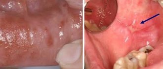
There is a common name for inflammatory diseases of the oral mucosa - stomatitis. When the pathological process is localized on the tongue, they talk about glossitis, on the gums - about gingivitis, on the lips - about cheilitis. When the mucous membrane of the mouth thickens, becomes horny and peels, they speak of a special type of disease - leukoplakia. A characteristic manifestation of stomatitis is the appearance on the oral mucosa of foci of redness, blisters, erosions (afts) or ulcers covered with plaque. These lesions are most often detected on the mucous membrane of the cheeks, floor of the mouth, hard palate, and tip of the tongue. Often there is pain at the location of erosions and ulcers, enlargement of nearby lymph nodes, and sometimes an increase in body temperature. The average duration of the disease is 7-14 days. Stomatitis can recur with decreased immunity, poor diet, hypovitaminosis, infectious diseases, and exacerbations are more common in spring and autumn.
DIAGNOSIS OF ORAL CAVITY DISEASES
Diagnosis of stomatitis and other diseases of the oral cavity is based on a thorough clinical examination of the patient by a dentist, which makes it possible to determine the stage of the pathological process and its prevalence, and the presence of a general reaction of the body to inflammation. It is very important to establish the true cause of the disease (trauma, infection, allergy, pathology of internal organs, hypovitaminosis, etc.), because the effectiveness of treatment and the absence of exacerbations in the future will depend on this.
PRINCIPLES OF TREATMENT OF DISEASES OF THE ORAL MUCOSA
Etiotropic and pathogenetic therapy aimed at eliminating the cause of the disease (antiviral, antibacterial therapy for the infectious nature of stomatitis, glossitis, cheilitis, vitamin therapy for hypovitaminosis, treatment of the underlying disease that caused the appearance of a pathological process on the oral mucosa); Local treatment aimed at eliminating local traumatic factors, the main symptoms of the disease and the rapid healing of existing erosions and ulcers; A general strengthening treatment that stimulates the body's defenses. An early visit to a dentist when identifying the first signs of pathology in the oral mucosa is the key to a speedy recovery!
STOMATITIS
Stomatitis is a general concept of inflammatory diseases of the oral mucosa. This pathology usually occurs against the background of a general and local decrease in immunity. Depending on the cause of occurrence, the following types of stomatitis are distinguished: • Chronic recurrent aphthous • Herpetic • Ulcerative-necrotic • Candidal
Chronic recurrent aphthous stomatitis - manifests itself in the form of characteristic painful aphthae on the mucous membrane of the lips, cheeks, palate or tongue. The main causative agents of this disease are viruses and bacteria. The disease manifests itself against the background of an imbalance in the body of vitamins such as B1 and B12. Most often this can be observed in chronic diseases of the liver and gastrointestinal tract (gastrointestinal tract).
Herpetic stomatitis
The causative agent of herpetic stomatitis is the common herpes virus. Herpetic stomatitis most often occurs in children aged 1 to 3 years. At the same time, in children, at the very beginning of the disease, general symptoms of intoxication begin to appear: • General malaise occurs • Body temperature rises • Lymph nodes enlarge • Nausea and vomiting • Diarrhea
Then, on the oral mucosa, as well as on the red border of the lips, peculiar bubbles begin to form, which quickly open and form erosions with characteristic so-called scalloped (uneven) edges. After about 8-10, healing occurs.
Necrotizing ulcerative stomatitis is characterized by necrosis of the gingival margin. More often, inflammation begins with the interdental papillae and adjacent mucous membrane, namely the cheeks. Then painful, easily bleeding ulcers form, which very quickly merge and form quite large defects in the mucous membrane. As a result of the active process of necrosis, a characteristic putrid odor occurs from the mouth. This picture can be observed with poor oral hygiene. This type of stomatitis is most common in adults aged 17 to 30 years. Necrotizing ulcerative stomatitis can be associated with diseases such as influenza, tonsillitis, acute respiratory infections, various blood diseases, AIDS, tuberculosis and give quite unpleasant and serious complications. In addition to rashes, with this pathology of the mucous membrane, general symptoms of intoxication are also noted - increased body temperature, general malaise, as well as enlarged and painful lymph nodes. Candidal stomatitis
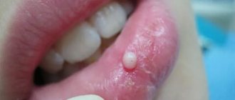
Candidal stomatitis
Candidal stomatitis is a fairly common disease of the oral mucosa, caused by fungi of the genus Candida. In the oral cavity, the following manifestations are noted: • Dryness • Burning • Formation of a white cheesy coating (when this plaque is removed, the mucous membrane bleeds profusely)
Treatment of stomatitis
First of all, at the first symptoms of one or another type of stomatitis, you need to contact a dental clinic. In this case, you should not self-medicate! Indeed, depending on the type of disease, the doctor prescribes a specific treatment for stomatitis. Therefore, the main task of the dentist is, firstly, competent diagnosis, secondly, elimination of the cause of the disease, and only, thirdly, symptomatic therapy for the complete and final treatment of stomatitis. The main prevention is, of course, high-quality and regular individual oral hygiene and careful attention to your health.
Clinical manifestations of stomatitis According to clinical features, the following types of stomatitis are distinguished: catarrhal, ulcerative, aphthous.
Catarrhal stomatitis is the most common lesion of the oral mucosa. The cause of its occurrence is considered to be local factors: poor oral hygiene, dental disease, dental plaque, oral dysbacteriosis. Diseases of the gastrointestinal tract, such as gastritis, duodenitis, colitis, can also cause catarrhal stomatitis. The cause of catarrhal stomatitis may be helminthic infestation. With this disease, the oral mucosa becomes swollen, painful, hyperemic, and may be covered with a white or yellow coating. Hypersalivation (increased salivation) is noted. Bleeding gums and bad breath may occur.
How is catarrhal stomatitis treated?
Treatment comes down to eliminating local causes - removing tartar, treating dental diseases. The mucous membrane is treated with antiseptic rinses - 0.05% and 0.1% chlorhexidine solution. During the day, you can rinse your mouth with a warm solution of chamomile and calendula decoction. A gentle diet is required. With this treatment, the symptoms of stomatitis disappear after 5-10 days. If the symptoms of stomatitis do not disappear, then it is necessary to establish a common cause - as a rule, these are diseases of the gastrointestinal tract or helminthic infestation. In this case, local treatment should be combined with general treatment.
Ulcerative stomatitis is a more severe disease than catarrhal; it can develop either independently or be an advanced form of catarrhal. Most often, this disease develops in patients suffering from gastric ulcers or chronic enteritis. It also often occurs in patients with diseases of the cardiovascular system and blood, infectious diseases and poisoning. Unlike catarrhal stomatitis, which affects only the superficial layer of the mucous membrane, with ulcerative stomatitis the entire thickness of the mucous membrane is affected. The initial symptoms of catarrhal and ulcerative stomatitis are similar, but subsequently with ulcerative stomatitis there is an increase in temperature, weakness, headache, enlargement and tenderness of the lymph nodes. Eating is accompanied by severe pain. If such symptoms appear, you should consult a doctor.
Aphthous stomatitis is characterized by the appearance of single or multiple aphthae (ulcers) on the oral mucosa. Aphthae are oval or round in shape, no larger than a lentil grain, with clear boundaries in the form of a narrow red border and a grayish-yellow coating in the center. The causes of this variant of stomatitis are considered to be diseases of the gastrointestinal tract, allergic reactions, viral infections, and rheumatism. The disease begins with general malaise, increased body temperature, and the appearance of pain in the mouth at the site of aphthae formation. This disease should be treated by a doctor.
Leukoplakia is a chronic lesion of the oral mucosa, which is based on increased keratinization of the epithelium (hyperkeratosis). It affects mainly men after 40 years of age and is localized on the mucous membrane of the cheek, in the corners of the mouth and the lateral surfaces of the tongue. The causes of leukoplakia can be mechanical injuries to the mucous membrane: cuts from hooks from a denture, burns from hot or spicy food, etc. This disease most often does not have pronounced symptoms, only sometimes the patient may feel mild itching and burning. But the danger of the disease is that it can develop into malignant forms, so the patient needs to consult an oncologist.
GLOSSITIS is an inflammation of the tissues of the tongue. It can be superficial or deep. Most often, glossitis is a symptom of some general disease of the body, but it can also occur independently.
The main causes of glossitis are: carious teeth, difficult teething, tartar, injuries to the mucous membrane of the tongue and oral cavity, smoking, alcohol abuse, poor oral hygiene, poisoning with heavy metal salts, burns, too hot food, hot spices, allergic reactions etc. Superficial glossitis is often a sign of diseases of the gastrointestinal tract and infectious diseases. It is characterized by the presence of plaque on the tongue, its swelling, hardening, and limited mobility. The tongue becomes bright red, there is a burning sensation in the tongue, pain, loss of taste, and excessive salivation.
Treatment of superficial glossitis is based on the use of local anesthetics and anti-inflammatory drugs. For oral administration, multivitamins, desensitizing agents (antihistamines), and immunostimulants are prescribed. Oral debridement (the process of cleaning an open wound by removing foreign material and dead tissue from it so that nothing impedes its healing) is of great importance.
With deep glossitis, everything is much more complicated. The inflammatory process in this form of the disease is localized in the thickness of the tongue and manifests itself in the form of an abscess (a limited accumulation of pus that occurs during acute or chronic focal infection). Deep glossitis can spread to the floor of the mouth and cause inflammation in the chin and neck. For this form of glossitis, surgical treatment is indicated.
In addition to the above, there are also non-inflammatory forms of glossitis, namely:
— desquamative glossitis (geographical tongue) This form of the disease occurs during pregnancy, damage to the gastrointestinal tract, blood diseases, metabolic disorders, some infectious diseases, helminthic infestations, and rheumatism. Desquamative glossitis is characterized by focal destruction of the red epithelium on the back and lateral surfaces of the tongue. The alternation of lesions with restored and destroyed epithelium makes the surface of the tongue look like a geographical map. In addition to external changes, burning and pain in the tongue may occur. Therapy for desquamative glossitis is based on the treatment of the underlying disease that provoked the development of glossitis.
— rhomboid (median) glossitis Rhomboid glossitis is a congenital anomaly of the tongue as a result of disruption of fetal development processes.
- villous glossitis: this form of glossitis is characterized by the proliferation and keratinization of filiform papillae.
- folded glossitis: glossitis of this form is a congenital anomaly and is characterized by the formation of folds on the back of the tongue, the deepest of which runs longitudinally along the midline. Folded glossitis usually does not cause complaints and does not require treatment.
- Gunter's glossitis: this form of glossitis is one of the signs of anemia caused by a lack of vitamin B12 and folic acid. It is characterized by the absence of papillae and a smooth (varnished) surface of the tongue.
— interstitial glossitis: a similar form of glossitis develops with syphilis in the tertiary period. The tongue becomes denser, its mobility is limited.
villous glossitis geographic language Glossitis Prevention of glossitis includes: oral and dental hygiene, regular visits to the dentist, reducing the consumption of aggressive and spicy foods, avoiding smoking and alcohol abuse.
Treatment of diseases of the oral mucosa
The basis of treatment for diseases of the oral mucosa is the elimination of the causes that provoked them. The oral cavity is subject to sanitation, the sharp edges of the teeth are processed, and the denture is correctly adjusted. The patient is advised to stop smoking and eating spicy and hot foods.
Tartar in case of stomatitis is removed, and the teeth are subject to treatment. It is necessary to rinse the oral mucosa with antiseptic agents. Folk remedies are also used: infusions and decoctions of chamomile and calendula. If signs of stomatitis persist after 5-10 days, most likely their cause is a disease of the gastrointestinal tract or helminthic infestation. Then local treatment is combined with general treatment.
The dental clinic diagnoses and treats a wide range of diseases of the oral mucosa. These diseases are diverse, variable and often cause a lot of suffering to patients, despite the fact that they cannot be correctly diagnosed and treated correctly everywhere. In addition, diagnostics using the mucous membrane of the oral cavity and tongue helps to clarify the condition of internal organs and systems, which is important because it does not require additional complex laboratory methods.
Therapeutic measures
Treatment of oral diseases requires an integrated approach. It is important not only to treat the pathology, but also to prevent its recurrence in the future. This is achieved through timely professional oral hygiene, as well as maintaining an appropriate level of hygiene at home.
Treatment of diseases of the oral mucosa involves the prescription of certain pharmaceutical agents with antibacterial, antiseptic and wound-healing effects. For example, for stomatitis, the use of Stomatidine has a good effect. Drugs for the treatment of the oral cavity are selected exclusively by the attending physician.
If caries is present, a filling is indicated. If pulpitis occurs, then the carious tissue is removed, then the nerves are removed and the pulp chamber is cleaned. The cleaned tooth roots are treated with antiseptics and filled. Next, a filling or a crown made of metal ceramics or metal-free ceramics can be installed.
Causes of diseases of the oral mucosa
Diseases of the oral mucosa occur throughout life in an average of 5-10% of the population.
Their cause may be:
- Traumatic damage to the tissues of the oral cavity and other traumatic effects (chemical, thermal, etc.) with the development of traumatic erosion, ulcers, leukoplakia or leukokeratosis (keratinization of an area of the mucous membrane capable of malignant degeneration).
- Infectious diseases that affect the oral mucosa due to the penetration of viruses, spirochetes, bacteria, and fungi.
Quite often, the occurrence of pathological changes in the oral mucosa is associated with disruption of the functioning of various organs and systems of the body: allergies, dysfunction of the cardiovascular system, gastrointestinal tract, endocrine disorders, systemic connective tissue diseases, blood diseases, dermatoses, tuberculosis, AIDS and some other conditions. It is often quite difficult to identify the true cause of pathology of the oral mucosa - a lot of experience, high professionalism, and the ability not only to carefully collect information, but also to interpret it correctly and draw appropriate conclusions are required. Experienced medical dentists will quickly understand the intricacies of the existing manifestations of the disease in relation to a specific patient, determine the cause of the disorder and prescribe highly effective treatment.
Candidiasis
Sometimes it appears in a healthy person, but it can also be a complication of serious diseases that cause immunodeficiency. Most often, candidiasis occurs with a decrease in general and local immunity. Changes in the microbial flora inhabiting the mucous membranes (dysbacteriosis) sometimes lead to the development of candidiasis. Candidiasis can affect the skin, mucous membranes and internal organs. It is imperative to consult a dentist, as it is necessary to conduct laboratory studies of changes in the microbial flora. In accordance with the results obtained, the doctor prescribes adequate treatment.
Advantages of treatment at the VivaDent clinic
The main advantage of our dental clinic is that we employ high-class professionals with extensive experience and rich knowledge in the field of diagnosis and treatment. We are proud of our reputation as one of the leading clinics in Moscow, so we offer our patients only the best!
The VivaDent clinic is equipped with the most modern equipment, which allows you to quickly and accurately make a diagnosis and begin treatment measures in a timely manner. In addition, we offer affordable prices for all services. The clinic constantly holds promotions with favorable conditions, which make it possible to significantly save on dental treatment. For regular customers there is an individual discount system.
We have a cozy environment, patients do not feel discomfort within the walls of the medical facility. This is especially important for people with a panic fear of dental procedures and for children. We tried to create all the conditions so that our clients feel calm and confident in the clinic.
If you decide to undergo an oral cavity examination, contact the best specialists! The first visit to the dentist in our clinic is free for all categories of patients!
Inflammation of the oral mucosa
The oral cavity often suffers from inflammation and ulcers.
Lesions vary in size, appearance, and localization area. Pathologies can occur in any area, including the palate, lips, and tongue. In such a situation, a person complains of pain, the tissue turns red and swells. When the top layer of tissue is destroyed, ulcerative holes form. They may be whitish, take the form of a blister, and fill with liquid. Inflammation of the oral mucosa is primarily manifested by a burning sensation. Therefore, you should consult a doctor in time. Periodontology in St. Petersburg uses complex technologies to treat the tissues that surround the chewing organs. Thanks to modern equipment and advanced techniques, the loss of units is reduced to zero. The main task of the dentist is to maintain reliable fixation of teeth in the gums and jaw bones. To do this, bleeding, discomfort, bad breath, and receding gum tissue are eliminated.
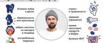
Causes of inflammation
The inflammatory process often begins when the rules of oral hygiene are ignored. The risk of disease increases with the following factors:
- Poor nutrition, when the body is deficient in microelements and vitamins, overeating fatty and sweet foods.
- A decrease in the body's defenses appears as a result of excessive loads and stress.
- Colds, chronic diseases, gastrointestinal pathologies, and blood diseases have a negative effect on the oral cavity. Patients note that under such circumstances the mucous membranes in the mouth and tongue hurt.
- Passion for alcoholic beverages and smoking.
- Taking medications that have a negative effect on periodontal tissue.
- Anomalies of the jaw bone that prevent the complete cleaning of accumulated plaque.
In older patients, gingival tissue dystrophy may be diagnosed, which provokes periodontal pathologies. In addition, abnormal phenomena are caused by infections, chemical and physical irritants, allergies, herpes, and systemic disorders. If normal salivation is disrupted, the risk of disease increases, as the mucous membranes dry out. Learn more about the most common provoking factors.
Stomatitis
The symptom of stomatitis is an inflammatory process; patients have pain in the oral mucosa. More often a child may get it, although adults are not immune from this pathology either. In fact, this disease refers to defensive reactions to irritation factors. Pathology actively develops when:
- decreased immunity;
- the body is affected by viruses;
- an excessive amount of pathogenic bacteria has appeared;
- mouth tissues are injured;
- hygiene rules are not observed;
Adults more often suffer from herpetic pathology with the formation of multiple blisters that turn into ulcers. In addition to this form there are:
- allergic form, which is characterized by swelling, tissue hyperemia;
- Vincent's disease with ulcerative-necrotic anomalies of the gingival papillae;
- thrush, which is caused by yeast-like fungi that create cheesy layers;
- aphthous form, which often indicates diseases of the gastrointestinal tract;
Also, Koplik spots may appear on the mucous membrane in measles, exudative erythema, lesions in scurvy, a “crimson” tongue in scarlet fever, tissue hyperemia in Kawasaki disease.
Gingivitis
The initial stage of periodontitis in medicine is called gingivitis. This is inflammation of the gums. The most common causative agents of the disease are gram-negative anaerobic bacteria. Sticky deposits are located along the edges of the gums, as well as in areas that are difficult to clean. Then the soft coating compacts, forming stones. The disease also develops:
- for malocclusions;
- with incorrectly placed filling material;
- for pathologies of oral breathing;
- with endocrine changes;
- for diabetes mellitus;
The disease is diagnosed in pregnant women because their hormonal levels have changed. The swollen tissues bleed and pyogenic granulomas form. Pathology often appears during menopause, because at this time an insufficient number of special epithelial cells are formed. They are very vulnerable, they bleed and hurt. Without treatment, gingivitis gives rise to periodontitis, gum pockets deepen, persistent bad breath appears, the supporting apparatus of the masticatory organs weakens, and bone destruction begins. All this leads to thinning of the bone tissue, loosening of the units, and atrophy of the gum mucosa. In the later stages of the disease, teeth fall out.
Oral injuries
If the oral cavity became inflamed, most often the patient had injury and damage to the mucous membranes. Scratches and accidental bites from uneven or broken crowns or poorly ground dentures first cause the appearance of blisters, and after their ruptures, ulcers. A number of products, as well as chemicals, cause irritation and allergies. Ulcerations form in these places. Fruit acids, flavorings, astringents, some toothpastes, sweets, and mouthwashes are especially irritating to mucous tissues.
How to treat at home
When inflammation of the mucous membrane in the mouth begins, you need to consult a doctor. Gum disease can be eliminated when its cause is identified. Home therapy is carried out in case of minor pathologies when the provoking factors are known.
- For irritation, you can use antiseptic rinses.
- Pharmacy antimicrobial agents or decoctions of medicinal herbs help well.
- Dentists recommend light massage movements with a medium-hard brush. The massage is performed carefully to avoid injury.
- It is necessary to exclude too cold and hot foods from the diet to avoid unpleasant sensations.
- Stress damages the health of gum tissue. After all, the body actively produces cortisol, which causes inflammatory processes.
When and which doctor to contact
If the pathology does not go away after 3-5 days, such patients require consultation with a periodontist. Treatment begins with a medical history and examination of the oral cavity. Tests are prescribed according to indications. Blood tests, x-rays, and bacteriological studies may be required. After identifying the cause, a treatment plan for the pathology is drawn up. Local treatment includes:
- Anesthetic solutions;
- Applying protective applications;
- Use of corticosteroids;
- Cauterization with laser beams;
- Use of chemicals;
Anesthetic liquids can be used. Pain is relieved by applications of sucralfate. Many dentists add lidocaine. An alternative option is to use paste. Some types of ulcers are treated with low-power laser, which prevents relapses.
Prevention of inflammatory processes in the mouth
Inflammation of the oral cavity is easier to prevent than to cure. You should visit your dentist regularly so that he can detect the disease at an early stage.
- If there are bite defects, they must be corrected.
- You should properly care for your oral cavity, clean your tongue, crowns, and clean soft tissues.
- There should be a balanced diet with all the necessary microelements and vitamins.
- It is imperative to treat chronic diseases.
Visits to the dentist for preventive examinations are carried out twice a year. If necessary, professional cleaning is performed. Visits to a periodontist should be scheduled at the first periodontal problems.
+7 St. Petersburg, Maly Prospekt V.O., building 4
You can ask any questions you may have and find out the available time for making an appointment with the administrators of the ARTES clinic! Call and come for an appointment!
Zaeda
A disease of the mucous membrane of the skin of the corners of the mouth, caused by streptococci or yeast-like fungi of the genus Candida. The occurrence is promoted by vitamin B2 deficiency, diabetes mellitus, irritation of the skin and mucous membrane of the corners of the mouth with saliva. Candidiasis (yeast-like) infection can occur with long-term treatment with antibiotics, cytostatics and corticosteroids. Zaeda causes a lot of inconvenience to a person: pain in the corners of the mouth when brushing teeth, eating, yawning. Don't self-medicate! Contact your dentist! It is necessary to determine the cause of the disease in order to properly treat it.
References
- Pomazanov, V.V. Microecological status of a person and its definition. — Prospects for the implementation of innovative technologies in medicine and pharmacy, 2021. — pp. 167-184.
- Gostry, A.V., Simonova, A.V., Mikhailova, A.N. and others. Chronic pharyngitis: etiology, pathogenesis, treatment. New approaches to assessing etiopathogenesis. - Archives of Internal Medicine, 2021. - No. 1 (45). — P.32-43.
- Man, W., Clerc, M., Pieters, W. et al. Loss of microbial topography between oral and nasopharyngeal microbiota and development of respiratory infections early in life. – American journal of respiratory and critical care medicine, 2021. – Vol. 200(6). — P. 760-770.
Main manifestations
There is a common name for inflammatory diseases of the oral mucosa - stomatitis. When the pathological process is localized on the tongue, they talk about glossitis, on the gums - about gingivitis, on the lips - about cheilitis. A characteristic manifestation of stomatitis is the appearance on the oral mucosa of foci of redness, blisters, erosions (afts) or ulcers covered with plaque. These lesions are most often detected on the mucous membrane of the cheeks, floor of the mouth, hard palate, and tip of the tongue. Often there is pain at the location of erosions and ulcers, enlargement of nearby lymph nodes, and sometimes an increase in body temperature. The average duration of the disease is 7-14 days. Stomatitis can recur with decreased immunity, poor diet, hypovitaminosis, infectious diseases, and exacerbations are more common in spring and autumn.
What is professional oral hygiene?
Professional oral hygiene is a set of measures aimed at removing accumulated deposits from the surface of the gums and teeth. Comprehensive oral hygiene includes:
- removal of tartar;
- enamel cleansing;
- grinding and polishing of teeth;
- applying a protective coating containing fluoride to the teeth.
All hygiene procedures are absolutely painless; there is no need for anesthesia. Experts recommend professional hygiene at least once a year. If the patient has braces or crowns, hygiene measures should be carried out more often. In some cases, the doctor draws up an individual schedule for oral hygiene, taking into account the intensity of carious processes and the presence of various pathologies.
Periodontal disease
Dental periodontal disease is a serious disease in which the last stage of gum inflammation occurs. This is often the cause of the development of infectious diseases, gastritis, stomach ulcers or cirrhosis of the liver. Even more often, the patient’s teeth simply fall out, and he cannot lead his usual lifestyle or eat his favorite food.
How to recognize periodontal disease
The signs of this dental disease are unclear and blurred. The patient is most often concerned about:
- exposure of the necks of the teeth;
- presence of tartar;
- burning gums;
- discomfort when eating.
There are 3 stages of periodontal disease:
- Easy. The patient has no complaints; very rarely there is a reaction to cold or hot food. The presence of periodontal disease can be determined during a dental examination. The mild stage of the disease is best treated.
- Average. The roots of the teeth are exposed by an average of 4-6 mm. The patient begins to experience a burning sensation in the mouth, and there is an acute reaction to eating hot, cold or sour foods.
- Heavy. The roots of the teeth are exposed by 8-10 mm. Chewing food causes severe pain.
Treatment methods
Diagnostics
Before starting treatment for periodontal disease, the dentist conducts an initial examination, during which he determines the extent of damage to the teeth and gums: which teeth can be restored and which will have to be removed. This is necessary in order to draw up an algorithm for further actions. The patient is then sent to the diagnostic room to take targeted and panoramic X-rays. Using them, the periodontist determines the depth of the pockets and the condition of the bone tissue.
Removing plaque and tartar
Inflammation of the gums, which is always observed with periodontal disease, mainly occurs due to soft plaque, subgingival and supragingival calculus. The main reason for their appearance is poor oral hygiene. Therefore, the specialist’s task is not only to treat the disease, but also to teach the patient proper hygiene.
General and local therapy
To increase immunity, the patient is prescribed a complex of vitamins and anti-inflammatory drugs. If the inflammation is minor, the dentist will prescribe a course of local therapy, which can be done independently at home.
Splinting teeth
An increase in tooth mobility indicates that the jawbone and soft tissue around them have begun to rapidly deteriorate. To prevent the teeth from changing position and falling out (for example, they may fan out), they are held together with fiberglass tape and filling material. This is also necessary before surgical treatment.
Surgical operations
If periodontal pockets reach 5-10 mm, it is impossible to prevent the progression of the disease without surgical intervention. First, the pockets are cleaned of granulations and food deposits. This procedure is called curettage. It comes in two types - open and closed.
Closed is carried out with special instruments, curettes. It is carried out only for periodontal disease at the initial stage (pockets reach 3 mm), when there is slight inflammation of the gums.
Open curettage is necessary in advanced stages of periodontal disease. With its help, all granulations and food deposits are completely removed. This operation is more difficult to perform. To completely clean out the pockets, incisions are made in the gum. Flaps of the mucous membrane are peeled away from the bone and the root surface is cleaned with curettes and an ultrasonic scaler. To restore bone tissue, the periodontist implants synthetic bone.
Next, the patient undergoes flap surgery to prevent receding gums. The doctor removes a 1.5 mm marginal strip of gum, since after prolonged inflammation the gum changes in such a way that it can no longer adhere normally to the tooth. After this, the mucous membrane flaps are pulled to the neck of the tooth.
Timely diagnosis and choosing the right treatment will help stop periodontal disease and maintain healthy teeth!