Cheek cancer is a malignant neoplasm of the oral cavity.
Examination of patients with symptoms of cheek cancer at the Yusupov Hospital is carried out using modern diagnostic equipment from leading companies in the USA and European countries. Oncologists provide comprehensive treatment for cancer of the buccal mucosa. The medical staff is attentive to all the wishes of the patient. Chefs provide special dietary meals. Professors and doctors of the highest category develop tactics for managing patients with cheek cancer at a meeting of the expert council. Early diagnosis of the disease and multidisciplinary therapy improve the prognosis and five-year survival of patients after treatment.
Causes of cheek cancer
Cancer of the buccal mucosa develops under the influence of the following provoking factors:
- Use of tobacco in any form (cigarettes, cigars, pipes, chewing tobacco);
- Alcohol abuse (the risk of developing cancer increases when the use of alcohol and tobacco is combined);
- Infection with carcinogenic forms of human papillomavirus.
One risk factor is exposure to sunlight. Both family history and genetic predisposition, as well as exposure to mutagenic environmental factors, play a role in the development of cheek cancer. The formation of a malignant tumor occurs in several stages. The most important is the disruption in the functioning of oncogenes and genes that inhibit tumor growth. The development of malignant neoplasms of the cheek is associated with inactivation of the p16 gene, mutations in the p53 gene, and the introduction of the human papillomavirus.
The mechanism of development of cheek cancer
Malignant tumors of the cheek occur against the background of precancerous changes in the epithelial and subepithelial layers (leukoplakia or erythroplakia). The risk of leukoplakia degenerating into invasive cheek cancer is about 4-6%, with erythroplakia it reaches 30%. Subsequently, dysplasia transforms into “cancer in situ”, which penetrates into surrounding tissues and metastasizes to local and regional lymph nodes.
In some patients, even those cells that do not initially raise suspicion of dysplasia upon initial microscopy may gradually become malignant. As the tumor process progresses, distant metastases occur in the bones, lungs, and liver. Buccal cancer can grow through the skin.
About papilloma
Papilloma is considered the most common form of growths that form in the oral cavity. It occurs after the body is infected with papillomavirus. Pathogenic microflora penetrates in the following ways:
- contacts with a carrier of the virus (kissing, etc.);
- use of utensils and hygiene products after the carrier;
- when carrying a child (the virus enters the fetus through the placenta).
Characteristic growths on the mucous membrane occur only against the background of a weakened immune system. A decrease in the body’s natural defenses occurs under the influence of the following factors:
- smoking;
- hormonal imbalance ;
- infectious pathologies;
- malfunction of internal organs;
- chronic pathologies;
- pregnancy;
- severe fatigue;
- taking certain medications.
The above factors provoke the appearance of papillomas, but they are not the absolute reasons for the appearance of such neoplasms.
Symptoms of cheek cancer
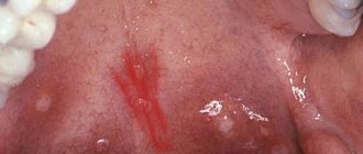
Cheek cancer in the initial stages of the tumor process is asymptomatic. Any ulcer that is not prone to rapid healing and any area of hyperkeratosis should be regarded as an early stage of cancer. In the early stages of cheek malignancy, there is little or no pain. As the size of the cancer tumor increases, the following symptoms appear:
- Pain;
- Induration and infiltration of underlying tissues;
- Enlargement of regional lymph nodes, including damage to the lymph nodes of the submandibular triangle, jugular-digastric group and deep cervical lymph nodes.
What does cheek cancer look like? Any non-healing oral mucosal ulcer should be assessed as a potentially malignant tumor and consultation with an oncologist should be sought.
Why do my gums or cheeks swell?
In most cases, swelling of the gums or cheeks is caused by purely dental reasons:
- Flux
During gumboil, the gums become swollen and painful, and the temperature rises. The development of a tumor begins with an ordinary carious cavity. It gets infected, which eventually leads to tooth decay. Pus accumulates and begins to look for a way out. The cheek swells greatly and a white spot appears on its surface. Without treatment, a fistula forms in this place, and the cheek turns into a huge purulent wound. If you do not seek help from a doctor in time, blood poisoning may occur.
- Removal of a tooth
This operation inevitably involves damage to soft tissue, and swelling is a natural phenomenon. After a while it subsides. If this does not happen, and the gums swell more and more, then you need to urgently visit a doctor. Causes of severe swelling can be:
- a large accumulation of pus,
- incipient periostitis (inflammation of the periosteum),
- infection in the wound.
If you notice swelling of the cheeks and gums, or tissues of the oral cavity, then you need to consult a dentist.
The doctor will determine the exact cause, begin treatment or refer you to another specialist.
Leave your phone number. The clinic administrator will call you back.
By leaving a request on the site, you consent to the processing of personal data
Make an appointment
Initial consultation with a dentist
For free!
- Inflammatory infiltrate
It usually occurs against the background of pulpitis or periodontitis. Pus begins to accumulate in the soft tissues, which causes the appearance of phlegmons and abscesses. It is considered a very dangerous disease because it can lead to big troubles.
- Periodontal disease
Swelling is accompanied by aching or sharp pain. There is no conservative treatment; surgical intervention is necessary. Often it is necessary to remove some teeth and install dentures.
- Teeth chips
The sharp edges of a destroyed tooth begin to scratch the inner walls of the cheek, which leads to inflammation and severe swelling.
- Wisdom tooth growth, pericoronitis
The eruption of the figure eight often leads to inflammation of the gingival hood. Swelling, as a rule, extends to both the cheek and gum.
- Carious teeth
Advanced caries often causes not only the gums to swell, but also the tonsils and cheeks. After eliminating the cause, the swelling gradually goes away. Folk remedies are useless here; urgent medical help is needed.
- Cyst
Depending on the type of cyst, its shape, and location, the doctor decides on conservative or surgical treatment. In some cases, a consultation with an oncologist may be required.
- Gum disease
Swelling is one of the main signs of inflammation of periodontal tissue. Without treatment, the disease quickly becomes chronic and difficult to treat. There is a high chance of losing a tooth.
In addition to the reasons listed, swelling of the gums and cheeks can be caused by:
- inflammation of the facial nerves;
- pathologies of the maxillofacial skeleton;
- allergies;
- disruption of the immune system;
- inflammation of the submandibular and parotid lymph nodes as a result of an infectious disease;
- malignant neoplasm;
- facial injury;
- diseases of internal organs, such as the heart;
- eye diseases;
- blood pathologies.
Diagnosis of cheek cancer
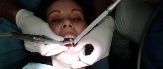
If a tumor of the mucous membrane of the cheek is suspected, oncologists at the Yusupov Hospital conduct an examination using a mirror, palpation of the tumor and lymph nodes. If a long-term non-healing ulcer is detected, a biopsy is performed. If a negative result of histological examination of the material obtained during the biopsy is obtained, the suspicion of the malignant nature of the tumor remains, the biopsy is performed again.
Oncologists clarify the stage of the tumor process, assess the extent of spread of the malignant tumor from the buccal mucosa to adjacent tissues, and find out whether there are metastases to regional lymph nodes and distant organs. If the presence of metastases is suspected, computer and magnetic resonance imaging, ultrasound, and scintigraphy are used. Distant metastases are found in 20% of patients at the time of diagnosis.
Computed tomography and magnetic resonance imaging make it possible to assess the condition of the deeper anatomical structures of the oropharynx and surrounding tissues. If there is a suspicion of metastases to the lymph nodes or tumor infiltration of the floor of the mouth, a cytological examination of the aspirate obtained under ultrasound guidance is performed. To exclude distant metastases, a chest x-ray in two projections and an ultrasound examination of the abdominal organs are done.
Considering that the prognosis for cheek cancer is serious, a tomography of the neck, chest and upper abdominal cavity is performed. Using bone scintigraphy, I exclude bone metastases. Positron emission tomography allows one to identify the source of metastases in cases of undetected primary tumors.
Causes
It is possible to determine what a lump on the mucous membrane is after determining the cause of its occurrence. This growth often occurs as a result of injury. The tissues inside the oral cavity are soft, so any mechanical impact often leads to the appearance of tumors. The lump appeared as a reaction of the body, which in this way protects structural elements from damage.
A lump on the cheek indicates the development of atheroma papilloma, mucocele or cancer of the salivary glands. Symptoms depend on the ongoing pathological process.
Diagnosis includes external examination, computed tomography and magnetic resonance imaging, tissue biopsy if a malignant tumor is suspected.
Treatment for each case is individual, selected taking into account the type of compaction.
Another cause of growths on the mucous membrane is hormonal imbalance. In this case, the lump should be understood as a granuloma. It also occurs due to wearing improper dental appliances or having an incorrect bite.
The mucous cyst should resolve on its own within a few weeks.
Doctors recommend avoiding biting the inside of your cheek. If the cyst has grown so large that it interferes with speaking and eating, you should consult a dental surgeon. The specialist will carefully puncture the mucocele with a sterile needle.
Dentist
Novikova Olga Alexandrovna
8 years of experience
Due to the abnormal structure of the jaw, the sharp edges of the teeth constantly injure the tissues of the mucous membrane, which leads to the slow formation of growths on its surface.
Granulomas grow quickly. They reach a diameter of two centimeters in about one week.
A lump on the inside of the cheek may be a so-called wen, or lipoma. This is a harmless neoplasm. It occurs when a large number of fat cells accumulate in a particular area, caused by metabolic disorders.
This neoplasm can be distinguished from other growths by palpation. When pressed, the fatty tissues begin to move.
Other growths that appear on the oral mucosa are classified into several types.
Papilloma
The papilloma has a stalk on which the growth is attached. Basically, several similar neoplasms appear on the mucous membrane. Moreover, they affect not only the inside of the cheek, but also the tongue. In rare cases, papilloma is formed in a single copy.
Atheroma
Atheroma has a spherical shape. It occurs due to blockage of the sebaceous glands. Atheroma is a cyst filled with secretion. To the touch, this neoplasm resembles a small ball.
Mucocele
A mucocele is a mucous cyst. The neoplasm is formed against the background of regular damage to individual tissues of the inner part of the cheek or due to damage to the salivary gland. Mucocele is most often diagnosed in young patients.
Salivary gland cancer
The cancerous tumor reaches a large size, resulting in deformation of the facial part of the head. With such a neoplasm, the clinical picture is characterized by a variety of symptoms.
Treatment for cheek cancer
Malignant tumors are successfully cured using radical radiation therapy while preserving the function of the oral cavity. Radiologists implant radiation sources because it is possible to irradiate a small volume of tissue at a high dose. Radioactive isotopes of cesium, gold, radium, iridium, tantalum, which have the same efficiency, are used as sources.
For small tumors, the size of which does not exceed 1 cm, they are limited to implantation of a radiation source, without resorting to additional external influence. In cases of slightly larger tumors that are not suitable in size for implantation, external irradiation is used along with source implantation.
Traditionally, large cheek tumors are treated with external beam radiation. Recently, oncologists have been using a combination of radiation and chemotherapy. Mobile lymph nodes are radically excised. During prophylactic removal of lymph nodes without signs of damage, in a significant number of cases, foci of micrometastasis are found in them. For this reason, radiologists prefer to perform prophylactic irradiation of the neck in patients without evidence of lymph node involvement by the external beam, sometimes in combination with surgical treatment.
After surgery for cheek cancer, a cosmetic defect is formed. The results of surgical intervention are improved using the technique of microvascular free skin grafting. If a patient develops dry mouth after radiation therapy, he is prescribed oral pilocarpine. The drug increases salivation, which is usually accompanied by minor side effects - sweating and increased urination. For primary or secondary treatment of tumors of the buccal mucosa, patients are prescribed chemotherapy drugs.
Early diagnosis of a cheek tumor allows for effective treatment. If the tumor process is at a late stage, the prognosis worsens. If you experience unpleasant sensations in the oral cavity or identify ulcers of the buccal mucosa, undergo an examination at the clinic by calling the Yusupov Hospital.
Diagnostic features
Due to the fact that almost all growths that appear on the inside of the cheek do not cause discomfort, diagnostics are necessary to determine the appropriate treatment regimen. It involves the following activities:
- collecting information about the patient’s condition, previous injuries and current illnesses;
- external examination of the growth;
- CT and MRI, if external examination did not allow diagnosing a neoplasm;
- biopsy for suspected cancer.
If a growth on the inside of the cheek does not go away on its own after appearing within a few days, it is recommended to undergo examination by a specialist.