Lichen planus
The main morphological element of the lesion is a keratinized papule of round or polygonal shape up to 2 mm in diameter. On the skin, papules are usually flat, with a waxy sheen, and have a pinkish or bluish-red color. On the oral mucosa, due to keratinization of the epithelium and constant maceration, they acquire a whitish-gray color, standing out against the background of normal or hyperemic mucous membrane. A characteristic feature of lichen planus is the tendency of papules to merge in the form of a pattern resembling a lace mesh, snowflakes, tree-like branches, sometimes rings, stripes. Papules slightly rise above the level of the mucous membrane, giving it some roughness. On the dorsum and lateral surface of the tongue, papules, merging, often form hyperkeratic plaques of various sizes, reminiscent of leukoplakia; the papillae in this area are smoothed.
In smokers, the papules are more pronounced and larger, sometimes overlaid with leukoplakia spots. On the red border of the lips, papules can merge, forming a whitish stripe, in some cases taking on a star-shaped shape. Lichen planus on the red border and mucous membrane of the lips sometimes leads to glandular cheilitis. The most typical localization of lichen planus is on the mucous membrane of the cheeks at the junction of the large molars with the capture of transitional folds, on the lateral surfaces of the tongue and the back with a transition to the lower surface in the area of the large molars. Less commonly affected are the lips, gums, palate, and floor of the mouth.
Due to the variety of clinical manifestations of lichen planus in the oral cavity, the following forms are distinguished: typical (simple), exudative-hyperemic, erosive-ulcerative, bullous, hyperkeratotic.
The typical form is more common than others. Whitish-pearly papules are located separately or in the form of patterns, lace, fern leaves, rings, stripes on the apparently unchanged oral mucosa. With such a typical picture of lichen planus, subjective sensations are minimally expressed and can be manifested by a feeling of burning, tightness, roughness, and dryness of the oral mucosa. Quite often, the disease is asymptomatic and can be detected accidentally during a dental examination.
The exudative-hyperemic form is less common. Papules are located on the hyperemic, edematous mucous membrane. This form is accompanied by more pronounced pain:
burning, pain, aggravated by eating spicy food or talking. Against the background of an inflamed, hyperemic mucous membrane, the pattern of papules may lose the clarity of their outlines and even partially disappear, but in the process of reverse development, when the swelling and hyperemia of the mucous membrane decrease, the pattern of papules reappears.
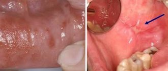
The erosive-ulcerative form is the most severe of all forms. It can occur as a complication of typical or exudative-hyperemic forms as a result of erosion of the area of the affected mucous membrane by various traumatic factors (sharp edges of teeth, dentures, galvanism, etc.). In this form, on the hyperemic and edematous mucous membrane of the mouth there are erosions, sometimes ulcers, around which, against a background of pronounced inflammation, papules typical of lichen planus are arranged in a pattern. Erosions or ulcers of irregular shape are covered with fibrinous plaque, after removal of which bleeding easily occurs. They can be single, small, slightly painful, but they can also be multiple with pronounced pain. Such erosions and ulcers last a long time, sometimes for months, even years, without epithelialization. Often, under the influence of treatment, they partially or completely epithelialize, but soon recur again in the same or another area of the mucous membrane, sometimes even immediately after cessation of treatment. Sometimes, at the site of long-term erosions and ulcers, areas of mucosal atrophy appear. In some cases, long-term erosions and ulcers can become malignant.
The bullous form is rare. It is characterized, along with typical rashes of whitish pearly papules, by the appearance of blisters with a diameter of 2-3 mm to 1 - 1.5 cm with a dense cover.
The blisters can have serous or hemorrhagic contents and open quite quickly. Their lifespan ranges from several hours to 2 days. The erosions that form at the site of the blisters quickly epithelialize, which distinguishes the bullous form of lichen planus from the erosive and ulcerative form. The duration of the course of the bullous form can be different; sometimes blisters can appear over many months and form on the oral mucosa simultaneously with papules or join them later. In some cases, blisters precede the appearance of papules, which creates difficulties in diagnosing lichen planus.
The hyperkeratotic form is observed very rarely. It is characterized by the presence of various shapes and outlines of hyperkeratotic plaques, rising above the level of the mucous membrane with sharp boundaries. Around the foci of hyperkeratosis there are papular rashes, typical of lichen planus. Most often, this form is localized on the mucous membrane of the cheeks and the back of the tongue.
Existing forms of lichen planus can transform into one another. Thus, as a result of complications, the typical form can turn into exudative-hyperemic and (or) erosive-ulcerative. The transformation process is determined by the influence of general (somatic diseases) and local factors. The presence of sharp edges of teeth and dentures, dissimilar metals, dentoalveolar anomalies and deformations, periodontitis, and tartar provoke the transformation of lichen planus from a typical form to a more severe one.
Lichen planus is a chronic disease characterized by a persistent long-term course. It can last for decades with alternating exacerbations and remissions, the duration of which is influenced by the course of common somatic diseases and the effect of local traumatic factors in the oral cavity.
Lichen planus on the oral mucosa can become malignant, most often in elderly people who have suffered from erosive-ulcerative or hyperkeratotic forms for a long time. Signs of malignancy of lichen planus are the formation of a compaction at the base of the lesion, increased keratinization processes, and the appearance of vegetation on the surface of long-term non-healing and untreatable erosions and ulcers.
Pathohistology .
Pathohistological changes in the epithelium are characterized by the presence of hyper and parakeratosis, as well as acanthosis. In some cases, the formation of a granular layer is detected. In the stroma, edema is detected, a diffuse inflammatory infiltrate (mainly from lymphocytes and plasma cells), the cells of which penetrate into the epithelium (exocytosis) and through the basement membrane, as a result of which it has unclear contours. The infiltrate almost never penetrates into the deeper layers and the lamina propria of the mucous membrane. It comes close to the epithelium, as if supporting it from below. Each form of the disease has its own characteristics. In case of erosive and ulcerative lesions, in addition to the phenomena of hyperkeratosis and parakeratosis, ulceration and destruction of the epithelium, hemorrhages in the mucosal layer proper are diagnosed. The infiltrate is detected directly under the basement membrane. Bullous lesions develop beneath the epithelium as a result of extensive swelling in the connective tissue. The blisters are located subepithelially, beneath them there is a massive round cell infiltration into the lamina propria of the mucous membrane. Changes in the epithelium characteristic of lichen planus in erosive, ulcerative and bullous forms are found in areas bordering the defect.
Causes of the disease
Lichen planus occurs under the influence of a number of factors, among which the most common is immuno-allergic. When the body's immunity decreases under the influence of external and internal factors, T-lymphocytes cannot cope with their protective function.
Factors that provoke the development of oral lichen planus include:
- Chronic stress;
- Strong shocks;
- Diseases of the gastrointestinal tract;
- Diabetes;
- Hypertension;
- Grynszpan syndrome;
- Injuries to the oral mucosa;
- Incorrectly installed dentures;
- Fillings that injure the oral mucosa;
- Prostheses made of dissimilar materials;
- Long-term use of tetracycline;
The risk group also includes patients whose work involves developing camera film or who come into contact with paraphenyldiamine.
Treatment
For different forms of the disease, a different course of medications is prescribed. If the disease is asymptomatic, drug treatment may not be necessary. If erosions and ulcers have already formed, it is necessary to begin treatment as quickly as possible to avoid worsening the condition.
Inflammatory processes are controlled by the use of corticosteroids. Local treatment is carried out to relieve pain and accelerate tissue regeneration. To avoid the development of candidiasis, antifungal drugs are prescribed.
Electrophoresis and phonophoresis are also effective. In rare cases, when erosions do not heal for a long time, surgical intervention is indicated.
In all cases, without exception, the oral cavity is sanitized and the source of inflammation is removed (for example, periodontitis, caries or pulpitis).
Signs on the skin
Lichen planus is characterized by a homogeneous rash consisting of small shiny reddish nodules with a purple tint with a slight depression in the middle. In the case of larger rashes, a mesh pattern of the rash becomes noticeable. The nodules can occur individually, can be arranged linearly, or can form plaques or rings. The rash with lichen planus is sometimes accompanied by severe itching; after the papules disappear, hyperpigmentation of the affected areas occurs. Favorite habitats of lichen planus are the flexor surfaces of joints, forearms, anterior surfaces of the legs, and the sacral area.
Symptoms of the disease
There are six types of oral lichen planus. This is a typical, exudative-hyperemic, hyperkeratotic, erosive-ulcerative, bullous and atypical form. They are united by the formation of foci of inflammation on the oral mucosa, the appearance of defects and erosions. In some particularly severe forms of the disease, the patient may complain of itching and burning sensation in the mouth and difficulty eating. In milder forms, the disease may not manifest itself at all.
Typical lichen planus is the most common form, occurring in half of patients. White nodules appear on the mucous membrane of the oral cavity. Merging, they resemble lace.
The esscudative-hyperemic form is characterized by the appearance of gray papillomas on the edematous mucous membrane.
The hyperkeratotic form is one of the least common forms, which is characterized by the formation of gray spots that rise above the affected surface. Over time, these plaque spots become rougher and flake off.
Erosive-ulcerative is the most severe form of the disease, occurring in 23% of cases. Multiple erosions and ulcers covered with fibrinous plaque appear on the lining of the oral cavity. There is a strong burning sensation in the mouth, and when trying to remove plaque, bleeding and pain occur.
The bullous form is a relatively rare variety, occurring in 3% of cases. Accompanied by the appearance of dense blisters, inside of which there is hemorrhagic exudate. After the bubbles open, erosions form.
Atypical form - occurs slightly more often than bullous - in 4% of cases. This form is characterized by dilation of the mouth of the salivary gland, as well as inflammation of the lips and gums.
Quite often, one patient can develop several forms of lichen planus in parallel. Often one form can flow into another. Ringworm is a chronic disease prone to relapse.