MOUTH, ORAL CAVITY
[
mouth, oral cavity
;
os (oris)
(PNA), os (JNA, BNA),
cavitas oris
(PNA)] - the initial section of the digestive tract, consisting of the oral opening and the oral cavity. The oral opening is limited by the upper and lower lips (see). When the lips are closed, the mouth opening has the shape of a mouth slit (rima oris); when the lips are open, it has a rounded shape. The size of the oral fissure varies and averages 6-8 cm in adults. In men, the oral fissure is usually larger than in women.
In the oral cavity, mechanical grinding of food occurs and its chemical processing begins, preparing the food for further digestion into the gastrointestinal tract. tract. In addition, the oral cavity takes part in breathing, as well as in the formation of speech and singing sounds.
Embryology
During embryogenesis, the formation of the oral cavity is closely related to the development of the face (see). At the head end of the embryo, an invagination of the ectoderm is formed, which grows towards the blind end of the foregut. The so-called the oral fossa, or bay, which is the rudiment of the primary oral cavity, as well as the nasal cavity. During the 6-8th week. During embryonic development, the primary oral cavity is divided into the final oral cavity and the nasal cavity, which is associated with the formation of the hard and soft palate. The formation of the vestibule of the mouth is closely related to the development of the lips and cheeks. At approximately the 7th week of embryonic development, epithelium grows along the upper and lower edges of the primary oral fissure, followed by its immersion into the underlying mesenchyme in the form of an arcuate plate. Along the course of the plate, a gap soon appears; the edges separate the rudiments of the upper and lower jaws from the rudiments of the lips. Initially, the oral opening of the embryo is very wide and reaches the rudiments of the outer ear. Its reduction in size occurs due to the fusion of the edges of the oral fissure and the formation of the cheeks.
Anatomy and histology
The oral cavity is limited in front and on the sides by the lips and cheeks; its upper wall is the hard and soft palate (see), and the lower wall is the floor of the oral cavity. The basis of the floor of the mouth is the diaphragm of the mouth, the edges of which consist of the paired mylohyoid muscle (m. mylohyoideus). Above it are the geniohyoid muscles (mm. geniohyoidei), as well as the muscles of the tongue (see). At the back, the oral cavity is connected to the pharyngeal cavity through the pharynx (see).
Rice. 1. Median sagittal section of the head at the level of the oral cavity and pharynx. Rice. 2. Oral cavity, front view (the corners of the mouth are cropped). Rice. 3. Frontal cut of the head at the level of the molars, 1 - hard palate; 2 - teeth; 3 - upper lip; 4 - oral fissure; 5 - lower lip; 6 - vestibule of the mouth; 7 - lower jaw; 8 - sublingual gland; 9 - genioglossus muscle; 10 - geniohyoid muscle; 11 - mylohyoid muscle; 12 - hyoid bone; 13 - pharynx; 14 - tongue; 15—soft palate; 16—the oral cavity itself; 17—frenulum of the upper lip; 18— gums; 19— palatoglossal arch; 20—palatine tonsil; 21—uvula; 22—frenulum of the lower lip; 23—velopharyngeal arch; 24 - transverse palatal folds; 25 - anterior belly of the digastric muscle; 26 - buccal muscle; 27 - fatty body of the cheek.
Anatomically, the oral cavity is divided into the anterior section, or vestibule of the mouth (vestibulum oris), and the posterior section, or the oral cavity itself (cavitas oris propria). The vestibule of the mouth looks like a gap enclosed between the lips and cheeks (in front and outside) and the teeth and gums (back and inside). With the help of interdental spaces and retrodental spaces, the vestibule of the mouth communicates with the oral cavity itself (color. Fig. 1 - 3).
The oral cavity itself is separated from the vestibule of the mouth by teeth and gums (see). When the teeth are closed, it looks like a gap; when the mouth is opened, the oral cavity takes on an irregular ovoid shape. There are individual and age differences in the shape and size of the oral cavity: in brachycephals it is wider and shorter than in dolichocephals. In newborns and children up to 3 months, the oral cavity is very small, short and low. In the oral cavity there are teeth (see), tongue (see), the excretory ducts of the large and small salivary glands open into it (see).
Both the vestibule of the mouth and the oral cavity itself are lined with a mucous membrane that is resistant to various mechanical, chemical and thermal irritants, has high regenerative capacity and is relatively resistant to infection.
Rice. 10-15. Microscopic specimens of the oral mucosa are normal. Rice. 10. Buccal mucosa: glycogen (indicated by an arrow) in the superficial layers of non-keratinizing stratified squamous epithelium, detected using the PAS reaction, cells of the basal layer do not contain glycogen; x 200. Fig. 11. Non-keratinizing stratified squamous epithelium of the buccal mucosa; coloring with azan; x 400. Fig. 12. The mucous membrane of the hard palate with underlying adipose tissue, covered with keratinizing stratified squamous epithelium: the stratum corneum is intensely red in color; coloring with azan; X 140. Fig. 13. Keratinizing stratified squamous epithelium of the mucous membrane of the hard palate: the stratum corneum is intensely red; coloring with azan; x 400. Fig. 14. Gingival mucosa, covered with keratinizing stratified squamous epithelium (the stratum corneum is indicated by an arrow); hematoxylin-eosin staining; x 200. Fig. 15. The mucous membrane of the red border (transitional section) of the lip, covered with keratinizing multilayered squamous epithelium: the stratum corneum is purple; aldehyde-fuchsin staining; x 140.
Throughout its entire length, the oral mucosa is covered with stratified squamous epithelium. The epithelium covering the mucous membrane of different parts of the oral cavity differs. In the area of the cheeks, lips, soft palate, and floor of the mouth, the epithelium under normal conditions does not keratinize. A characteristic feature of the non-keratinizing epithelium of the human oral cavity is its ability to accumulate large amounts of glycogen in the cytoplasm (color Fig. 10 and 11). In the area of the hard palate and gums, the epithelium shows a pronounced tendency to become keratinized. In these areas, on top of the layer of spinous cells there is a granular layer consisting of elongated cells, which contain keratohyalin grains in their cytoplasm. Above, the granular layer passes into the stratum corneum, consisting of completely keratinized and nucleated cells. In the keratinizing epithelium, glycogen is usually absent (color fig. 12-15).
The epithelium of the oral mucosa has a high level of activity of enzyme systems, including enzymes of the tricarboxylic acid cycle (see Tricarboxylic acid cycle), glycosyltransferases.
The lamina propria of the oral mucosa, on which lies a layer of epithelium, forms numerous projections, or papillae, protruding into the layer of epithelium. The distribution of cellular elements (fibroblasts, mast cells, plasma cells, segmented leukocytes) in different parts of the oral mucosa is uneven: the lamina propria of the mucous membrane of the cheeks and lips is richest in cells. Fibrous structures are represented by bundles of interwoven collagen fibers, between which are located argentophilic and elastic fibers. The largest number of elastic fibers is observed in the lamina propria of the mucous membrane of the cheek and palate.
The lamina propria of the oral mucosa, without a sharp boundary, passes into the submucosal layer (submucosa, T.), which is especially well developed at the bottom of the oral cavity. The submucosal layer contains numerous small salivary glands. The muscular plate of the mucous membrane characteristic of the digestive tract is absent here. In the gum area, in the lateral parts of the hard palate and in the area of the palate suture, the submucosal layer is completely absent. In these areas, the mucous membrane is tightly connected to the periosteum of the corresponding bones.
Blood supply, lymphatic drainage and innervation of the walls of the oral cavity are closely related to the vascular and nervous systems of its constituent formations (see Throat, Teeth, Palate, Jaws, Tongue).
Blood supply and innervation of the maxillofacial area
The blood supply to the brain and cervical spine is provided by the common carotid arteries. The common carotid artery, as a rule, does not form branches. The blood supply goes through paired terminal branches: the internal and external carotid arteries. The bottom is penetrated by blood vessels filled from the external carotid artery. The blood supply to the teeth occurs thanks to the maxillary artery.
All oral organs have nerve endings: 12 paired and 5 nerves connected to the cerebral cortex. The hypoglossal, lingual and mylohyoid nerves approach the floor of the mouth. The innervation of the teeth, masticatory muscles, skin and forebrain is created by the ternary nerve. The innervation of part of the facial muscles is carried out by the facial nerve. The innervation of part of the tongue, pharynx and parotid gland is created by the glossopharyngeal nerve. The vagus nerve is connected to the palate.
INTERESTING: innervation of teeth and blood supply to jaws
Microflora of the oral cavity
Over 100 different types of microbes have been found in the oral cavity. The oral microflora includes aerobic and anaerobic bacteria, yeast-like fungi, mycoplasmas, and protozoa. According to S. Neychev (1977), the concentration of aerobic bacteria in 1 ml of saliva is 107, anaerobic - 108.
Among the permanent flora of the oral cavity, streptococci, veillonella, lactic acid bacteria, and actinomycetes predominate. In addition, the permanent flora includes saprophytic Neisseria and diphtheroids. bacteroides, fusobacteria, leptotrichia, spirochetes, etc.
Non-permanent, or random, microflora includes gram-negative bacteria, including Escherichia, Klebsiella, Pseudomonas, Proteus, and Clostridia. The detection of these microorganisms in the oral cavity indicates dysbacteriosis (see).
The microbial flora of the oral cavity, as well as the normal flora of other body cavities (see Human microflora), is a consequence of mutual adaptation of the body and microbes. Despite the known constancy, there are fluctuations in the quantity and composition of the microbial flora associated with hygienic skills, age, dental condition and other factors. It should also be noted that microorganisms are distributed unevenly in the oral cavity. Most bacteria are found on the root of the tongue, on the surface of the gingival margin and in dental plaque (plaque). According to V.G. Petrovskaya and O. Marko (1976), there is a certain specificity in the distribution of flora in the oral cavity, so. for example, Streptococcus salivarius is more often found on the mucous membrane of the tongue, Str. mitis - on the mucous membrane of the cheeks and on the surface of the teeth, Str. sanguis and Str. mutans is isolated primarily from dental plaque (see Teeth, dental biochemistry).
The microflora of the oral cavity performs a number of physiological functions. In a healthy body, due to its antagonistic properties, microflora acts as a “biological barrier”, preventing the proliferation of random microorganisms, including pathogenic ones, entering the oral cavity from the environment. The beneficial value of oral microflora is also associated with its participation in the decomposition of organic substances (food residues), i.e., in the self-cleaning of the oral cavity. In addition, the oral microflora is a constant stimulator of local immunity.
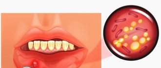
A decrease in the resistance of the oral mucosa and a change in the body's reactivity (see), caused by various factors, can lead to a persistent change in the composition and properties of the oral flora, or dysbacteriosis (see). The altered microflora loses its protective functions and often becomes a source of autoinfection (see). Disruption of microbial balance under the influence of certain therapeutic effects (irradiation, antibiotics, immunosuppressants, dental prosthetics, etc.) can lead to diseases of the mucous membrane such as stomatitis (see), glossitis (see), gingivitis (see) , which are more often of a fungal nature. The generalization of the process is possible - visceral candidiasis (see Candidiasis).
The microflora of the oral cavity is important in the development of dental caries (see Dental caries), periodontal diseases (see). In caries, the most significant role is given to acid-forming microorganisms (streptococci, lactobacilli, actinomycetes) that form dental plaque. In the development of periodontal disease, the most important is given to gram-negative anaerobic bacteria (bacteroides, fusobacteria, spirochetes, veilonella, etc.). It is believed that the endotoxins produced by this flora, which have antigenic activity, stimulate immune responses that support hron. inflammation in periodontal tissues. For example, in the pathogenesis of such pathol. processes such as pulpitis (see), periodontitis (see), often developing as a complication of dental caries, sensitization of the body by microbial metabolic products plays an important role. Chronic inflammatory processes in the oral cavity cause allergic changes in the body and can contribute to the development of foci of infection, sometimes developing into sepsis (see).
The vital activity of microorganisms in the oral cavity is largely determined by the state of local protective factors. Some of them are not directly directed against microorganisms, but have a negative effect on their development. Such nonspecific resistance factors are: pH of saliva, bacteriostatic properties of the secretion of the salivary glands, tissue metabolic products, regular desquamation of the epithelium in the oral cavity, lysozyme (see), etc. Specific factors of protection of the oral mucosa - immune mechanisms directed directly against microorganisms - represented by humoral and cellular immunity. In a healthy body with an intact oral cavity, protective factors prevent the excessive proliferation of microbes, keeping them in certain quantitative proportions.
Oral cavity examination methods
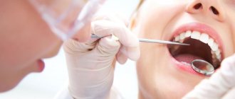
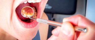
Methods for examining the oral cavity come down, first of all, to a thorough examination using directional (preferably shadowless) lighting and special instruments (see Dental Instruments) - a spatula, wide hooks for retracting the cheeks, lips, tongue and stomatol. mirrors for inspecting hard-to-reach areas. Sometimes, to identify luminescent compounds in the oral mucosa (see Luminescence), an inspection of the oral cavity is performed under UV light. During the examination, pay attention to the presence of bad breath. With the help of palpation, the mobility, density, consistency and soreness of various parts of the mucous membrane and patol are determined. formations.
To study the organs surrounding the oral cavity, various X-ray diagnostic methods are used (see). Research methods such as ultrasonic echolocation (see Ultrasound diagnostics) and thermography (see) are also used. According to indications, Citol is produced. examination of swabs and prints from pathologically changed areas of the mucous membrane (see Cytological examination), as well as the study of the microflora of the oral cavity. According to strict indications, a biopsy is performed (see).
In some cases, there is a need to conduct immunological and biochemical studies, as well as to determine various types of sensitivity: tactile, pain, temperature, taste (see Esthesiometry).
See also Patient examination, dental examination.
What parents should pay attention to and be wary of
- Breathing through the mouth if the child does not have a runny nose.
- No gaps between teeth by 4-5 years.
Baby teeth are small and there is usually always room for them, even if the jaw has not developed enough. As the child grows up and prepares for a mixed dentition, so-called trema should appear between the baby teeth - small but distinct distances. When baby teeth fall out, permanent teeth will erupt, which are much wider than baby teeth, and they should have enough space for normal growth and the formation of an even, beautiful dentition.
- Observe the child’s swallowing of saliva; normally, swallowing occurs imperceptibly, without tension in the facial muscles.
Usually, during an appointment, a pediatric dentist draws the attention of parents to the emerging problems of forming a correct bite and refers such children to an orthodontist.
If this does not happen, it is necessary to show the child at the age of 4-5 years to a pediatric orthodontist, preferably one who takes into account the functional approach in his work.
Pathology
Pathologies of the oral cavity include malformations, diseases of the oral mucosa, innervation disorders, diseases of the organs surrounding the oral cavity, and tumors.
Developmental defects
may refer to congenital defects of the lips (see Lips), palate (see), jaws (see), tongue (see); Congenital facial clefts are rare (see).
Diseases of the oral mucosa
Diseases of the oral cavity include, first of all, lesions of its mucous membrane, characterized by a variety of morphols. violations and wedge, manifestations, which often presents serious difficulties in differential diagnosis. There are several main groups of diseases of the oral mucosa.
Traumatic damage to the oral mucosa can be caused by mechanical, chemical, thermal, and radiation factors. The severity and duration of the course depend on the size and depth of the lesion, however, wounds and damage to the mucous membrane heal faster and are less likely to be accompanied by complications than similar skin damage. Long-term exposure to irritating factors can lead to the formation of traumatic erosions, hron. ulcerations, decubital ulcers. The cause of mechanical trauma can be the crowns of incorrectly erupted or displaced teeth, sharp edges of carious cavities, incorrectly applied fillings and artificial crowns, uneven edges of dentures, their clasps, tartar deposited on the surface of the teeth (see). Irritation and damage to the mucous membrane can occur as a result of ingestion of excessively hot, hot, spicy food, strong alcoholic drinks, especially often smoking, as well as as a result of certain traditional bad habits: chewing tobacco, betel leaves, etc. Often the effect of chronic irritants and traumatic factors leads to disruption of the process of keratinization of the epithelium of the mucous membrane, hyperkeratosis (see), leukoplakia (see).
Inflammatory diseases of the oral mucosa - stomatitis (see) are distinguished by the location of the lesion, etiology, morphol. changes and wedge, flow. According to localization, inflammation of the mucous membrane of the lips, their red border is distinguished - cheilitis (see), tongue - glossitis (see), gums - gingivitis (see).
Allergic reactions (see Allergy) are a relatively common cause of damage to the oral mucosa. Some of them are classified as infectious-allergic, for example, chronic recurrent aphthous stomatitis; others are caused by chemicals, especially drugs, or are local manifestations of general allergic reactions.
The oral mucosa can react to various patols. processes and functional disorders in many body systems. Its characteristic changes are sometimes the earliest symptoms of diseases of the digestive, excretory, circulatory systems, blood diseases, as well as hypovitaminosis and many inf. diseases. Characteristic changes in the oral cavity are accompanied by certain dermatoses (see). Changes in the oral mucosa are important for diagnosing professional and household chronic intoxication with various chemicals, for example, heavy metals (See Mercury, Lead).
Tumors of the oral cavity can develop both from the mucous membrane and spread from deeper tissue structures and organs. Of the benign tumors, the most common are papillomas (see Papilloma, papillomatosis), fibromas (see Fibroma), cystic formations of small salivary glands located in the thickness of the walls of the oral cavity, the so-called. mixed tumors of the glands (see Mixed tumors). Vascular tumors - hemangiomas (see Hemangioma), much less often lymphangiomas (see Lymphangioma) can be localized in different parts of the oral cavity.
Among malignant tumors, cancer is of great importance in the pathology of the oral cavity. Cancer of the oral cavity, including cancer of the lips (see Lips) and tongue (see), accounts for about 10% of all cancers. It is generally accepted that cancerous lesions often develop in areas of the mucous membrane that have chronic. damage, ulceration, cracks, as well as in areas affected by hyperkeratosis, leukoplakia, and certain other so-called. precancerous diseases (see Precancerous diseases). Timely detection and elimination of precancerous diseases of the oral cavity is the most important part of cancer prevention.
Disturbances in the innervation of certain areas of the oral cavity can manifest themselves in the form of loss of sensitivity (analgesia), the occurrence of distorted and unpleasant sensations (paresthesia) and various pain syndromes associated with neuritis or neuralgia of individual branches or branches of the nerves involved in the innervation of the oral cavity and its organs. One of the typical and relatively common types of such a disorder is glossalgia (see), manifested in the form of pain or a burning sensation in the tongue.
Diseases of other organs related to the oral cavity
Along with diseases of the mucous membrane, one of the most common types of oral pathology are dental diseases: caries (see Dental caries), pulpitis (see), periodontitis (see), periodontal disease (see), as well as dental anomalies (see .), dentition, bite (see).
Severe forms of odontogenic inflammatory processes are periostitis and osteomyelitis of the jaws (see), abscesses (see Abscess) and phlegmon of the surrounding soft tissues (see Phlegmon), in particular phlegmon of the floor of the mouth (diffused purulent inflammation of the tissue of the intermuscular and interfascial spaces between the body of the mandible and the hyoid bone), as well as Ludwig's angina (see Ludwig's angina). Treatment of phlegmon consists of opening through wide incisions all possible areas of accumulation of pus and draining them in combination with intensive measures of general anti-inflammatory therapy.
Among diseases of other organs associated with the oral cavity, it should be noted diseases of the salivary glands that affect the condition of the oral mucosa and disrupt the functions of the oral cavity (see Xerostomia, Sialadenitis, Sialolithiasis).
For a number of diseases of the oral mucosa of an inflammatory or necrotic nature (ulcerative necrotic stomatitis, gingivitis, etc.), dental diseases (caries, periodontal disease, pulpitis, etc.), chronic. tonsillitis, for diseases of the upper respiratory tract (ozena, decaying tumor), lungs (bronchiectasis), gastrointestinal tract. tract (anacid gastritis, esophageal diverticulum), metabolic disorders (diabetes mellitus, scurvy, etc.), bad breath (foetor ex ore) may be observed, to eliminate which treatment of the underlying disease is necessary.
Operations
Small-scale surgical interventions in the oral cavity are performed, as a rule, by dental surgeons, most often on an outpatient basis. Before any operation in the oral cavity, the oral cavity is sanitized (see).
The most common are tooth extraction operations (see Tooth extraction), as well as interventions associated with diseases of the periodontal tissues and odontogenic processes. They are usually performed under local (in the lower jaw, mainly conduction) anesthesia (see Local anesthesia, maxillofacial area). Opening of gingival abscesses and purulent foci during periostitis is carried out using incisions to the bone followed by drainage.
Incisions for cysts (see Dental cyst), benign neoplasms, and pathologically altered areas of the mucous membrane are made within unchanged tissues, followed by suturing with catgut. Wound suturing for oral injuries is performed tightly. Features of surgical treatment of wounds for wounds, including gunshots, affecting the oral cavity - see Face, Jaws.
More extensive interventions in the oral cavity are performed in an inpatient setting under local anesthesia with premedication or general anesthesia. Plastic surgery is performed in the presence of congenital malformations (cleft lip and palate, “double lip”, shortened frenulum of the upper lip and tongue, etc.), as well as for the consequences of injuries and diseases (scars, defects).
In cases of cicatricial deformities in the area of the corners of the mouth, to eliminate the narrowing of the oral fissure, the so-called. microstomy, the edges of the oral fissure with their end-to-end scar change are dissected and epithelialized, everting the mucous membrane from the cheek (Evdokimov’s method). If the strip of the red border is preserved, it is cut off with a through incision, keeping it in the form of a bridge between the upper and lower lips, and then, after dissecting the scars and tissues, the cheeks are pulled up to the required level, where they are secured with sutures, thus forming a new corner of the mouth (Vasiliev’s method).
It is very rare to resort to plastic surgery for an excessively wide mouth gap, the so-called. macrostomy, resulting from a unilateral transverse cleft of the face of a congenital nature (see Face, malformations).
In the postoperative period, careful hygienic care of the oral cavity is necessary (excessive rinsing of the mouth, rinsing), as well as the administration of liquid or softened food, introducing it through a sippy cup if chewing is impossible.
Anatomical defects of the lingual frenulum
The most common anatomical anomaly of the frenulum of the tongue is its incorrect location - attachment to the lower part of the tongue is not in the middle, but much closer to its tip. Even with a normal anatomical length, the frenulum in this case is considered pathologically short. Often this defect does not cause noticeable inconvenience, unlike ankyloglossia - perhaps the most serious defect of the frenulum of the tongue. It is characterized by a combination of three anatomical pathologies: the area of attachment of the frenulum is shifted from the middle of the tongue to its tip, the length of the frenulum is significantly less than normal, the elasticity and mobility of the tongue are noticeably reduced. The negative impact of ankyloglossia on the dental system is very significant. Thus, babies suffering from it cannot properly latch onto their mother’s breast, and therefore are not sufficiently saturated 2.
In older children, ankyloglossia “inhibits” the development of the lower jaw, causing tooth displacement. In general, this pathology causes malocclusions, difficulties with breathing and swallowing, contributes to the appearance and development of speech defects, and makes it difficult to eat comfortably.
Ankyloglossia has three main varieties, which differ in the presence of certain anatomical features:
- the frenulum is thin, translucent, attached close to the tip of the tongue, hindering its movements. When the tongue rises upward, its tip acquires a characteristic heart-shaped shape;
- the frenulum is short and thick, “powerful”, restricts the movements of the tongue. When the tongue sticks out forward, its back takes on a hilly appearance, and the tip is lowered down;
- the frenulum is greatly shortened, has a whitish color, the fibers of the frenulum are fused with the muscle fibers of the tongue. This pathology is often combined with clefts of the upper lip or palate, the so-called “cleft palate”, “cleft lip”.