Wisdom teeth, or “eights,” erupt at a late age – after 20 years. According to statistics, they are the ones who most often suffer from caries. And all because the teeth are “far away”, it is inconvenient to brush them, and so they begin to deteriorate.
Should I treat or remove a wisdom tooth due to caries? A pressing issue for patients. In 80% of cases, doctors advise immediately getting rid of carious “eights”. After all, filling them takes a long time, is painful and often does not give results.
Clinical picture
Caries of eights is often asymptomatic, especially in the early stages. But when a deep carious “hole” is formed, acute pain appears when exposed to cold, hot, or sweet food. The discomfort goes away immediately after the irritant is removed.
As a rule, conventional instrumental examination is used for diagnosis. When probing the cavity, sensitivity occurs at the enamel-dentin boundary. At the bottom of the “hole”, softened, dead brown dentin accumulates. Tapping on the enamel does not cause pain.
Wisdom tooth to be removed due to caries
In addition to location, dentists classify caries according to the depth of the pathology:
- The spot stage is considered the mildest. This type is easy to clean and saves the tooth from further damage.
- The superficial type of caries is characterized by the process of demineralization of tooth enamel with further destruction.
- Deep caries is characterized by irreversible destructive effects on the tooth due to the development of carious microflora. During the examination of the affected tooth, you can see a layer of dentin located between the pulp and the bottom of the carious cavity.
Based on the types of anatomical location, caries of dentin, enamel and cement is distinguished.
What to do if caries appears on the “eight”
Any type of caries must be treated, otherwise the infection will spread throughout the entire oral cavity, affecting healthy teeth. Therefore, the problem cannot be ignored, even if an unnecessary and invisible wisdom tooth is sick.
Contact your dentist promptly. Before visiting the clinic, rinse your mouth with antiseptic liquids: chlorhexidine, soda-saline solution or chamomile decoction at room temperature.
For severe pain, you can take an analgesic tablet - Analgin, Ketanov, Nurofen, etc.
Stages of treatment
- Drilling out carious tissues with a drill.
- Rinsing the cavity with an antiseptic solution, such as hydrogen peroxide.
- Applying an insulating and therapeutic pad to the bottom.
- Layer-by-layer filling of the cavity with a photopolymer composite.
- Restoration of the anatomical shape and masticatory cusps on the surface of the molar.
The procedure lasts about 30 minutes and is performed under local anesthesia.
Restoration of wisdom teeth
Fissure, interdental, cervical caries of wisdom teeth
If we compare the classification of caries of “eights” with other teeth, no special differences are visible. The types of carious lesions are distinguished depending on a number of factors, for example, location, degree of damage to the tooth by caries, as well as the area of the pathology on the tooth.
Depending on the location of the pathology, the following types are distinguished:
- Fissure caries is the most common. This type of caries is the most difficult to diagnose. However, the hidden danger is that over time the disease spreads to the internal parts of the tooth. The term “fissures” among dentists refers to the grooves visible on the chewing surface of the figure eights. If the furrows are deep, it can be difficult to clean them using traditional methods.
- As the name suggests, a form of interdental caries occurs in the space between the teeth. It is in this area that a large number of bacteria often accumulate. Visually the presence of such caries can be quite difficult to determine. It is for this reason that most patients turn to the dentist already at the stage of pathology transition to the root part of the tooth.
- Cervical caries is characterized by the location of the lesion in the area of contact between the gum and tooth. The main danger of this type is its long-term course without any symptoms, which is why clients often develop the disease. This type of caries can reveal itself by the presence of sharp pain in the lower part of the teeth.
Treat or remove
Treating a wisdom tooth is an extremely difficult task due to its inaccessibility. Some instruments do not even reach the carious lesion. In addition, the patient should sit with his mouth wide open at all times. Therefore, dentists suggest immediately removing the problematic tooth.
Why do wisdom teeth need to be removed in most cases?
- Firstly, the third molar has completely lost its function in the process of evolution and is now considered a vestige. It's not at all a shame to get rid of it.
- Secondly, it is almost impossible to clean a carious lesion efficiently. There is a high risk of developing secondary caries. Is it worth spending money and time on treatment?
The decision is made by the doctor on an individual basis; first you need to take an x-ray and conduct an instrumental examination.
3 indications for removal
- The advanced stage is when the wisdom tooth has crumbled and cannot be restored.
- Absence of an antagonist tooth. The antagonist is the closing molar, which is located on the opposite jaw. If it is missing, the wisdom tooth decays faster due to lack of chewing load.
- Retention or dystopia - if the tooth grows horizontally, at an angle, or remains in a semi-erupted state, then this is a reason for removal.
If, due to anatomical features, a person cannot open his mouth wide or has a pronounced gag reflex, it is also not possible to carry out full therapy.
Of course, if the wisdom tooth has grown correctly, in its place, then it can be treated if desired. It is recommended to leave the “eight” if it is needed to install a prosthesis. It often happens that a patient is missing the seventh and sixth teeth in a row, so the third molar becomes a support for fixing a bridge, clasp prosthesis or other orthopedic structure.
What to do if your wisdom tooth gets sick during pregnancy?
Pregnancy is a contraindication to removal due to the high risk of complications. In this case, the doctor decides: to treat wisdom tooth caries or remove the “eight” later (after the birth of the child). As a rule, it is better to carry out therapy, because any infection in the body, even caries, is harmful to health. And if there is a deep carious “hole”, it is necessary to put a filling.
Wisdom tooth hurts after treatment
Often, patients who have undergone wisdom teeth treatment for caries complain of pain for a long time. Pain syndrome is normal, since figure eights have a more complex structure and have increased sensitivity.
After conservative treatment, pain is considered normal, which decreases in intensity within 5–7 days and then disappears completely. After extraction, due to the persistence of a large wound and traumatic manipulations, pain in the area of the extracted tooth may be felt for 10–14 days. After two weeks, the pain should be eliminated or almost not felt.
If the pain lasts longer or the sensations cause severe agony, you should immediately contact a specialist. Complications may develop that require immediate dental attention.
Wisdom teeth are difficult to treat; due to the lack of their functional significance in the presence of acute processes, dentists do not recommend saving the tooth. To avoid the consequences of caries of eights, experts advise visiting a dental clinic every 6 months for a preventive examination and professional teeth cleaning.
Causes
“Eight” is the outermost tooth in the row; it is difficult to reach it, even with a toothbrush. Therefore, the quality of daily hygiene care deteriorates. Bacterial plaque accumulates on the enamel, containing a large number of cariogenic microbes - Streptococcus mutans.
Bacteria destroy the enamel, starting the carious process. This is why third molars deteriorate more often than other teeth.
There are cases when a wisdom tooth has partially erupted. The gums cover part of the figure eight, which prevents normal cleaning. Food particles and plaque get under the formed gingival hood, which contributes to the process of suppuration. Therefore, even an unerupted tooth is often already sick.
Complications of wisdom tooth caries
How does the removal work?
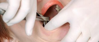
If there are indications for the removal of wisdom teeth with caries, the procedure is performed exclusively by a dental surgeon. Depending on the condition of the tooth and the characteristics of its development, simple or complex extraction can be performed.
Simple removal involves the usual manipulation of tooth extraction using forceps. But such a procedure is only possible if the only pathology is caries.
Most often, third molars require complex removal, which involves dental surgery and surgery. Complex removal is necessary for pathology of the root system, destruction of more than 50% of the tooth surface, dystopia or retention. During the procedure, the dentist may resort to cutting soft tissue, breaking the tooth and removing it in parts and other manipulations.
After removal of wisdom teeth with caries, an x-ray diagnosis is required to exclude remnants of parts of the tooth or root. After this, stitches are applied.
Complication of caries
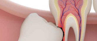
With prolonged caries, the infection penetrates into the deep tissues and affects the dental nerve (pulp). An inflammatory process begins inside the tooth - pulpitis, which is accompanied by spontaneous aching pain. In this case, it is necessary to fill the root canals; this is a difficult task, given that the roots of the “eights” are usually very curved and have an irregular shape.
If pulpitis is not cured, sooner or later more serious forms of complications will arise - periodontitis or granuloma. Therefore, do not delay your visit to the dentist; treat caries in a timely manner.
Prevention
To ensure that wisdom teeth hurt as little as possible, they must be thoroughly cleaned. In addition to paste and brush, purchase a mouthwash, or better yet, an irrigator. It is able to remove fresh plaque and food debris even in hard-to-reach corners of the dentition. Just remember that the irrigator is considered an additional means of oral hygiene and is used only after brushing with a toothbrush. You should also not neglect preventive examinations at the dentist. Remember, only a professional doctor can detect a disease in the early stages of its development. Take care of your teeth!
Treatment of deep caries
Before treatment, a diagnosis must be carried out and only after that, based on the identified violations, the doctor draws up a treatment plan for deep dental caries. At this stage, the carious process is treated only with the help of invasive (preparation and filling) methods; non-invasive methods (for example, ozone therapy) are of auxiliary value.
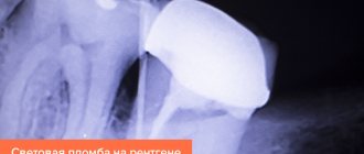
Diagnosis of deep caries
The dentist questions the patient, identifies complaints about the nature and duration of pain and other symptoms. Then he examines all the patient’s teeth, identifying both advanced and initial stages of caries. He examines the diseased tooth especially carefully, using a dental probe to identify the carious cavity.
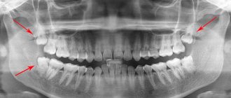
Further diagnosis of deep caries includes an orthopantomogram (OPTG) - a panoramic X-ray of the jaws. The study reveals the carious cavity, its size and the degree of penetration into hard tissues during a deep carious process. Treatment of caries is carried out only after a final diagnosis has been made.
Caries on x-ray
Crown cleaning
Before starting treatment, the crown must be cleaned of soft plaque and tartar. To do this, the crown is treated with special antiseptics, and tartar is removed with an instrument.
Anesthesia
Treatment of deep dental caries is a painful process. But modern local anesthetics (Articaine, Ultracaine, etc.) allow it to be performed without pain. Depending on the location of the tooth and the nature of its damage, anesthesia is carried out by infiltration (impregnation of soft tissues with anesthetic) or conduction (switching off the nerve trunk innervating the area of interest) method. The injection is made with a carpule syringe with a very thin needle and the ability to control the dosage of the medicine. In case of high pain sensitivity, double anesthesia is performed: first, application (anesthetic spray or gel is applied), and then an injection is made into the gums.
It is also possible to carry out treatment during sleep. The anesthesiologist-resuscitator puts the patient into a medicated sleep using:
- short-term mask anesthesia - the drug for inhalation anesthesia Sevoflurane (Sevoran) is most often used;
- short-term controlled intravenous anesthesia - a drug for intravenous anesthesia is injected intravenously; Usually Propofol (Diprivan, Pofol) is used for this purpose; special equipment allows you to clearly control the dose of the drug.
These types of anesthesia have almost no contraindications, since gentle short-acting anesthetics are used. But a preliminary examination of the patient is required.
Preparation
To prevent deep caries from spreading, the carious cavity is expanded using a bur, removing all affected tissue. It is very important to do this as carefully as possible: if the affected tissue remains, the disease will recur, moving deeper under the filling and affecting the pulp, periodontium, soft tissues and even the jaw bones.
Preparation of a carious tooth
After giving the cavity a clear shape (this is necessary for proper filling), it is dried and checked to see if there are any pathologically altered tissues left in it. For this purpose, a dental probe is used (it slides over healthy tissues and destroys changed ones) and special caries markers - dyes that stain only necrotic tissues.
The cavity is washed with water, dried and an antiseptic is applied - a 2% solution of chlorhexidine. Then the cavity is etched with orthophosphoric acid gel, washed and treated with an adhesive - a substance that enhances the adhesion of the filling material. Some modern adhesive systems have self-etching properties. If the disease is complicated by pulpitis, the pulp is removed from the tooth cavity and root canals and only then filling is performed.
Filling for deep caries
The final stage of treatment is filling. Depending on the degree of risk of developing pulpitis, a temporary or permanent filling is installed.
A permanent filling is immediately installed only if there is no severe pain and the dentin layer covering the dental cavity is in satisfactory condition. In such cases, an insulating lining is placed at the bottom of the prepared cavity, reliably protecting the dental cavity, and then a filling is installed (treatment in one visit).
But more often the patient has to visit the dentist twice. On the first day, he is fitted with a therapeutic pad impregnated with calcium, and then a temporary filling. After a few days, if there is no severe pain, the temporary filling is removed and an insulating lining with a permanent filling is installed. The filling is ground (excess filling mass is removed) and polished (dental plaque does not form so quickly on an absolutely smooth surface).
Installing a temporary filling
To install temporary fillings, materials that are sufficiently tight, hypoallergenic, and drug resistant are used. They should harden quickly and be easily removed. These are different types of cements: artificial dentin (zinc sulfate cement), zinc eugenol, polycarboxylate, glass ionomer cements, etc.
They are sealed with the installation of permanent fillings using the following materials:
- glass ionomer cement (GIC) – its distinctive feature is the constant enrichment of dental tissue with calcium;
- metal, including alloys (amalgam - an alloy of various metals with mercury) fillings are strong, but toxic, and often lose their tightness over time;
- light-curing fillings (photopolymers) – paste-like material for layer-by-layer application and rapid curing with an ultraviolet lamp; strong, durable and aesthetically attractive material;
- Compomers - a mixture of GIC and composite - are the most durable and beautiful material, but expensive.
If the crown is completely destroyed, the following is used to restore it:
- stump inlays - a mini-prosthesis installed on molars when they are destroyed by no more than a third and healthy roots;
- artificial crowns - their installation is indicated in cases of significant destruction of the crown; they completely restore the function of the tooth.
Stump tab for future crown
Painless wisdom tooth treatment at the Beryozka dental clinic
Wisdom teeth (or as doctors also call them - “eights”, third molars) often cause people a lot of trouble.
Moreover, according to medical expert statistics, it is they who are at risk of carious lesions more than any other area on the jaw. The causes of caries on the “eight” are no different from the causes of the disease in other teeth - poor heredity, neglect of hygiene and an excessive amount of carbohydrates in the daily diet. A specific factor that increases the risk of developing caries on the surface of a wisdom tooth is its location. Third molars that erupt in the gums, as a rule, not completely, are “embedded” in the jaw at an angle, which contributes to the accumulation of food debris in the tissue “hood.” It is difficult to clean the hidden corners of distant areas, and bacteria developing due to food debris contribute to active carious lesions of the “eight”.
Cracked tooth: treatment or removal
Cracking of teeth is a common reason for visiting a dental clinic. The category of “mechanical” diagnoses is numerous. These include:
- the appearance of cracks - vertical, horizontal, diagonal, root;
- tooth decay, fillings, crowns.
Damage differs in intensity, location, and difficulty of recovery.
In the past, the fate of cracked teeth was a foregone conclusion. The patient walked with the damaged structure for as long as he could, then went to the surgeon, who used forceps to radically eliminate the damage. Along with the tooth.
Today, the patient does not have to panic when damage appears. It is enough to come to a good doctor with the question of a crack or a broken tooth with a filling - what to do, in order to find out about alternative treatment options. If you have any doubts about your dentist’s recommendations, you should seek independent advice from other clinics. Because – with a high degree of probability – it is possible to save a cracked tooth!
Rescue Cracked Teeth
The complexity of dental procedures determines the scope of work - the location of the damage, the depth of the cracks, the consequences of the injury:
- Removal of surface microcracks. The doctor schedules several visits and sequentially covers the enamel with a medicinal composition with a high content of fluoride and calcium. Gradually, the microcracks are filled and the tooth is restored.
- Darkening of the enamel due to minor local damage. What is required is not a strengthening composition, but veneers. Neat plates will give your teeth an aesthetic appearance and strengthen the enamel.
- Cracks in the tooth surface and resulting chips. They do not require treatment if they do not reach the pulp tissue. The tooth is restored to the desired aesthetics and functionality with extensions or a crown.
- Complex damage that permeates the entire structure is often the cause of inflammatory processes. The tooth is restored sequentially - through depulpation, disinfection, crown.
- The tooth is split, part of it is loose. Until recently, such objects were immediately subject to removal. And today many public clinics do not offer alternatives. In fact, the tooth can be saved. The doctor will formulate the principles of treatment after viewing the x-ray, since it is important to see the surviving root base. If there is a root, after removing the moving part, the missing section is built up onto the pin. If the pin cannot be placed, the structure is restored with a photopolymer filling and the tooth is covered with a crown.
- Crack at the root. Why root cracks occur in a living tooth, let the diagnosticians deal with this; for the patient, it is important to save the damaged object. It's real. Examining the dental structure using a dental microscope will show how far the process has progressed. If the breakage concerns only part of the root and the tooth is not damaged, endodontic surgery can be used to remove the broken area and save the rest.
There are no uniform algorithms in dentistry. Each diagnosis requires separate study and selection of treatment. And the better the equipment of the clinic, the higher the professionalism of the doctors, the greater the likelihood that the patient will save his teeth and return them to their original health.
Why is it still not worth treating wisdom teeth?
The appearance of pain signals complications of caries such as pulpitis or periodontitis. This means that the nerve of the tooth is damaged, which is why unpleasant sensations appear. In this case, work with root canals is necessary. If it is not possible to promptly seek help from a dentist, you can take painkillers. But it’s worth noting right away that you don’t need to take pills on the day of visiting the clinic. Otherwise, the anesthesia may not work. Typically, a problematic wisdom tooth that is causing pain to the patient is removed. But in rare cases, doctors provide treatment. First, an x-ray must be taken to study the structure of the root canals and the degree of damage to the tooth. After this, the dentist carefully removes carious tissue, cleans the canals and fills them with medication. Most often, treatment of pulpitis or periodontitis takes place in 2-3 visits. It all depends on the complexity of the particular case. If on the first visit a medicinal product is placed in the root canals, then on the second visit they are tightly sealed. After which the doctor restores the natural shape of the tooth. At each stage of working with wisdom teeth for pulpitis or periodontitis, X-ray diagnostics are performed. It is important to make sure that the inflammation has passed and treatment can continue.
When is it possible and advisable to save the eight?
Your doctor may recommend not removing your wisdom tooth if:
- caries was detected at an early stage, its treatment is not associated with great difficulties, costs and will not cause discomfort to the patient;
- removal is associated with more serious risks than treatment - for example, in case of superficial caries and individual intolerance to drugs for local anesthesia, an enamel defect can be removed without pain relief;
- the tooth is needed as a support for a bridge prosthesis or a solid (clasp) prosthesis;
- The figure eight has a normally growing, fully erupted antagonist, which allows for a uniform load on this area of the upper and lower jaw when chewing.
Should resorcinated teeth be removed?
Patients of Soviet dentistries are aware of what resorcinol fillings are. Beginning in 1912 and for almost 100 years, caries was resorcinolized - eliminated using the resorcinol-formalin filling method. At the same time, the dentists did not inform the patients that subsequently the teeth turn yellow, decay, become increasingly fragile, and cannot be refilled.
Times have changed, and seemingly harmful fillings can be replaced with modern filling compounds. However, this turned out to be practically impossible - it is impossible to clean the channels from the resorcinol-formalin mixture. During the hardening process, the filling acquires the density of glass and is soldered to the walls of the root canal. The verdict of most doctors when dealing with resorcinated teeth is the same - removal.
But there is no need to rush in this case either. Today, modern dental equipment allows you to drill out and remove even the strongest glassy material and bring the tooth into excellent condition. Yes, not all clinics have the necessary equipment, but it is there. And finding “your” doctor after several consultations will completely solve this problem.