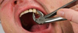
Teeth are bone formations whose main purpose is the primary mechanical processing of food. In most cases, an adult has 32 teeth. In addition to performing the chewing function, they influence the formation of human speech. The loss of baby teeth and the appearance of permanent teeth occurs after the baby’s fifth birthday - there are four types: incisors and canines, molars and premolars.
On one side of the upper jaw there are the following teeth: 1 canine and 2 incisors, as well as 2 premolars and 3 molars. The opposite side is similar to the upper one, and the arrangement of the lower jaw teeth is identical to the upper row of teeth. The condition of teeth indicates a person’s health, and if it is unsatisfactory, this may indicate some kind of disorder in the body.
READ ALSO: location of teeth in adults and their numbers
Human dental formula
Many people have heard that dentists call teeth by different numbers. Thus, each unit is assigned a specific number, which guarantees maximum accuracy in records of dental conditions or treatment procedures. The count is made starting from the middle of the dentition to the right and left sides. The anterior units - incisors - are No. 1 and 2, 3 - canines, 4-5 - premolars, 6, 7 and 8 - molars, the last of which is the wisdom tooth.
To determine exactly where the tooth is located, the jaw is figuratively divided into segments. The beginning of the numbering is the right side of the upper jaw of the teeth. Units are written as two-digit numbers, where the first determines the serial number, and the other determines the so-called segment. The upper right half of the teeth is designated by numbers 11-18, the left - 21-28, and the lower left segment - 31-38 and 41-48. Regarding primary teeth, the count is made starting from the front units on the left in a clockwise direction. They are assigned the numbers 50, 60, 70 and 80.
READ ALSO: structure of the human upper jaw and functions of its processes
There are several types of numbering that dentists use:
- With the square-numeric Zsigmondy-Palmer method, Arabic numerals are used for radical units, and Roman numerals are used for dairy units. Often used by orthodontists and surgeons.
- Haderup's method is based on the symbols "-" and "+", where the first denotes the lower and the other the opposite arc. Units are written in Arabic numerals, and in the case of dairy units, 0 is added.
- The Viola system uses Arabic numerals 1-8 for units and two-digit numbers for segments. This type is very convenient, which is why it is widely used by dentists around the world.
- The last system is universal. Each group of units is assigned a letter, number and segment number.
Fusion of the lateral and central incisors with congenital edentia of one of the incisors
Dental fusion is a form of embryological disorder in which two separately developing primordia form a single structure by the synthesis of dentin and/or enamel tissues. The pulp chamber and canals can also be fused or located separately - it all depends on the complexity of the pathology. Although the etiology of this disorder is not fully known, one of the factors is physical pressure, which leads to the fusion of teeth and provokes necrosis of the tissue separating them. Genetic factors also play a role. The degree of fusion depends on the degree of formation of teeth: the earlier the generalization of teeth occurs, the more complete it is. As a result, a person may develop one large tooth.
Tooth duplication, on the other hand, occurs as a result of incomplete division of a single tooth germ and results in the formation of a complete or partial crown with a single root canal and root system. Fusion can be clinically distinguished from duplication by the number of teeth present in the dentition, unless, of course, fusion occurs involving supernumerary teeth. With a physiological number of teeth, doubling can be suspected, but with insufficiency, fusion can be suspected. During fusion, the crown of the tooth appears divided by a deep groove like a bicuspid crown or cutting thickening, while during duplication, the parts of the crown are usually mirror identical. Radiographically, fused teeth often have a double pulp chamber, whereas duplication teeth show a single pulp and root space.
There is also often no interdental dentin between the fused roots. The frequency of these anomalies is higher in primary dentition than in permanent dentition (0.5% and 0.1%, respectively). During the analysis of 3517 plaster models, it was found that in 57.2% of cases of registration of such anomalies, it was the fusion of teeth that was observed, and only in 42.9% - their doubling. The fusion of teeth is more often observed in the frontal area and on the lower jaw. Central incisors are affected by pathology with a frequency of 3.6%, demonstrating the greatest tendency to this type of change. It was not possible to establish a gender predisposition to fusion or duplication of teeth. In addition to the above, people can often experience dental agenesis, the prevalence of which is higher in the permanent dentition than in the primary dentition, and varies in the range of 1.6-10%, if third molars are not taken into account. The premolars and maxillary lateral incisors are most commonly affected. Maxillary lateral incisors are missing in approximately 1-2% of the total population, and account for almost 20% of all cases of dental agenesis. These teeth are also the most genetically variable, which often provokes significant violations of the aesthetics of a smile. In addition, in cases of fusion and agenesis, a violation of the symmetry of the dental arch, the presence of diastemas, changes in occlusion, crowding of teeth, protrusion, and also a sign of carious lesions are often observed. Due to the presence of fissures and grooves between fused teeth, plaque accumulation progresses in these areas, which subsequently provokes periodontal problems. Treatment in such situations should be carried out through a multidisciplinary approach.
This article presents a case of 10-year follow-up of a patient in whom both the fusion of the central and lateral incisors and congenital edentulism of the second lateral incisor were recorded. During the analysis of this case, the entire complex treatment algorithm will be described.
Clinical case
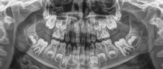
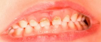
A 9-year-old girl was referred to a dentist by her primary care pediatrician with the chief complaint of being teased by other children because of an overly large front tooth. During clinical and radiological diagnostics, a fusion of the central and lateral incisors (7 and 8 teeth), as well as the absence of the 10th tooth germ (photo 1-3) was discovered.
Photo 1-2. View before treatment: patient age – 9 years.
Photo 3. X-ray of the fused central and lateral incisors (7 and 8 teeth), edentia in the area of the lateral incisor (10 tooth).
To restore the 10th tooth, it was proposed to carry out implantation after the completion of general somatic growth of the body. The wax reproduction was used to simulate the treatment capabilities of fused teeth, so as to somehow simulate the area of the papilla between them, and create the appearance of two separate teeth. To close the diastema between teeth 8 and 9 and create a space for a future implant, the patient was referred for an orthodontic consultation. To assess the area of prospective implantation, the patient was additionally referred for a consultation with a periodontist. In order to assess the risk of damage to the pulp chamber during the preparation of fused teeth for a new construction, the doctor personally discussed this case with the endodontist. Using the wax reproduction as a template, a composite restoration was made for 7-8 fused teeth. This fixation was carried out after preparation, etching and adhesive treatment of the surface of their enamel. A portion of the composite in shade A1 was applied to the mesial and distal surfaces of the teeth, using the darker shade of the material to restore the cervical contact area. The teeth were contoured to closely mimic the shape and size of two separate incisors, based on the normal adjacent central incisor (Figures 4 and 5).
Photo 4-5. Contouring of fused teeth to simulate both incisors.
The patient and her family were very pleased with the results, and the girl did not experience any postoperative sensitivity after the intervention. The patient was constantly monitored by her pediatrician and periodically underwent repeated diagnostic examinations by the orthodontist. At the age of 12 years and 1 month, she began to undergo orthodontic bite correction, which lasted for 2 years and 7 months. The purpose of this intervention was to create a place for the implant in the area of the 10th tooth, as well as to close the diastema between the 8th and 9th teeth (photo 6). After completion of orthodontic treatment at 14 years and 3 months, she received an adhesive retainer design to stabilize the position of the teeth bordering the defect (Figures 7 and 8).
Photo 6. Fixation of orthodontic equipment.
Photo 7. View of the adhesive retainer.
Photo 8. View from the palatal side.
At the age of 17 years and 5 months, the patient underwent a CBCT scan, as a result of which it was discovered that the thickness of the buccal part of the alveolar ridge did not exceed 4.5 mm. In order to correct these parameters, the periodontist recommended performing lateral bone tissue augmentation to increase the ridge parameters by another 3-4 mm. During the analysis of CBCT images, a pathological concavity of the root was also discovered in the area of fused 7-8 teeth, which compromised the condition of the soft tissues in this area. Taking into account the recommendations of specialists, bone tissue augmentation was performed in the area of the 10th tooth. 6 months after the manipulation, the orthodontist analyzed a repeat cephalogram to confirm the fact that the patient’s somatic growth had ceased. Alginate impressions were obtained to make a surgical guide. Considering the previous aesthetic appearance of the composite restoration in the area of 7-8 teeth, it was decided to produce the final structure in the form of a lithium disilicate veneer using pink ceramic for imitation in the gingival area. A dental implant was planned to be installed in the area of the 10th tooth, and a veneer restoration was planned for the 9th tooth (at the patient’s personal request). Implant surgery was performed after the patient reached the age of 18 without any subsequent complications (Figures 9 and 10).
Photo 9. Installation of an implant in the area of the 10th tooth.
Photo 10. Installation of an implant in the area of the 10th tooth.
The position of the implant was checked after three months using a targeted radiograph, the torque of the structure was also assessed, and only after that the gum former was fixed (3 mm by 4 mm by 4 mm). After 2 and a half weeks, the orthopedist took an impression from the implantation area using the open tray method, which he sent to the laboratory for the production of a customized abutment, a zirconium cap and a provisional crown. The abutment was fixed with a torque of 15 Ncm, and its tightness was checked using a radiograph. The role of the provisional crown was to form an adequate and adapted gum profile, based on the prosthetic conditions of the situation. After filling the screw access hole with PTFE tape, the temporary restoration was secured, carefully cleaning the cement used. After the maturation of the soft tissues, the shades of the final restoration were selected, based on the ratio of the parameters of pink and white aesthetics, as well as the preparation of 9 and fused 7-8 teeth for future constructions (photo 11).
Photo 11. Preparation of teeth 7, 8 and 9 for veneers.
During the preparation, special attention was paid to ensuring that on the front side the tissue reduction boundary runs somewhat deeper than on the distal and incisal sides, since under such conditions it will be easier for technicians to recreate a structure that imitates two separate crowns. A thread was used to retract the gums, after which a zirconium cap was tried on, and then impressions were taken from the areas of prepared teeth 7-8 and 9, including the same zirconium cap (photo 12).
Photo 12. Taking an impression.
When the patient reached the age of 19, lithium disilicate veneers and a ceramic crown were tried on. After modification, the veneers were fixed using adhesive cement, having previously silanized their inner surface. The inner surface of the implant crown was treated with a special etching agent and silane, and then fixed with carboxylate cement. The cement was applied with a brush to minimize excess material. Each time, any fixing material during cementation of veneers and crowns was carefully removed, after which the parameters of the occlusal relationship were checked. In order to monitor hygiene, the patient came for a follow-up visit 2 weeks after the intervention (photo 13 - photo 18), and on the same visit she was given a night guard. Subjectively, she was more than satisfied with the results of the treatment.
Photos 13-16. View after treatment.
Photo 17-18. X-ray after treatment.
Discussion
This article describes a clinical case of conservative restoration of aesthetic, functional and occlusal parameters for the treatment of dental fusion in conditions of congenital edentulous lateral incisor. Alternative methods for treating the absence of a lateral incisor involve orthodontic intervention with movement of the canines, replacement of the defect with a bridge, an adhesive or partial removable denture, a cantilever part of a fixed denture, an autotransplantation procedure, and implantation. Canine relocation may be chosen as a therapeutic algorithm in cases of class 2 malocclusion with crowding of teeth in the lower jaw and the need for iatrogenic extraction of dentition units. In such cases, the disposition of the canines in place of the lateral incisors is quite reasoned. Even taking into account that the preparation protocol for adhesive dentures involves only minimal reduction of hard tissues, in such cases, in order to maximize the area of adhesive connection with the abutment tooth, it is sometimes sufficient to modify its occlusal parameters.
The use of such structures is contraindicated in case of mobility of supporting teeth, presence of parafunctions, deep bite or excessive inclination of teeth. The clinical failure rate with adhesive prostheses reaches 54% at 11 months and 10% at 11 years, most often due to debonding of restorations. Considering the length of the root and the size of the canine crown, a cantilever prosthesis, replacing the edentulous area of the lateral incisor with support on the canine, is often the most promising type of design. This method of treatment does not depend on the amount of inclination of the supporting tooth, and additional retention in the area can be achieved using additional pins (taking into account, of course, pulp tomography). The success of a cantilever partial denture depends on the control of occlusal forces in the overhang area, so it is desirable to remove all occlusal contacts at the site of the defect-replacing restoration. When introducing contacts in the area of a cantilever prosthesis, the risk of restoration failure during operation, displacement of the abutment tooth, or even the risk of its fracture increases.
Premolar autotransplantation is another treatment alternative. But this approach is extremely sensitive to execution and time parameters. The ideal period for autotransplantation is when the root length ranges from 2/3 to 3/4 of the physiological size, which is observed in patients 9-12 years of age. Under such conditions, periodontal healing and further development of the tooth root is observed in 90% of patients. Research results indicate that the success rate of this procedure ranges from 79-90%, but it should be remembered that any damage to the periodontal ligament can lead to ankylosis.
The least conservative method of treatment is the production of a partial fixed denture supported on an adjacent tooth. This approach is not advisable in young patients if the supporting teeth are completely healthy without signs of any restorations or carious lesions. Ideally, the therapeutic approach for the treatment of congenital edentulous lateral incisors should be the least invasive while still providing adequate functional and esthetic results. Restorations supported by dental implants are characterized by a survival rate of up to 96.8% five years after installation of superstructures. In this case, it is possible not only to maintain the level of the alveolar ridge, but also to preserve the integrity of adjacent teeth. The installation of intraosseous supports should be carried out after completion of somatic growth: 16-17 years in men, 20-21 years in women. When implantation is carried out during a period when adjacent teeth continue to erupt, a number of complications can be provoked with a violation of the periodontal and occlusal parameters of the dentofacial apparatus, as well as compromise of the level of the gums in the implant area. When a canine erupts at the site of the lateral incisor, it can be orthodontically distalized in order to form a place for a future titanium support. When the canine erupts in its place, then in the area of the lateral incisor there is a deficiency of the bone crest, for the correction of which it is simply impossible to do without an augmentation procedure. CBCT scanning can help to objectify the implantation site, which also allows you to plan the entire range of future surgical and prosthetic measures.
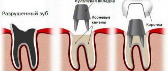
As for the fusion of teeth, this pathology can be corrected by extracting these teeth, followed by retention of this area and installing implants in a delayed period; endodontic treatment and periodontal surgery followed by restoration using crowns supported by pin structures; In addition to crowns, you can use veneers, simulating the desired area of the gums using pink ceramics. When fused with a supernumerary tooth, an extraction procedure can be performed, followed by orthodontic correction, replacement of the necessary dental units with veneers, or the supernumerary tooth can be removed through flap separation. In special cases, extraction, separation and replantation can be performed. The latter approach carries the risk of periodontal inflammation, increased pocket depth, pulp necrosis, ankylosis, or internal/external root resorption. As an additional treatment, it is possible to perform a pulpotomy, hemisection of a supernumerary tooth, and perform a composite restoration, correcting the gum area using a soft tissue graft.
One of the problems of aesthetic restoration of the area of fused teeth is the need to imitate the gingival papilla. To solve such problems, there is a special pink composite for cervical areas. Additionally, endodontic treatment can be performed by creating access from the labial side, after which the gingival area can be imitated using a pink composite veneer. As an alternative, it is also possible to use a metal-ceramic crown with pink ceramic in the cervical area, in which case it is necessary to reduce the root diameter through periodontal intervention and narrow the connection interface through adaptation of the core inlay. The advantage of treatment using ceramic veneers is the ability to achieve improved aesthetic rehabilitation parameters without significant reduction of enamel tissue. The latter fact makes it possible to strengthen the adhesive bond between the restoration and the tooth tissues. Porcelain veneers also provide higher tensile strength due to the reliable bond of the composite cement to the enamel, which ensures high success rates of such structures. Lithium disilicate restorations are characterized by high flexural strength (from 350 MPa to 400 MPa), as well as excellent aesthetic parameters, and can be manufactured using CAD/CAM technologies. When using composite luting cements, the success rate of lithium disilicate restorations increases even further.
conclusions
A conservative interdisciplinary approach to the treatment of fusion of teeth and congenital edentia of the lateral incisor is a successful method of treating combined pathology. The rehabilitation protocol included the implementation of a composite pink restoration, adjacent orthodontic correction, bone crest augmentation and implantation, as well as a set of diagnostic measures such as cephalometry and CBCT to ensure the most effective results of iatrogenic intervention. To imitate soft tissues, special lithium disilicate restorations with a pink tint were used in the cervical area; a similar approach was implemented on the crown supported by an implant in the edentulous area of the lateral incisor. Such a treatment algorithm improves the occlusal, aesthetic and functional parameters of the dentofacial apparatus, and also increases the patient’s confidence in social interaction.
Authors: Leslie Stone Hirsh, DDS Peter M. Greco, DMD Jay B. Laudenbach, DMD Alan M. Atlas, DMD
The structure of the upper and lower teeth
The elements of a human tooth are: crown, neck and root. The crown is visible to the naked eye - it is located above the gum. The connecting link between the tooth root and the alveolus is connective tissue. The neck of the tooth is located between the crown and the root.
READ IN DETAIL: dental formula for adults and children
From a histological point of view, the constituent basis of the human tooth is dentin, hidden under the enamel. Near the root, where there is no enamel, there are fibers that seem to hold the periodontium together. In the center of the unit there is a pulp, which is penetrated by blood vessels and nerve endings. With inflammation or caries, it makes itself felt with severe pain.
Incisor and canine crown
Both the lower incisor and its antagonist have similar components: the root and the crown. Slightly above the alveolar layer is the enamel crown of the tooth. The root of the tooth is located in the alveolar cavity under the gum (we recommend reading: how many roots does a person have in his teeth?). The large dimensions of the upper incisor are about 21-23 mm, while the length of the crown is 9-95 mm, and the width of the teeth is 4-6 mm, so the pressure on them is distributed evenly. The enamel is protected from physical damage and mechanical stress. The lateral (or lateral) incisor, in comparison with the medial one, has a huge root and a wide crown.
Anatomical features of the upper teeth
- The central incisor (the largest of all eight) has a chisel-shaped crown with a convex surface and one cone-shaped root. The only root canal in 75% of cases is straight.
- The lateral incisor with the same chisel-shaped crown and the same convex surface has a characteristic depression in the enamel - a “blind fossa”. There is one root canal, but more often it is deviated to the side.
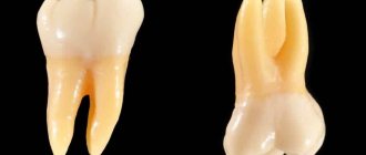
- The canines on the upper dentition are often larger than those on the bottom. They are characterized by a crown pointed on all sides and the longest cone-shaped root. Canines have a single root canal, which can be straight (45%), distally deviated (30%) or vestibular deviated (12%).
- The first premolar with a prism-like crown is characterized by a convex lingual surface. On the chewing side there are tubercles, and between them there is a fissure. In the upper dentition, these teeth are always larger than in the lower one. The root has extended longitudinal grooves, which divide it in 60% of cases into two parts - buccal and palatal. There are often also two root canals.
- The second premolar with the same prism-shaped crown has predominantly one straight cone-shaped root with expanded lateral surfaces. Sometimes the root bifurcates closer to the top. There are often two root canals.
- The first molar is the largest tooth in the dentition. The rectangular crown has a diamond-shaped chewing surface with four cusps and an H-shaped fissure between them. There are usually three root canals, but sometimes there are 4 (25%) or 5 (1%).
- The second molar with a classic cube-shaped crown has 4 cusps and an X-like fissure on the chewing side. The tooth has 3 roots and three (87%) or four (13%) canals.
What are baby teeth?
The formation of baby teeth begins when the baby is in the womb, namely at the 7th week of pregnancy. Teething begins when the baby reaches 6-24 months. The change from temporary units to permanent ones is carried out at the age of 5-13 years. The complete formation of a permanent dentition is completed by the age of 14. A deviation of no more than one year is acceptable. If the period of teeth replacement is exceeded, this indicates anomalies in the dental arch or jaw formation.
Structural features
Milk teeth are similar to permanent teeth, but are much smaller in size and have short, rounded roots. The thickness of dentin and enamel is also less. Due to low mineralization, teeth are susceptible to the rapid development of caries. It is worth noting that temporary units are characterized by a considerable volume of pulp and wide root canals, which facilitates the penetration of harmful microorganisms into the pulp.
The number of roots in baby and permanent human teeth is identical. When the time comes to replace temporary teeth with permanent ones, the roots of baby teeth dissolve, thereby making room for new ones. Their change is almost painless, since they are not strongly attached to the alveoli.
READ ALSO: The number of teeth of an adult healthy person is normal
Differences from permanent
Despite the similarities between milk and permanent units, there are a number of differences. First of all, this is the number of units - in contrast to permanent ones (32), temporary ones are only 20. In addition, children do not have premolars, and there is only one molar. There are also other differences:
- smaller crown
- thin enamel, white-blue color,
- dentin composition with a lower degree of mineralization,
- roots are branched and massive,
- deep fissures,
- the roots are bent closer to the lip, they are not so strong and shorter.
The structure of the medial lower incisor, right:
Rice. 1-54. a — vestibular surface; b — medial surface; c - lingual surface; d — vestibular-lingual section; d — mediodistal section; e - cutting surface; 1, 2, 3 - shapes of cross sections at the level of the crown, middle and lower third of the root, respectively
The angles between the incisal and medial , as well as lateral edges are almost the same, and the sign of the crown angle is difficult to recognize. The cutting edge of the crown has 3 tubercles, well defined on unworn teeth. On the vestibular surface of the tooth, from the tubercles, the edges go towards the neck of the tooth, three differently pronounced ridges. The medial and distal ridges are usually clearly visible. In the middle third of the crown, the ridges flatten and disappear. The enamel border forms an arc open to the cutting edge of the tooth. The sign of crown curvature is not expressed, so it is not always possible to determine whether a tooth belongs to a specific segment. On the lingual surface, marginal ridges are noticeable, extending from the corners of the cutting edge to the neck of the tooth. On the lower incisors they are less pronounced and sometimes absent. In the cervical part of the crown there is a median dental tubercle, from which a small flattened ridge can sometimes extend to the median tubercle on the cutting surface. The lingual surface of the crown can be concave, flat or slightly convex. The lateral, contact surfaces of the tooth (medial and distal) have a wedge shape. The contour of the vestibular surface of the crown is formed by a convex arch, and the lingual surface is formed by a concave arch. The enamel border is arched, convex towards the cutting edge. The root of the medial lower incisor is flattened in the mesiodistal direction. The contour of the vestibular surface of the root is convex or smooth, while the lingual surface is convex, smooth, or even concave. The root apex quite often deviates vestibularly. Signs of root angle are not expressed. The cavity is similar to the shape of a tooth; the root canal sometimes splits into two. The height of the crown of the medial lower incisor ranges from 7.0 to 9.5 mm, width 5.0-5.7 mm, vestibular-lingual neck size 5.5-6.0 mm, mesiodistal - 3.5-5.0 mm; root length 9.5-14.0 mm. Lateral lower incisor . In the vestibular norm, the crown of the lateral incisors is trapezoidal (Fig. 1-55).