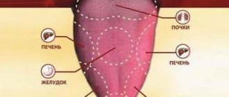
48647
Crooked, unhealthy teeth can ruin the appearance of an otherwise attractive person. Oral health is important not only for our attractiveness, because the main function of teeth is to grind food. The condition of the stomach and intestines depends on this, which directly affects health and life expectancy .
How to determine?
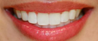
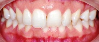
A distinctive characteristic is the tight contact of the upper and lower jaws when closing . The upper teeth are superimposed on a third of the lower crowns, leaving no distance between the upper and lower molars.
photo: this is what a correct bite looks like
The shape of the dentition and their relative sizes are also important. The top row has a semi-oval shape, the bottom row has a parabolic shape. The lower incisors are in contact with the palatal cusps of the upper ones. During chewing, the molars constantly touch without losing contact.
Correct bite forms a harmonious face , the lower and upper jaws are symmetrical, the vertical axis of symmetry of the face coincides with the junction line between the incisors.
Signs of a correct bite are also:
- no speech defects;
- comfortable biting and chewing of food;
- absence of uncomfortable sensations and clicking in the mandibular joint.
If you notice that plaque appears only on individual teeth, this means that they are not involved in the process of grinding food . In this case, it is worth visiting a dentist: this is a sign of a defectively formed dentition.
Anatomical and histological characteristics
Each tooth is an independent, full-fledged organ that performs a number of tasks assigned to it. Functionality is ensured thanks to a set of specific properties inherent in nature.
From an anatomical point of view, a tooth consists of three main components:
- crowns This is the outer, visible part of the organ. It is the strongest and hardest, which makes it possible to effectively resist external irritants, as well as intensively process food;
- root _ It is, on the contrary, an element hidden from view. Depending on the functions assigned to a particular organ, the root can be single or branched (for example, “wise” teeth of the lower jaw can have up to eight roots). The processes allow them to be securely fixed in the bone tissue of the jaw and provide stability;
- cervix. This component is strategically important and at the same time vulnerable. It is the neck that unites the radically different crown and root, guaranteeing optimal functioning of the organ.
From the point of view of histology (that is, the structure of the structure), nature “equipped” the tooth extremely successfully:
- The pulp is located in the central part of the organ. This is the tissue that connects numerous branches of nerves and blood vessels. The location of the pulp in the core of the tooth is ideal, because this is its softest and most defenseless part;
- nerves and vessels are surrounded on all sides by dentin. This tissue is much stronger due to its high mineralization, therefore it serves as a kind of protective shell for the pulp;
- The part of dentin that lies below the gum is surrounded by cementum. On the one hand, it promotes reliable fastening of the tooth in the bone tissue. On the other hand, cement serves as additional protection for dentin and, accordingly, pulp;
- in the crown part the dentin is covered with enamel. This fabric is phenomenal in its own right. Unlike all other compounds, it contains only 2-3% water, the rest is minerals. This specific composition makes the enamel the hardest and most durable in the human body. It serves as an indispensable shield between external irritants and the delicate contents of the pulp. It is the enamel that saves the tooth from death until the last moment, slowing down the spread of carious bacteria to the inner layers.
What types are there?
There are temporary and permanent bites. The bite in children before the eruption of permanent teeth is called temporary. 6-9 years – time of replacement bite. During this period, the child has simultaneous presence of permanent teeth and milk teeth.
Peculiarities of eruption influence the formation of the maxillofacial system. Any removal of replacement teeth earlier than a year before natural loss causes the risk of various deformities:
- asymmetrical dentition;
- midline shift;
- blocking chewing movements of the jaw.
A permanent bite is formed when a teenager reaches 15 years of age, when the formation of roots in relation to the position of the teeth ends. Their number during this period is 28 (32 - if the third molars have erupted).
Permanent dentition can be physiological or abnormal.
Physiological
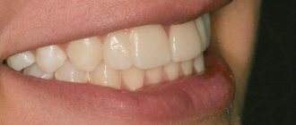
photo: this is what a correct bite looks like from the side
Physiological ones include types of occlusions that provide:
- the most efficient and comfortable chewing of food;
- correct speech formation;
- aesthetic appearance of the maxillofacial area.
There are several varieties:
- Orthognathic. This is the standard bite that provides maximum functionality of the jaws. Premolars and molars intersect with antagonists of the opposite row. The upper incisors and canines overlap the lower ones due to a slight outward slope.
- Straight. It differs from orthognathic in the type of closure of the incisors. The edges of the upper ones do not overlap the lower ones, but touch end to end. The molars have good tight contact. A big disadvantage is the rapid wear of the upper and lower incisors due to contact of the cutting edges.
- Progenic . The lower jaw is pushed forward. The dental arches close tightly, the chewing function of the jaw is preserved.
- Biprognathic. The incisors and canines have a greater angle of inclination forward than with orthognathic. The contact of the anterior teeth is good. The incisors meet at the edges.
- Opisthognathic . The upper and lower incisors are inclined towards the oral cavity. The molars and premolars are tightly closed.
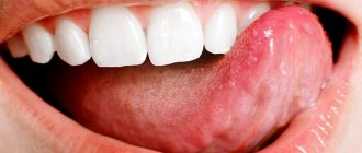
Abnormal
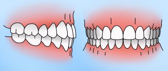
photo: malocclusion
Malocclusion is characterized by a complete or partial absence of tooth contact in occlusion. Such a bite distorts the face, disrupts the chewing process and can affect the formation of speech defects.
There are several types of abnormal bite:
- Deep. Its peculiarity is that the upper incisors overlap the antagonists by more than a third. The wear of the cutting edge is increased, causing the masticatory muscles to experience hypertonicity. This type of bite is also determined by constant sensations of tension in the temporomandibular joint and headaches.
- Open. A rather complex defect that entails disruption of several body systems at once. With the open type, some teeth do not have contact with the antagonists. There are lateral and frontal types.
Symptoms indicating the presence of an open bite are elongation of the mandibular part of the face, constant tension of the facial muscles, speech disturbances, difficulty swallowing, discomfort when chewing.This type may have a rachitic and traumatic origin. The rachitic variety is difficult to correct and usually requires surgical intervention. The traumatic variety is formed in childhood as a result of bad habits or trauma; at a young age it is quite easy to correct.
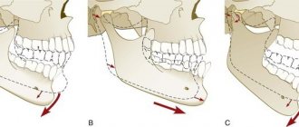
- Cross. The displacement of the lower jaw to one side forms a noticeable asymmetry of the face, the dentitions close like scissors crosswise.
A person with such a defect has difficulty chewing food and chews only on one side. Poorly chopped food provokes stomach diseases, discomfort in the jaw joint during eating, and the development of periodontal diseases. - Distal. The defect is formed as a result of good development of the upper jaw and at the same time underdevelopment of the lower jaw.
With this anomaly, the upper incisors stick out forward, leaving a large gap between the upper and lower incisors. In the formation of such an anomaly, heredity, poor posture, and removal of baby teeth (more than a year before the appearance of permanent teeth) become decisive. - Mesial . Characterized by a protracted lower jaw. With such a defect, the chin protrudes strongly and the facial profile is curved. The lower teeth are located in front of the upper teeth during occlusion.
Fundamentals of occlusion and biomechanics of the jaws: a new look at old concepts
A deep understanding of the basics of occlusion and biomechanics of the jaws is one of the most important and necessary components for providing comprehensive patient rehabilitation in dental practice. Knowledge of the principles of differential diagnosis of pain, planning of future iatrogenic intervention, as well as algorithms for the treatment of prosthetic disorders provides the doctor with all the necessary tools for further normalization of the patient’s dental status.
An orthopedic doctor simply cannot do without understanding how significant the concept of occlusion is not only in pathology, but also in a state of stable and adequate function. The formation of appropriate occlusal schemes is based on the redistribution of acting forces, because, in fact, it is precisely because of the excess of such indicators that diseases, pathologies and dysfunctions of the elements of the dentofacial apparatus arise.
Occlusal disorders can manifest themselves in the form of various structural damage to the dental status, such as pathological abrasion, fractures, and premature wear of restorative structures. In addition to the latter, functional pathologies are characterized by tooth mobility, loss of volume of soft and hard tissues, muscle pain, as well as pain and noise in the joints (the so-called clattering), limitation and impairment of movements of the lower jaw, remodeling changes in bone tissue in the structure of the temporomandibular joint . In such cases, patients form so-called parafunctional habits, the presence of which he himself does not know. Clinically, signs of such are manifested by excessive wear of one’s own teeth and various types of restorative structures present in the oral cavity.
There are different opinions regarding the relationship between the state of occlusion and disorders of the temporomandibular composition. According to the most extensive literature reviews, such associations are rather weakly expressed, as evidenced by the fact that when correcting occlusal relationships, it is not always possible to prevent the development and progression of joint pathologies. Based on the available data, the following conclusions can be drawn: only the absence of traumatic occlusal injuries, which are manifested by the action of excessively high parafunctional forces exceeding the adaptive capabilities of the body, ensures complete prevention of the occurrence of pathologies and dysfunctions, or the presence of such in the acceptable adaptation range. This conclusion is evidence-based, regardless of how ideal or non-ideal the occlusal schemes of each individual patient are. On the other hand, with prolonged exposure to excessive occlusal forces, the development of corresponding dysfunctions and diseases occurs regardless of the characteristics of a particular occlusal scheme. The corresponding pathological types of occlusion only further aggravate the course of related prosthetic diseases.
From the above it follows that if the doctor is fully familiar with the specifics of occlusal movements in a particular patient, and also understands their impact on the condition of soft and hard tissues, muscles and joints, then he can ensure the formation of such occlusal patterns that would be the most stable and least traumatic for each specific patient. In other words, understanding the basics of occlusion helps doctors not only plan future interventions, but also predict the functional rehabilitation of prosthetically compromised patients. The main connecting link between the pathology of the temporomandibular joint, the state of occlusion and the functional disorder of the dentofacial apparatus is the repeated action of excessive occlusal load, which goes beyond the adaptive range of the body. Based on this, the author considers it wrong to separate the dynamics of the application of force on human tissue from disorders and diseases developing in the same tissues - after all, in fact, these processes are of an indirect cause-and-effect nature.
The question is different: what is the true connection between the existing parafunction, the state of occlusion and functional deviations of the dentofacial apparatus. In order to understand how the jaw functions and where occlusion begins, you need to repeat in detail the anatomy of the masticatory muscles, the temporomandibular joint, and, of course, the teeth, taking into account the functional parameters of each of the above-mentioned components. After analyzing the anatomy, you should focus on how the relationship between the upper and lower jaws is generally formed, taking into account the occurrence of static and dynamic contacts between the surfaces of antagonist teeth. After this, the data obtained during the analysis can be implemented into a plan for future iatrogenic intervention aimed at eliminating structural disorders of the dentition and aesthetic problems, while ensuring not only the functional comfort of the dentofacial apparatus, but also the stability of the achieved results of complex rehabilitation.
In the course of analyzing the features of anatomy and intermaxillary relationships, doctors should look for key parameters of each of these components, on the basis of which they will subsequently make a decision regarding one or another possible treatment plan.
In this article, the author will refer to the concept of dental treatment planning, which takes into account changes in the facial profile during iatrogenic interventions, developed by Frank Spear. With significant destruction of the tooth structure, the main occlusal landmarks are simply lost, and the pathology goes beyond the boundaries of possible dental-alveolar compensation. Consequently, the clinician’s task is also to restore the supporting occlusal points of the intermaxillary relationship, and then, based on their stability, carry out further prosthetic rehabilitation. When implementing an approach to treatment taking into account changes in the facial profile, it is possible to ensure successful prosthetic reconstruction of the bite, based precisely on the position of the supporting occlusal landmarks.
Pankey rules and the concept of optimal occlusion
Dr. LD Pankey, being a pioneer and developer of comprehensive approaches to dentition restoration, proposed a specific concept that helps critically evaluate occlusion both during systemic dental rehabilitation and during everyday dental care:
- When the condyle of the jaw is completely in the glenoid fossa, all the posterior teeth exhibit equal and uniform contact, while the anterior teeth only lightly touch the opposing teeth.
- When the jaws are clenched, neither the teeth nor the lower jaw move.
- When the mandible moves in any direction, none of the back teeth contact faster or more strongly than the teeth in the anterior region.
Having analyzed these features, we can take a fresh look at the specifics of the anatomy of the dentofacial apparatus.
Anatomy of the temporomandibular joint
In Photo 1 you can see that the condyle of the mandible is in very close contact with the biconcave disc of the joint. These elements of the joint are located inside the capsule, which is protected behind by retrodiscal ligaments, and below, by means of capsular ligaments, is attached to the neck of the condylar process. Anteriorly, the superior portion of the lateral pterygoid muscle attaches to both the disc and the neck of the condylar process, while the inferior portion of the muscle attaches only to the neck of the condyle. Behind the joint is the external auditory canal. Anterior and superior to the condyle is the eminence, and directly above it is the glenoid fossa. The articular surfaces are covered by fibrocartilage, which is a smooth structure, and is supported by synovial fluid. The latter lubricates the surfaces of the joint, providing them with nutrients and oxygen, as well as ensuring the removal of possible debris. In the structure of the capsule, the number of blood tissues is very limited, or they may be completely absent.
Photo 1. Classic diagram of the anatomy of the temporomandibular joint.
When analyzing a joint, it is necessary to note the most important relationships of its individual components. First, there is the close condyle/disc/fossa connection. Essentially, they are in as much contact as possible, allowing the joint to withstand the necessary loads. On the other hand, this form of connection of the elements ensures the anatomical and functional integrity of this organ during dynamic movements of the lower jaw. In certain articular pathologies, this relationship is disrupted, which leads to permanent functional changes. It is obvious that the variation in the size, volume and shape of the temporomandibular joint is quite significant, and these differ greatly from person to person. Historically, we have assumed that the dimensions of the condylar process are relatively stable. However, recent research has established that the size of this anatomical structure can change and adapt over time and depending on the prevailing circumstances. A striking example is the increase in condyle size when using night guards. Due to the protrusion of the mandible to maintain patency of the upper airway, the condyle also remodels, increasing in size. Thus, it is obvious that this bone structure can not only adapt to functional conditions, but also change its shape, increasing in its geometric parameters. Consequently, the previously determined dimensional stability is very, very relative. As the first occlusal guideline, the doctor can use the position of the condyle during the registration of the centric relation, which is the most desirable. Firstly, the central ratio is a parameter that, with adequate execution technique, can be quite accurately and easily recorded. In addition, this position of the structures is repeatable, and it can be restored even if the position of the teeth changes or the contact between them is disrupted. A stable joint in this position has the ability to withstand significant loads, and the lateral pterygoid muscle can remain in a passive state even with strong clenching of the jaws (photo 2, 3).
Photo 2. Muscles that lift the mandible on the right side.
Photo 3. Muscles that lift the mandible on the left side.
Anatomy of the masticatory muscles
The function of raising the lower jaw is provided by three masticatory muscles. With parafunctional habits, such as bruxism, these masticatory muscles can develop significant force acting on all structures of the dentofacial apparatus. The masticatory muscle starts from the zygomatic arch and is attached to the lower edge of the lower jaw. The force vector of this muscle is directed upward and forward. The cross-sectional thickness of the masticatory muscles justifies the fact why they can develop the most significant strength indicators, located anterior to the temporomandibular joint. The temporalis muscle begins in the area of the temporal fossa and the deep part of the temporalis fascia. It is directed medial to the zygomatic bone and forms a tendon that is attached to the coronoid process of the mandible, and also passes into the region of the retromolar fossa distal to the last molar of the mandible. Since the muscle splits along its course, the action vectors of its force also diverge: the anterior component is directed upward and slightly anteriorly, while the posterior component is directed upward and posteriorly. This feature should be taken into account when diagnosing pain symptoms arising in the area of this particular muscle.
The medial pterygoid muscle consists of two heads: the main part of the muscle begins directly above the medial surface of the lateral pterygoid plate, while the superficial head begins from the maxillary tubercle and the pyramidal process of the palatine bone. The fibers of this muscle are directed downward laterally and posteriorly, and through the tendon are woven into the lower and posterior parts of the medial surface of the angle and ramus of the mandible. Insertable fibers connect this muscle with the masseter, forming common tendon slings, which allows both muscles to jointly perform the function of raising the mandible.
All of the above anatomical factors determine the direction vector of forces when activating the above-mentioned muscles. First, the directions of force of all three muscles shape the position of the condyle in the glenoid fossa: anteriorly and superiorly opposite the eminence and slightly medially, so that the medial pole of the condyle is the most load-bearing side of the joint. In other words, if we exclude the action of interdental contacts, then it is these muscles that determine the most superior position of the condyle, thereby ensuring close contact between the condyle, disc and glenoid fossa. The digastric muscle is one of the main muscles that lowers the lower jaw and opens the mouth. It begins in the area of the mastoid notch, forms a tendon along its path, and ends at the lower border of the mandible near the symphysis on the side of the digastric fossa. Since this muscle lowers the jaw, pain in this area is a rather unusual symptom. The lateral pterygoid muscle moves the lower jaw laterally and also forward. The upper head of this muscle starts from the infratemporal wing of the sphenoid bone, and the lower head starts from the lateral pterygoid plate. The muscle is woven in two bundles into the neck of the condylar process and the articular disc. Part of its function is to coordinate the position of the disc relative to the condyle to maintain the desired functional relationship, but in addition it provides support for the mandible in an eccentric position during intense clenching or bruxing when appropriate jaw movement is required to achieve maximum intercuspidation at existing tooth contacts . The three main levator muscles produce much more force than the lateral pterygoid muscle, therefore it must provide sufficient contraction to counteract the aforementioned elevator muscles. If these parameters do not correspond, painful sensations and even spasms may occur, which indicate a violation of muscle function.
Posterior teeth relationship
Pankey's first rule states that by positioning both condyles in centric relation, the posterior teeth form simultaneous and uniform contact, while the anterior teeth contact either slightly less or to a similar degree (Plates 4, 5).
Photo 4. Teeth contacts on the upper jaw.
Photo 5. Teeth contacts on the lower jaw.
Essentially, every type of occlusal design, regardless of condylar position, involves achieving multiple tooth contacts. With this ratio, the bite force is distributed more evenly over a larger number of tooth surfaces. In addition, maximal contact does not require activation of the lateral pterygoid muscles to maintain the mandible in the desired eccentric position. On the other hand, by ensuring simultaneous contact of the tooth cusps with flat bite pads, it is possible to guarantee the direction of the acting force down the long axis of the teeth, excluding the influence of deflecting lateral components (photo 6).
Photo 6. Areas of contact between the cusps.
It is the latter that provoke various structural damage to teeth, restorations, soft tissues and bone crest. And finally, with adequate contact of the posterior teeth, optimal distribution of the functional forces of the muscles that lift the mandible is ensured on both temporomandibular joints. When contact is ensured only in the area of the incisors, 60% of the total force generated by the muscles that lift the mandible is transferred to both joints, while with contact of the second molars this figure decreases to 5%. Posterior tooth contact is especially critical for painful and unstable joints.
Functional system assessment
Assessment of the functional system is a stage of diagnosing all conditions of the functioning of the dentofacial apparatus in each individual patient. The beginning of this stage is an analysis of how the patient talks to the doctor, and the idea of this approach was first proposed by Bob Barkley and later improved by Pankey. Barkley came to the conclusion that it is best when any disorders in a patient are diagnosed not only by the treating team of doctors, but also by the patient himself during diagnostic procedures. Therefore, a history review is a key initial aspect of treatment. A thorough and comprehensive patient examination algorithm was precisely described by William Lockard in his book “The Exceptional Dental Practice”.
Assessment of the functional state of the dental system includes diagnostics:
- temporomandibular joints
- masticatory muscles
- range and pattern of movements of the lower jaw
- analysis of static and dynamic tooth contacts.
The overall goal of a functional examination is to collect as much data as possible in order to determine whether the patient’s dental condition is stable or not. If the system is unstable, the doctor needs to determine where the structural damage occurred, what pathology could provoke it, and what type of dysfunction arose as a result. It is extremely important to consider all possible influencing factors before making a final diagnosis. Sometimes it happens that certain violations of functional parameters can be identified only at the end of the diagnostic process, or even between patient visits, based on his own complaints and comments that arose during a comprehensive examination. The order of diagnostic manipulations is determined by the attending physician himself, therefore the author developed the algorithm presented in the article himself, based on existing clinical experience. The first stage of diagnosis remains the assessment of joint function.
This step involves obtaining answers to the following questions:
- Do you feel pain when palpating your joints? If so, what is the nature of these sensations and how strong are they?
- Do you feel noises when moving your joints? If so, at what point in the movement and what is the nature of the sound? Is it painful?
- Is the movement of the lower jaw free and unrestricted? Are there any deviations? If so, how significant are they and in what direction are they observed?
- Can the joints withstand the forces or loads placed on them?
Positioning at the 12 o'clock position allows the doctor to examine the patient along the long axis of the head and at the same time analyze the existing deviations in movements and symmetrical relationships. In adult patients, full mouth opening exceeds 40 mm, and in some patients this parameter even goes beyond 50 mm. The lateral movement of the lower jaw is normally about 10 mm. In this case, the doctor must determine whether pain occurs during lateral movements? Do the ranges of lateral motion differ in any particular direction? The nature of the movements of the lower jaw is determined simultaneously by the state of the condylar process, disc, articular fossa and tubercle, as well as the stability of the lateral pterygoid muscles and the muscles that provide mouth opening. It is logical that movements in a damaged or unstable joint will be more limited than movements in a healthy state of the dental system. Therefore, assessment of the initial parameters of movements in the joint is a mandatory stage of a complex diagnostic algorithm. In addition, it is very important to record the position of the joint in the centric relation position. The author uses the bimanual centric relation technique developed by Dawson, as well as a leaf calibrator, a luchiu jig, and various types of frontal stoppers. The lack of contact in the area of the posterior teeth allows the muscles that elevate the mandible to position the condylar head more highly before the disc-fossa complex of the joint limits its position. The use of front stoppers is a fairly reliable method for recording centric relation. The patient is asked to stick the lower jaw forward and repeat this several times in the position of the frontal stopper - in this way it is possible to achieve activation of the muscles that understand the lower jaw. Initial attempts to determine the centric relation may be unsuccessful due to disc impairment, or fluid swelling within the joint capsule, not to mention possible spasms of the lateral pterygoid muscles. In such cases, the doctor will be able to determine only a preliminary central ratio. As the joints and muscles stabilize, more accurate centric relation parameters can be determined. Using the same three recording methods above, the doctor can also test the ability of the joints to tolerate force applied to them. In other words, the physician can determine whether the present condyle/disc/fossa condition is stable and healthy enough to withstand the forces of normal functional loading or even parafunctional conditions? In most cases, joint instability is caused by disorders such as inflammation, disc lesions, disturbances between the constituent surfaces, and pain in the area of attachment of the lateral pterygoid muscle to the condyle and disc. By repeated protrusive and retrusive movements of the patient using a sheet calibrator and a Lucia Jig, the condition of the joint and the lateral pterygoid muscle can be diagnosed. Adaptation to stress is tested using a bimanual technique, increasing the upward pressure force. After diagnosing the joint, they begin to evaluate muscle function. This part of the inspection is to determine the following:
- condition of the three muscles that lift the mandible. Is there any discomfort during palpation? What is the approximate cross-sectional thickness of the muscle? What level of tension is present when they are active? How does the patient react to palpation performed by the doctor?
- condition of the lateral pterygoid muscles. Does pain occur with palpation or the action of any other factor?
- condition of the muscles of the floor of the mouth.
- condition of the muscles in the neck and shoulders.
Palpation of each of these muscles is slightly different, but the doctor should palpate as many of them as possible. The physician should increase the pressure gently and slowly while observing the patient's response while rating their pain response on a scale of 1-2-3 (mild to moderate to severe). In addition, the clinician should analyze the relative cross-sectional thickness of the masseter and temporalis muscles, as this parameter is a reliable indicator of the patient's ability to generate loads of a certain force during jaw clenching or towing. Based on existing observations, it is known that patients with a shallower mandibular angle tend to have thicker masticatory muscles, which in turn allows them to generate more force during function.
And finally, the doctor needs to determine the nature of tooth contacts at different jaw ratios for a final understanding of the patient’s functional state. First, the first contact at centric relation must be determined, which will be considered the critical cutoff point, even if the patient has not reached the stage of restoration of centric relation. For this manipulation, the author uses a bimanual technique and a sheet calibrator. In addition, for the same purpose, you can use the “luchiu jig”, deprogramming the patient’s masticatory muscles, and then proceed to the bimanual technique with a progressive increase in the thickness of the calibrator sheets: this is how it is possible to evaluate the upper movement of the condyle deep into the articular fossa, and as a result, the area of occurrence of the most early contact in the dental area. This manipulation helps the clinician form an idea of the need to correct the corresponding vertical and horizontal components in order to achieve maximum contact between the antagonist teeth. To perform such a correction, it may be necessary to use various treatment methods, while at the same time it is necessary to evaluate whether such an intervention will be so effective as to compensate for all the changes in the dentofacial apparatus expected during its implementation, or whether performing it as a whole will be considered inappropriate.
conclusions
Understanding the anatomy of the joints, muscles and dentition, as well as their relationship, helps the doctor to objectively assess the parameters of the bite and the function of the dentofacial apparatus. This knowledge determines the adequacy of the choice of one or another treatment approach, based on the specific clinical conditions of each individual patient. Determining the forces generated during function and parafunctional states is a key step in diagnosing and solving the main clinical problems associated with occlusion and its constituent components. The following article will examine the features of the relationship of the anterior teeth and the dynamics of the movement of the lower jaw in case of malocclusion and various dysfunctions, including pathological abrasion of teeth. In addition, the concepts of treatment planning based on changes in the patient’s appearance remain promising in the structure of algorithms for restoring articulatory patterns, while helping to reduce the destructive influence of excessive forces on the state of the entire functional system.
Posted by Edwin A. McDonald III, DDS
Causes of anomalies
Let's look at the main reasons why defects occur:
- Genetic factor. Mesial and distal occlusion are most often inherited.
For parents, knowing about the high risk of such a defect occurring in their child, it is easier to control treatment in childhood, during the formation of the maxillofacial system. - Developmental anomalies in the prenatal period. Various pregnancy pathologies can often affect the formation of the fetal dental system.
- Birth injury. Mesial occlusion is caused by displacement or dislocation of the baby's lower jaw during difficult childbirth.
- “Wrong” habits in childhood. These include constant pacifier or finger sucking, improper nipple latching, and improper sucking during bottle feeding. If the hole in the nipple is too large, the child’s lower jaw practically does not work when sucking and remains undeveloped.
- Frequent sinusitis and rhinitis, due to which the child constantly breathes through his mouth. With such breathing, the development of facial bones is disrupted.
- Violation of tooth change. Early removal of baby teeth often causes abnormal maxillofacial development.
- Incorrect prosthetics, lack of prosthetics.
- Hypertonicity of the masticatory muscles due to stress provokes abrasion of the incisors and displacement of the jaws.
- Various injuries of the maxillofacial area.
Upper jaw
The upper jaw (in Latin - maxilla) occupies a central place among the bones of the facial part of the human skull. This bone structure has a complex structure and performs a number of vital functions.
INTERESTING : As their work activity developed, ancient people transferred some of the grasping functions from the jaw to their hands. As a result, the size of this bone structure has decreased significantly
Functions and purpose
The upper jaw bone performs a number of important functions. Below is a description of some of them:
- Shape-forming. Forms the nasal and eye cavities, the partition between the mouth and nose.
- Aesthetic. The size and shape of this bone will determine the oval of the face, the setting of the cheekbones, and the external attractiveness of a person.
- Respiratory. Forms an extensive maxillary sinus, in which the inhaled air is moistened and heated.
- Chewable . The teeth located on the jaw ensure chewing of the food consumed.
- Swallowing . The muscles and ligaments involved in the process of swallowing food (including the tongue) are attached here.
- Sound-forming. Together with the lower jaw and air sinuses, it takes part in the formation of various sounds. When this bone structure is damaged, a person’s diction is impaired.
IMPORTANT ! During the day, a person makes about 1.4 thousand chewing movements. When chewing bread, the jaw experiences a pressure of 15 kg, fried meat - 25 kg, maximum pressure - 72 kg
Structural features
The upper jaw bone has a complex structure. It consists of several segments and processes, shown in the following picture.
Below we will consider how the body of the jaw bone is structured and how many interconnected surfaces it consists of.
Jaw body
The anterior surface , located under the infraorbital margin, has a slightly curved shape. On it you can see the infraorbital foramen and the canine fossa.
The posterior surface consists of a tubercle and several alveolar openings for nerves and vessels. Next to the tubercle is the palatine groove.
The orbital surface consists of the lacrimal notch and the infraorbital groove, which passes into the infraorbital canal.
The nasal surface and the anterior surface are isolated from each other by the nasal notch. The main part of the nasal surface consists of the maxillary cleft.
REFERENCE : The fixed upper jaw bone is stronger than the movable lower one. Together with other bone structures of the skull, it protects the brain from injury and bruises.
Processes
The palatine process occupies a significant area of the hard tissues of the palate. It is connected to the second process, located on the opposite side, using a median suture.
The frontal process with its upper side is attached to the nasal region of the frontal bone, its anterior side to the new bone, and its posterior side to the lacrimal bone. The lower edge of the process connects to the body of the jaw. The process has a lacrimal groove and a ethmoidal ridge.
The zygomatic process begins at the outer upper corner of the body and has a lateral location. The upper part of the zygomatic process is adjacent to the frontal bone.
The alveolar process is a bone formation with a complex structure. It includes walls, dental alveoli, interdental and interradicular bone septa.
Mounds
The infratemporal part of the jaw has a convex shape. Its most prominent area is called the “maxillary tubercle” (in Latin - tuber maxillae). At the base of the tubercle there are alveolar openings for blood vessels and nerves. The oblique head of the pterygoid lateralis muscle is attached to the maxillary tubercle.
In international practice, the following abbreviations are used to designate tubercles: PNA (according to French nomenclature), BNA (according to Basel nomenclature) and JNA (according to Jena nomenclature).
Features of blood supply
The maxillary internal artery, or rather its four branches, is responsible for the blood supply:
- posterior superior alveolar;
- infraorbital;
- descending palatine;
- nasopalatine (see the following diagram).
The following table shows which areas the listed vessels supply blood to.
Blood supply to the maxillary bone
| Arteries | Areas to which blood is supplied |
| Posterior superior alveolar | Gums, teeth, alveolar process, mucous membrane of the maxillary cavity, bone walls (posterior, lateral) |
| Infraorbital | Inferior orbital canal, orbit, anterior gums, teeth |
| Descending palatine | Hard palate, pterygopalatine canal, soft palate and surrounding tissues |
| Nasopalatine | Nasal cavity, incisive canal, posterior parts of the nasal septum |
The venous network, which is responsible for the outflow of blood, does not always follow the pattern of the supply vessels. It is represented by parallel veins and venous plexuses. From the pterygopalatine ganglion, blood flows into the maxillary vein, and from there into the external jugular vein. From the plexus of the alveolar process it enters the facial vein, and then into the internal jugular vein.
Teeth
When studying the anatomy of the human upper jaw, one should dwell in more detail on the structure of the teeth. This bone structure contains incisors, canines, premolars and molars.
Below is a brief description of the structure of the teeth of a normal, healthy human upper jaw.
Teeth located on the human upper jaw
| Tooth name | Tooth shape | Number of tubercles | Root structure |
| Central incisor | Chisel-shaped | 3 | Single, cone-shaped |
| Lateral incisor | Chisel-shaped | 3 | Flattened from center to edge |
| Fang | Pointed | 1 | Single, powerful |
| First premolar | Prismatic | 2 | How many tubercles, so many roots |
| Second premolar | Prismatic | 2 | Cone-shaped, compressed front and back |
| First molar | Rectangular | 4 | With three branches |
| Second molar | Cubic | 4 | With three branches |
| Third molar | Cubic | 4 | Short, powerful |
Despite the fact that teeth differ in types (types) and shapes of crowns and roots, their internal structure is the same.
Diseases and pathologies of the upper jaw
Inflammatory processes in the oral cavity can provoke the appearance of cysts on the human jaw - hollow tumors filled with fluid. Cysts are treated in several ways, but surgery is considered the most successful. More information about the treatment of cysts can be found in the article “Treatment of jaw cysts: radicular, follicular, odontogenic and others.” Bone inflammation can lead to osteitis, periostitis or osteomyelitis, the characteristics of which are presented in the following table.
Inflammatory diseases of the human maxillary bone
| Name | a brief description of |
| Osteitis | Bone damage |
| Periostitis | Inflammation of the periosteum |
| Osteomylitis | Bone marrow inflammation |
Periostitis can occur in fibrous, purulent or serous forms, and osteomyelitis - in acute or chronic forms. The listed diseases can cause odontogenic sinusitis - a disease associated with the penetration of infection into the maxillary sinuses.
Among malignant formations of this bone structure, tumors of epithelial origin predominate.
How to form?
photo: diagram of the correct bite in a person
What do orthodontists advise to form the correct position of teeth:
- Breastfeeding is very important for a child; it lays the foundation for the correct development of the jaws.
If you had to feed your child with formula, it is best to purchase a pacifier that does not spoil the bite. The sucking hole should be made narrow so that the baby has to suck with effort. The lower jaw in infants is underdeveloped and muscle exercises (forceful sucking) are important for its proper functioning in the future. - Formation of correct chewing habits. You should not accustom your baby to a pacifier. From the moment of teething, the baby should be offered complementary foods for the habit of proper chewing.
- Parents should pay attention to how their child breathes. Physiological breathing through the nose. Breathing through the mouth is a sign of ENT diseases. They must be treated: in addition to general damage to health, this is a dangerous factor in the formation of a defective bite.
- From the moment of complete eruption of baby teeth, regular examinations by an orthodontist (every six months). When abnormalities are diagnosed at an early age, they are much easier to treat.