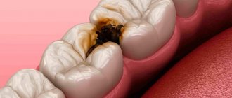
What does specific damage to tooth enamel look like?
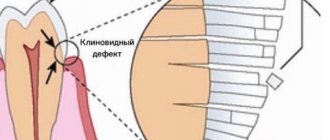
- At the initial stage, the defect looks like a small gap with a depth of only 0.1 mm on the front surface of the crown, so even when examining the oral cavity, it is almost impossible to notice it at this stage.
- As the disease progresses, the enamel is gradually destroyed, the teeth become hypersensitive, and the patient begins to experience serious pain when consuming hot and cold drinks, sweet and sour foods.
- At the third stage, the wedge-shaped defect in the photo already looks V-shaped: on the clouded, discolored enamel, a “wedge” measuring 2–4 mm can be distinguished, the pointed part of which is directed inside the dental crown.
- At the fourth stage, the depth of the defect exceeds 4 mm, and if the pulp chamber of the tooth is affected, there is a serious risk of crown chipping even with a slight mechanical load.
The disease more often affects men than women, and mainly affects symmetrically located premolars, canines and molars of only one jaw. There are also cases of damage to the frontal teeth and incisors, and then the defect also becomes a serious cosmetic problem. As a rule, V-shaped damage to the enamel coating occurs in people aged 30–40 years.
There is another interesting statistic. In right-handed people, wedges on the teeth form on the left side of the dentition, and in left-handed people - on the right, exactly where it is easier and more convenient to brush your teeth, and where greater mechanical force is applied. This is precisely one of the main reasons for the appearance of a wedge-shaped enamel defect: improper brushing technique, aggravated by the use of an excessively hard toothbrush. In the cervical area, the thickness of the enamel is minimal, and it wears off much faster under significant mechanical stress. Unfortunately, pathology arises not only and not so much due to the excessive “zeal” of patients.
What to do after surgery
The recovery period after removing an overgrown root, as a rule, is longer. This is due to the need to cut the gums. Medicines are selected by the doctor individually, depending on the initial condition and complexity of the operation. Typically prescribed:
- Anti-inflammatory drugs. The doctor may prescribe oral anti-inflammatory drugs and rinses.
- Painkillers. After any dental surgery, the patient feels pain. There is no need to take painkillers constantly. If discomfort occurs, you can take any analgesic.
- Healing agents. After surgery, it is important to ensure rapid healing of the wound channel. For better healing of the cut gum and tooth socket, the doctor may prescribe medications that accelerate tissue regeneration and promote rapid healing.
It is important to follow all the doctor’s recommendations, otherwise there is a risk of developing an inflammatory process. After final healing, the patient undergoes prosthetics.
Despite the fact that in the vast majority of cases the overgrown tooth is removed, there are exceptions. For example, if the crown was destroyed due to injury, the root is strong and healthy, a new crown can be built on it. In any case, the doctor makes the decision to remove or preserve the root based on the results of the examination.
What factors provoke the destruction of tooth enamel?
The integrity of tooth enamel also depends on the quality of hygiene procedures when using the “correct” brush. If food debris and soft plaque are not removed from the surface of the teeth in a timely manner, acid-forming pathogenic bacteria begin to actively multiply in them. In this case, the acidity of the environment increases sharply, and under the influence of acid, demineralization of the enamel coating occurs. The loss of calcium leads to the destruction of enamel and the looser and softer dentin located underneath.
It has been noted that a wedge-shaped defect is often formed:
- abuse of carbonated drinks and sour juices;
- in patients with malocclusion - due to non-physiological movement of the jaws chewing food and increased chewing load on individual teeth;
- when correcting the bite with braces;
- in patients with periodontal diseases, accompanied by subsidence of inflamed gums and, accordingly, exposure of the necks of the teeth.
Even properly selected treatment helps to avoid undesirable consequences only if the problem is identified at an early stage. Therefore, patients should promptly seek dental care when the very first signs of pathology are detected.
Treatment of depressions in tooth enamel
As long as the enamel is not seriously damaged, the process can be stopped with the help of fluoride and calcium-containing preparations, as well as sealants. In more complex cases, treatment of depressions in tooth enamel and dentin requires preparation and filling:
Restoring the anatomical shape involves reproducing the tubercles and depressions on the tooth enamel for the full functioning of the organ.
What symptoms of a wedge-shaped defect should you pay attention to?
Painful sensations during caries appear when destroyed dental tissues (the so-called carious cavity) are no longer able to protect the neurovascular bundle located in the pulp chamber. With a non-carious defect, the affected area and decaying dentin are covered by the gum, and the walls of the formed wedge-shaped cavity remain smooth and hard. Therefore, the patient does not begin to experience unpleasant sensations immediately, but only in the third or fourth stages. Treatment of advanced pathology turns out to be more complex, costly, and in some cases ineffective.
Therefore, you should definitely see your doctor if you have:
- individual teeth have become hypersensitive and react to external irritations with sharp, piercing pain;
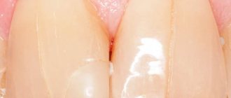
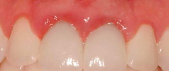
- the enamel has become cloudy, pale or more pigmented;
- The gum line has changed and the necks of the teeth are exposed.
Fillings that destroy teeth
Most people, in fact, do not even remember when and which tooth was treated and turn to the dentist only when the old filling breaks or falls out.
It is believed that modern “light” fillings are the pinnacle of modern dentistry technology, a quick and reliable way to restore even severely damaged teeth.
However, it is not.
Even the most modern filling is a polymer. In essence, it is a technologically advanced type of plastic that is matched to the color of the tooth and cured with light. There are filling materials of different properties, some are more aesthetic, some are more durable, and so on. But at the core, each filling remains a piece of plastic.
And everything would be fine if it weren’t for the hopes for “eternal life” that we place on this miracle of modern dentistry.
Demand always creates supply. Unfortunately, this also applies to dentistry: When a patient comes to the doctor with a broken tooth, the patient asks the doctor to carry out all the treatment quickly, efficiently and with a guarantee.
And if you manage to save money, it will be absolutely wonderful!
All filling materials have significant limitations in their use and service life.
- The size of the filling cannot exceed 50% of the chewing surface of the tooth - even the strongest fillings cannot fully withstand the chewing load, which in an adult can reach 150 - 200 kilograms.
- The filling can be hermetically glued only to that part of the tooth on which there is enamel - often due to exposure of the roots, people develop sensitivity in the necks of the teeth and wedge-shaped defects are formed. It is important to know that in these cases fillings cannot be placed, but other treatment is necessary.
- Only a living tooth can be restored with a filling. When the tooth has been depulped (removal of the nerve) and the canal is hermetically sealed, this tooth needs increased protection, because to access the nerve it is necessary to create a deep cavity in the tooth. This access breaks the tooth frame. In this case, it is imperative to strengthen the tooth with ceramic restoration. Without such protection, the tooth will break due to a chip or crack.
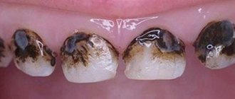
- The seal has a limited service life. On average, every 3-5 years the filling must be replaced due to wear and loss of tightness. Microbes begin to get under the filling and Secondary caries is formed in the tooth - this is a carious process that occurs under the existing restoration. Such a process can develop asymptomatically for a long time, revealing itself only as a darkened filling on the tooth. But in fact, this darkening is tooth decay. Often such destruction leads to tooth loss; the process goes under the gum.
- Fillings disrupt the bite - this is due to the fact that when treating a tooth using filling materials, it is not possible to fully restore its morphology (the anatomical shape of the chewing cusps of the tooth). This leads to incorrect distribution of loads when closing the jaws. Some teeth begin to wear out faster and become overloaded. Ultimately, overload can lead to jaw displacement and/or injury to the temporal joint.
What to do? The answer is simple - get treatment correctly!
To do this, you do not need to graduate from a medical university on your own.
It is important to remember a few rules:
- If you have large fillings, they need to be replaced with ceramic onlays and restorations. This will protect your teeth and bite. It will also restore the beauty of your smile. Today there are even “Crown in an hour” technologies that allow such treatment to be carried out as quickly as possible.
- Do not place fillings near the gums, as they cannot be sealed there. Gum recession is treated by gum grafting (microsurgical treatment).
- Promptly undergo a dental check-up with an inspection of fillings. The inspection is carried out using a special contraster that detects a violation of the tightness of the seals.
- In order for the filling to be installed correctly, the doctor must use a rubber dam to isolate the tooth from saliva and moist air. Before installing the filling, the tooth surface is checked using a caries detector and treated with a special sandblasting machine to enhance the adhesion of the filling. If root canal treatment of a tooth is necessary, you cannot do without a dental microscope and 3D x-ray.
- Regularly undergo preventive examinations with a personal hygienist and follow the recommendations.
Health is the main treasure we have. You need to trust it to professionals who treat each patient with due care and attention.
Smirnov D.V. Ph.D.
Surgical treatment of gum granuloma
Intervention is necessary in cases where conservative medicine has not had the desired effect. It is required when diagnosing destruction processes in the acute stage. Local or general anesthesia can be used for the operation. The simplest option is gingival excision to expose the purulent sac. The pus flows out, the cavity formed is washed and medicine is placed into it for disinfection and pain relief.
Root resection
During the procedure, the dental surgeon acts as follows:
- The gum tissue is peeled away to gain access to the top of the root system.
- The dental canals are opened and cleaned.
- The formed passages are filled with drugs.
- The granuloma is excised along with the upper part of the tooth.
- The place where the inflamed node used to be is filled with a special bone substitute.
- Filling is being done.
Interradicular hemisection of the root
It is necessary in case of severe damage, when it is no longer possible to cut off only the top while maintaining the main size. Key steps of the operation:
- Removal of the area located inside the alveolus, while the plate itself can be saved.
- Cleaning of periodontal tissues.
- Filling the cavity with a special compound.
- Implant installation.
For preventive purposes, it is necessary to monitor the operated area by regularly taking x-rays. This is an effective and fairly simple method that allows you to preserve a living dental plate, and a neat crown can be installed on the remaining areas in the future.
How to cure granuloma on the root of a tooth with cystotomy
This is a gradual elimination of a large focus of inflammation. To remove all the purulent exudate, an artificial canal is created that connects the inflamed node directly to the oral cavity. After removing the infected fluid, the area is treated with antibiotics and the wound is sutured. The resulting holes can be filled with artificial biomaterial - bone cells. This is a long operation, but it is usually performed without complications.
Removal
It is prescribed as a last resort if other procedures have shown to be ineffective, for example, when:
- Complications arise, an advanced stage.
- An external purulent pocket began to form on the gum.
- A longitudinal crack is formed - the dental plate simply splits into two parts.
- Bone tissue is destroyed (the tooth crumbles).
- Origin of root perforation.
- The canals are destroyed.
During the operation, the root system is completely removed, leaving a hole in the periodontium. It is filled with pus that needs to be washed out. 2-3 months after surgery, a pin can be placed on the healed area to place the implant. Additionally, the doctor prescribes rinsing solutions (for example, Chlorhexidine), gels for local anesthesia and disinfection (Cholisal), as well as antibiotics against bacterial infections and painkillers in tablets or injections.
Sealing fissures on the surface of teeth
This procedure is mainly performed on children and consists of sealing the grooves with special fluid materials. Before filling the fissures on the surface of the teeth with sealant, the doctor thoroughly cleans them. A brush, paste, sandblaster or ultrasound can be used for this. If necessary, the grooves are slightly enlarged to ensure that there is no plaque left inside.
The next steps are treatment with a solution of phosphoric acid and filling the recesses on the surface of the teeth with a sealing agent. The material hardens under the light of a curing lamp.
The final point is grinding and polishing. The resulting protective layer remains on the teeth for five to ten years.
Prices for fissure treatment depend on the severity of the problem and the technique used. In general, sealing is less expensive than preparation followed by filling.