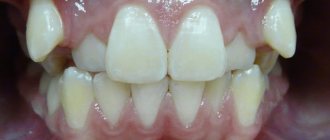
All human teeth seem to be similar to each other. But the structure of the eye teeth, or fangs, as well as their appearance are very different from other species. For this reason, braces are often placed on fangs.
As for other teeth, each person has 4 types:
- front incisors - 2 pieces in one row of teeth;
- fangs - 1 in a row;
- premolars - 2 pieces;
- molars - 2 pieces.
So 4 dentitions make up 28 teeth; subsequently, in most people, 4 wisdom teeth are added to them.
What are each type of teeth?
Let's take a closer look:
- The incisors are the front teeth in the middle of the jaw. They only have one root. They have an external convex and internal, slightly concave surface, as well as small tubercles at the base. They are intended primarily for tearing food into separate pieces (biting off). Due to their sufficient weakness, they are practically not suitable for chewing.
- The canines follow in a row behind the incisors, located in the corners of the jaws. They are equipped with the longest root and crown, so they are quite strong.
- Premolars are located behind the canines. Their chewing surface is much wider and has 2 tubercles for chewing. Children do not have such teeth.
- Molars are the largest size. They are designed to grind food. There are three to five chewing tubercles on the surface.
Anomalies in the development of the dentition
Most often, if we consider all types of teeth, anomalies of an orthodontic nature occur in the canines. Many patients turn to specialists to correct the incorrect position of these teeth. Curvature of the fangs is observed in every third patient of orthodontists.
Types of curvature (dystopia) of fangs:
- fangs protruding from the jaws;
- hidden behind other teeth;
- very short or long;
- expanded;
- did not cut through completely.
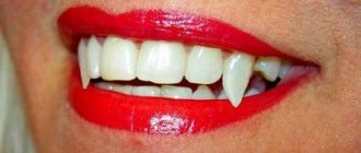
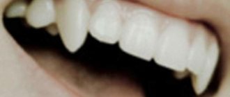
The anomaly of the fangs “vampire smile” not only spoils the aesthetic side of the smile, but also interferes with the correct implementation of the function of eating. In this case, they seem to support the corners of the mouth. Braces can be a great solution to this problem.
Violations of the correct position of the fangs
The most common reason for visiting an orthodontist is the need to straighten incisors . Every third patient needs correction of these teeth.
The manifestation of canine defects occurs in the following situations:
- Covering with other teeth.
- Rotate in different directions.
- Incorrect height or small dimensions.
- Insufficiently cut fangs.
Some call this arrangement of fangs a vampire smile. Meanwhile, the problem is not even an aesthetically unpleasant appearance, but serious disturbances in eating. The corners of the mouth are raised by the fangs, and treatment is only possible with the installation of braces.
Treatment options for canine dystopia
It is quite easy to determine canine dystopia. The complexity of its treatment depends on how advanced the disease is, the age of the patient and the nature of the anomaly.
Main methods of treatment:
- remove a wisdom tooth (when its growth is the cause of the anomaly);
- the use of braces (often you have to remove one of the teeth, usually a premolar, and then the canine is moved to the right place);
- installation of a prosthesis, or transformation of the first premolar into a canine (if it is missing in the row);
- reposition in more severe situations.
Note: The earlier treatment begins, the greater the likelihood that the patient will retain all teeth. For children under 12 years of age, removable plates are used for alignment, and teeth that are not fully formed are quickly aligned (in three to six months).
Teenagers aged 14-15 years old can benefit from braces very well without the need for surgical intervention, but in most cases such intervention is necessary for adults.
Correction methods
The fangs themselves will no longer fall back into place; urgent help from an orthodontist is required and the installation of braces is the only effective means for treating fangs.
Features of treatment, duration, and techniques used will depend on the circumstances of each specific case (age, degree of anomaly, condition of the oral cavity).
When treating adult patients, the canine location is corrected using the following methods:
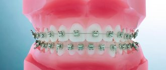
- Braces are installed only on fangs . With the help of the physical impact of the brace design, the fangs will independently move in the desired direction and give evenness to the dentition. Such treatment can accompany a person for many years.
- A faster option is to remove adjacent teeth , after which braces are placed, forcing the fangs to take the correct position within a short period of time. A short treatment period is achieved by creating free space.
During the process of straightening a tooth, patients may experience an unattractive gap between their teeth. This will require bite correction, which will affect the final duration of treatment.
In some cases, only reposition will help, i.e. wisdom tooth removal.
When prescribing a treatment regimen, the orthodontist will take into account the age characteristics of the patient, and timely treatment will allow the alignment course to be completed faster and more efficiently.
Patients are advised to learn in advance about the details that characterize the growth of permanent teeth. The jaw bone is not yet formed until the age of 12. This allows the use of removable plates in the treatment of small patients. As a rule, treatment at a young age is short-term, no more than six months. The fangs can be easily moved, being installed in the right place.
Why can such teeth be crooked?
The canines are the last to erupt. They appear in a child at the age of 9-12, when all the other teeth have already erupted and taken a certain place. Sometimes there is simply no place for fangs in the dentition; it may already be occupied by another dental organ. Then the eye teeth can grow outside the dentition, dystopia is formed.
Another reason for the curvature of the fangs is their discrepancy with the size of the jaw. When a child inherits large teeth from one parent and a small jaw from another, canine anomalies occur quite often.
Anomalies can also be caused by untimely replacement of baby teeth with permanent ones.
Process Features
In most cases, the fangs return to their place within a two-year course of treatment. As the position of the fang is corrected, the face will change, with and without the installation of braces. The most important thing in the treatment process is to find free space for the insertion of fangs. The solution to the problem is to remove the extra tooth.
More often, space is freed up by removing 4-6 teeth, depending on the specific structure of the dentition. If the external protrusion of the canine is slight, it is possible to do without repositioning. The tooth will be moved by the braces, widening the jaw and moving the canine in the desired direction.
Metal and aesthetic types of braces (sapphire, ceramic, etc.) are more common for treating fangs.
How does alignment happen?
It usually takes 2 years to return the fang to its place with the help of braces.
It is important in such a situation to free up space for the fang by widening the jaw or getting rid of a poorly functional tooth.
If the second and fourth teeth are too close together, the quads are most often removed. When the fang only slightly protrudes from the dentition, removal is not required; in this case, it is enough to widen the jaw and at the same time move the dental organs to the right place.
Both regular metal braces and more aesthetic ones (lingual, ceramic, sapphire) can be used to straighten fangs.
It is important to start treatment as early as possible to avoid unpleasant consequences.
Removal of impacted teeth
If there are no complaints at all about the impacted tooth, and it does not cause any inconvenience (cysts do not form near it, the mucous membranes of the mouth are not injured), then removal may not be necessary. But if the impacted tooth is not removed, then it is necessary to undergo an X-ray examination of the mouth 2 times a year.
However, there are a number of cases when it is necessary to remove an impacted tooth:
- Retention causes constant inflammatory processes in the oral cavity;
- The occurrence of various paradental or follicular carpal formations, abscess, osteomyelitis in the gum tissue;
- Injuries and scratches of the oral mucosa that are caused by retention;
- An impacted molar or canine is also dystopic (improperly positioned in the dental arch) towards the cheeks or tongue.
The main factors on which the decision is made to remove impacted teeth are:
- The likelihood of injury during surgery,
- The location of the canine or molar and ease of access to them,
- Possibility of complications.
In most cases, the operation is painless and eliminates the possibility of pain even after the anesthesia wears off.
The removal of an impacted tooth itself takes place in several stages:
- pain relief by local anesthesia,
— an incision in the gum above the impacted canine and removal of a flap of tissue of the mucous membrane and periosteum,
- sawing out the bone to the dental crown using a drill,
- removal of a fang or molar from the bone using forceps (special biomaterials are left in its place).
Removal of premolars located on the upper jaw, canines and molars of the lower jaw has its own characteristics:
- Due to the fact that the roots of the teeth of the upper jaw are sometimes located close to the sinus, their removal must be carried out with the utmost care.
- In the case where the maxillary sinus has nevertheless been opened, medical treatment of the hole is not carried out, and the place where the bone has been broken is immediately sutured.
- Then a biomaterial is placed inside, which will promote rapid and painless overgrowth of bone tissue.
- The previously removed flap of mucous tissue and periosteum is returned to its place and sutures are applied.
- When removing impacted teeth in the lower jaw, the dentist must determine their location as accurately as possible, calculating their proximity to the mandibular canal. The safest way to access such teeth is through the vestibule of the mouth.
Is it possible to fix fangs without braces?
Many patients refuse to use braces. Reasons for refusal, in addition to the aesthetic component of this treatment method, may be the following:
- high cost of design and procedures related to treatment;
- long duration of treatment;
- difficulty to predict the result;
- long period of rehabilitation.
Fangs can also be corrected using other methods. Which one is better suited in a particular situation depends on the complexity of the anomaly and the age of the patient.
Correcting an overbite for children is much easier than for adults. Therefore, there are more ways to achieve this goal.
Alternatives to correcting fangs with braces in children:
- using mouth guards at night;
- the use of soft plates that are removable are quite easy to maintain, but treatment with them will take longer;
- trainers - have a targeted effect on problem teeth, are easy to use, and quite effective (since they are made for a specific defect, which can be corrected much quickly).
For an adult who has serious bite problems, it is better to use braces. Only in cases where the pathology is minor can other treatment methods be used. Such as:
- the use of removable aligners - does not affect the appearance of the smile, they need to be used for 1.5-3 years;
- veneers - used for minor pathology affecting only the appearance, if necessary, quickly correct it; These are thin plates attached to the teeth, do not cause discomfort, are almost invisible, but do not solve the problem itself, improving only the appearance.
The doctor decides whether to use braces to treat the bite or use alternative methods. After a thorough examination and obtaining the necessary results, he applies one or another method.
Whatever structures are used by the patient for treatment, he needs to constantly visit a doctor who will advise and monitor the correct course of the treatment process.
Retention and traction of canines
Impacted teeth are teeth that either have not grown at all or have only partially erupted. Most often, this problem occurs with wisdom teeth. Then the surgeon simply removes them.
But what to do if, for example, fangs could not erupt? A good doctor will try to avoid removal and will prescribe traction.
Why can a canine tooth be impacted?
- Small jaw, which means there is not enough space for teeth
- Early loss of milk precursor
- Abnormal position of the permanent tooth germ
- Consequences of pulpitis of a baby tooth
A tooth is completely impacted when it is difficult to detect by palpation. Then an x-ray helps. The semi-retained tooth is not completely hidden under the gum. It can be detected during a routine examination and through diagnostics. A noticeable gap between the teeth or a “mound” on the gum is a strong argument in favor of contacting a therapist, and then an orthodontist.
Treat immediately!
An impacted canine may not cause any discomfort, but may disturb the bite. It cannot be ignored. The “hidden” tooth tends to erupt, so over time it begins to pose a danger. It puts pressure on other teeth, destroying them, and can cause inflammation.
Stages of treatment
- Surgical. The doctor carefully opens the tooth, making a small incision in the gum.
- Orthodontic. A special system is attached to the exposed tooth, which is fixed to the braces and gradually extends the fang.
An impacted tooth must be removed if it has become a source of infection. But in other cases, the doctor tries to preserve it. It is important to carry out the necessary treatment in a timely manner, otherwise the neighboring teeth will have to be removed over time.
Surgeon's work
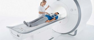
Proper surgical opening of the tooth is a very important stage of treatment. First, a thorough examination is carried out, including a computed tomography scan. The doctor takes into account the patient’s age, structural features of the dental system and the availability of space in the dentition.
What does the surgeon study before surgery?
- Location of blood vessels
- Tooth depth
- Proximity of nerves to the tooth
- Placement of impacted tooth
The problem requires complex treatment. Therefore, the orthodontist and surgeon plan subsequent procedures together.
Bite correction
An impacted canine is rarely the only dental problem. It occurs in conjunction with other malocclusions. The doctor faces a difficult task - to straighten the teeth and integrate the impacted tooth into the dentition. The result of his work should be not only aesthetic, but also functional. The ideal solution would be a braces system.
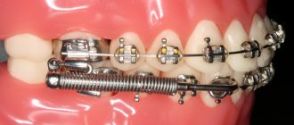
Locks are attached to the teeth to hold the archwire. It is she who moves the teeth, gently influencing them. At first, soft arches are used, and over time they are replaced by harder ones. It is necessary to create additional space for the impacted tooth and only then pull it out.
In addition to the main brace system, additional devices will be required: elastic chains and rods, metal springs and straps, buttons. With these tools, the impacted tooth begins to be pulled out with a very small but constant impact. By the end of treatment, the doctor details the position of the canine and eliminates other dental anomalies. This treatment takes some time. But the result is worth the effort. This is not only a beautiful smile, but also good health.
Commenting system SigComments
Back to section
Correcting the position of fangs with braces
Placing the fangs in the right place and aligning them takes 1-2 years of treatment with braces. This procedure sometimes requires removing one tooth from a row or widening the jaw, thus freeing up the necessary space. For minor curvatures, these procedures do not have to be resorted to.
You can use different braces to straighten your fangs: metal, ceramic, lingual or sapphire. It is better to carry out this treatment in adolescence. This way, the result will be achieved much easier and faster.
Braces can be installed only on the fangs, if there is no curvature of other teeth. It will take several years of treatment to straighten the fangs in such a situation.
You can straighten protruding fangs, depending on how severe the pathology is and its characteristics, in different ways.
Let's consider the average treatment option:
- First you need to cure all organs of the oral cavity, get rid of caries, plaque and stone.
- Then they examine the oral cavity, make impressions, and take x-rays. With their help, the structure is made.
- In order to free up space, the jaw is expanded if necessary or some teeth are removed.
- Next, the braces are installed.
- After the result is achieved, the braces are removed and retainers are selected for the patient. To secure the result, he will need to wear them for some time.
Aesthetic objectives: canines in the lateral incisor position
Very often, dentists do not think about the long-term consequences of their treatment. An incomplete treatment plan or poor treatment choices (often dictated by the patient) usually lead to more serious problems years later. If only we could know what will happen in the future, it would make the task much easier.
However, dentistry can be a predictable subject if approached with proper function and aesthetics in mind. Each dental student receives a phantom - a model that shows the ideal condition and position of the teeth. All dentists are taught the principles of balanced occlusion and the importance of reducing destructive forces.
This article will demonstrate what happens when the factors of time and aging are not taken into account when choosing a treatment method. Very often, what patients themselves do not notice or consider unimportant, about which they do not complain, can turn into a real problem only several years later. If we, dentists, approach the patient from the standpoint of individuality and at the same time as a component of one population, if we carry out careful planning and elaboration of parameters, striving for that same phantom model, then we come to a predictable and long-term quality result. The clinical case discussed here concerns a woman with congenital absence of lateral incisors. When she was a teenager, the maxillary canines were orthodontically moved into position as lateral incisors. In her younger years, her appearance was quite harmonious and attractive, but over time the picture began to change.
Absence of lateral incisors
It is reported that approximately 2% of the population are congenitally missing one or two lateral incisors. Paired absences are more common, and if only one is present, it is usually a microdent. Performing OPTG at an early age makes it possible to find out which of the permanent teeth have not formed.
It is very important to learn about the congenital absence of a tooth at an early age, so that the entire sequence of therapy can be correctly coordinated to restore aesthetics and function. Treatment of congenital absence of lateral incisors is an orthodontically and therapeutically complex task, which is based on the tooth-dental arch size relationship. Numerous studies have been published comparing the method of mesial repositioning of canines and distalization of canines followed by prosthetic restoration of missing incisors.
From a modern point of view, the most logical way is, of course, to open up space and replace missing teeth with prosthetics. The phantom from student life, at least, definitely had his own lateral incisors.
Moving the canine to the lateral incisor position may have several aesthetic and functional disadvantages:
- The canine, a fairly wide tooth, begins to replace the naturally narrow incisor. The color of the canines is usually somewhat darker than that of the lateral incisors, so they stand out from the general plan if they are not in the corners of the smile.
- The level of the gingiva of the canines is approximately similar to that of the central incisors, so moving the canine to the place of the lateral incisor causes visual disharmony.
- Insufficiency appears in the processes of occlusion and articulation, since after movement the canine guidance of the lower and upper canines is not carried out together.
- In the absence of canine protection, the risk of abrasion of the remaining teeth increases, usually consisting of small cracks and microfractures. Over time, periodontal problems and increased sensitivity may appear.
- Long-term retention with an orthodontic retainer will be required to retain the canine in the lateral incisor position.
- The patient may begin to experience TMJ discomfort, muscle tension, grinding, and headaches. This may occur due to effects on muscles that are not anatomically intended.
- The tissues of the vestibule of the oral cavity and the bony prominence in the area of the canine root do not look natural in the area of the lateral incisor. Without the presence of a tubercle in the corners of the mouth, the tissues do not have sufficient support, which leads to recession of the cheeks and narrowing of the buccal corridors. Over time, fewer and fewer teeth become noticeable when smiling.
Orthodontic vision
Traditionally, orthodontists have not encountered people in need of restorations, as they have primarily worked with younger patients. Young people rarely need major restorations. However, in the 21st century, orthodontists often began to take on patients with the need for restoration or due to periodontal disease in the latter. The absence of a lateral incisor is an aesthetic indication, so the orthodontist should treat such a case in this aspect.
The goals of orthodontic treatment often vary depending on the final goal and the need for restoration:
- Positioning of teeth can occur in order to improve the performance of any restoration, for example, to replace a lateral incisor or another tooth.
- There are some advantages to performing permanent or temporary restorations before, during or after orthodontic intervention. This restoration allows you to create the desired shape and at the same time gives an idea of the required space and dimensions. Tooth wear, fractures, underdevelopment in the form of barrel-shaped incisors, etc. are the main indications for restoration before orthodontic treatment.
- Orthodontists sometimes need to reposition a tooth to improve oral hygiene
- Orthodontic treatment may be performed due to periodontal problems, such as insufficient gingival margins, absent gingival papilla, or bone loss.
Today, the goals of orthodontic treatment have diversified significantly, as the aesthetic and functional vision of problems has expanded. Properly planned orthodontic treatment can achieve stable and functional occlusion, improve the health of periodontal tissues and improve dental and facial aesthetics. Orthodontists simply need to study facial aesthetics. Modern specialized literature, research and training always have a positive effect on a specialist and, accordingly, his work results. However, in the past, very little attention was paid to facial aesthetics and periodontal tissue health. A successful orthodontic treatment can be considered an intervention that results not only in the ideal articulation of the models (as well as the achievement of ideal cephalometric relationships and sizes), but also in the restoration of facial aesthetics and harmony in a given position of the teeth.
A smile analysis should include the following: the vertical position of the teeth at rest and when smiling; the transversal (horizontal) dimensions of the smile; the characteristics of the smile arc; and the vertical relationship of the gingival margins to each other. Taking into account all the data, it becomes desirable to move the canines to their natural position and replace the lateral incisors through prosthetics.
Interdisciplinary treatment planning
Innovations in aesthetic dentistry have led to developments in all dental specialties. In today's standard of dental rehabilitation, specialists proceed, first of all, from the individual characteristics of the patient's face and his needs. Each treatment plan begins with an aesthetic assessment. During the analysis, the patient's lips, skin, and cheeks are examined. We must always refer to the position of the teeth in the upper jaw and the level of the gums in relation to the face, and then determine what corrections need to be made for a given appearance. We cannot achieve correct occlusion until our final vision of aesthetics is determined. Aesthetics dictates where the teeth should be located, what the vertical position, guidance and relationship should be.
The main person in the team is the restorative dentist, and the success of teamwork is achieved through detailed discussions of each problem. The main specialist must achieve real therapeutic goals (take into account the economic component, expectations), create an aesthetic vision of the final result, determine the sequence of treatment and restore poorly formed teeth to ideal proportions. The restorative dentist acts here as a link among all specialists, uniting and controlling the manipulations carried out and the pursuit of the final goal.
Clinical case
Diagnosis and treatment plan
A 40-year-old woman came to the clinic (in 2003) in need of cosmetic correction of treatment performed in adolescence.
The patient had a congenital absence of lateral incisors, so she underwent orthodontic treatment at the age of 14 years to solve this problem. Relying on the orthodontist and unaware of other treatment options, the parents chose (or allowed) to move the girl's canines to the lateral incisor position. After the treatment, the smile did not look attractive enough, but the patient was able to get used to it. But as she reached middle age, the woman began to notice that her interlocutors were increasingly staring at her teeth. This made her insecure. The canine was darker, had a rounded contour and a gum level that was in disharmony with the rest of the teeth. The buccal corridors were narrowed to close the empty space, creating the appearance that the cheeks were somewhat sunken (Photos 1 and 2). The patient remained deeply dissatisfied with her appearance.
Photo 1: In 2003, a 40-year-old patient presented with an aesthetic problem.
Photo 2: The canines were moved to the position of the lateral incisors, which stood out from the overall appearance.
With the advent of the 21st century, there have been significant changes in dental technology and materials. Aesthetic manipulation has become common and routine. In this situation, should we have moved the canines to their correct position and replaced the lateral incisors? The patient was open to any options.
After weighing all our options, we decided not to move the canines back and place titanium implants. Although this would be the most common solution to the problem, this path did not seem entirely justified to us. Such treatment would take approximately 18 months, and there was also the possibility of resorption and the need for bone grafting. Since the patient clearly wanted to change the shape and color of her front teeth, an easier and more predictable alternative was in development.
After analyzing radiographs, photographs and working models, we decided that after flattening and narrowing the canine, we could use light orthodontic extrusion of the teeth, which would realign the ridge bone and create a new harmonious appearance at the soft tissue level. By leaving space distal to the canine, we can create porcelain veneers to correct the shape of the canines to resemble the lateral incisors and the first premolars to resemble the canines. Placing veneers on the side teeth will also be possible if the patient so desires. According to the plan, the treatment was supposed to take approximately 6 months. It is important to evaluate all the benefits and risks of any of the treatment options, and the chosen option already needs final approval by the patient himself.
Since the patient expressed a desire to install veneers on the teeth of the anterior zone, all we had to do was correct the position of the lateral incisors and canines.
Clinical protocol
The labial surface of the canines was flattened and the mesial and distal contours were tapered using a flame-shaped diamond bur (Dental Savings Club) followed by finishing with strips (Integra Miltex). Ormco Saphire (Ormco) braces are fixed on the upper jaw up to the first molar (Photo 3). Using Ni-Ti wires of increasing diameter from 0.13 to 0.16 with an extrusion force of 15 g, it was decided to carry out labilization of the central incisors and extrusion of the canines to correct the gingival level (Photo 4). The use of orthodontic extrusion to modify soft and hard tissues was first described in the literature by Heithersay and Ingber. Low-intensity forces (up to 30 g) stimulate marginal positioning of the bone crest, behind which movement of the gums occurs. Restructuring of the alveoli occurs along with the movement of the root.
Photo 3: Preparation for orthodontic treatment: the labial surface of the canines was slightly flattened, and the contours on the proximal sides were narrowed.
Photo 4: Ni-Ti arch with a force of 15 g is used to labilize the central incisors and extrusion of the gingival level of the canines.
Extrusion of the canines by 1 mm was carried out over 3 months, followed by stabilization for 3 months with a Ni-Ti arch 18x25. The total time spent on treatment coincided with the plan and amounted to 6 months. The gingival level of the canines is corrected and lowered, the canines are ready for restoration according to the proportions of the lateral incisors. For the formation of “fangs” from premolars, space is left distal to the true canines (Photo 5a-6). The braces are removed, leaving the teeth in an ideal position for further restoration with porcelain veneers (Photo 7).
Photos 5a and 5b. Reduced gingival level at the lateral incisor.
Photo 6: Space is left at the distal surface of the canine for restoration of the premolar in the shape of the canine.
Photo 7: After removing the braces, the teeth are left in an ideal position for further restoration.
A vinyl polysiloxane (VPS) impression was made, followed by a diagnostic wax-up of the anterior 6 teeth (Figure 8). At this stage, it was decided to evaluate the esthetics of the anterior sextant before widening the buccal corridor.
Photo 8: Diagnostic wax-up creating the ideal appearance of the 6 front teeth.
Conservative preparation (0.3 mm) was carried out (Photo 9) and the final VPS impression was taken (Honigum DMG America) using conventional and lightweight techniques. Bite registration was carried out by Luxabite (DMG America). Using the classic VITA scale, shade A1 was determined. Temporary structures made of bis-acrylic material (DMG America), made on the basis of a diagnostic wax-up matrix, are fixed to the prepared teeth. Fixation was carried out using Temp Bond Clear (Kerr). Next, the appearance of the structures was corrected with soft diamond and Flexi discs (Cosmedent) (Photo 10).
Photo 9: The teeth were prepared by 0.3 mm to install ceramic veneers.
Photo 10: Luxatemp temporary structures (DMG America), creating a natural smile appearance.
Models are cast from plaster (Photos 11a and 11b) and then porcelain veneers are made (Photos 12a and 12b).
Photos 11a and 11b. The Geller technique allows you to emphasize the contour and level of the gums.
Photos 12a and 12b. Veneers made from Creation Porcelain (Jensen Dental).
The veneers were fixed with colorless cement (Variolink Veneer Ivoclar Vivadent) in 2004. The patient was satisfied. Ultimately, she adjusted her smile to the desired result. The size and shape of the front teeth are made according to golden proportions, so it is almost impossible to determine the congenital absence of incisors. Premolars are shaped like canines. At this stage, the slightly reduced buccal corridor did not bother the patient at all.
After some time
Ten years later, in 2013, the patient returned to the clinic. Now the lateral teeth caused aesthetic dissatisfaction (Photo 15). The upper second premolars and molars on both sides were prepared (Photos 16a and 16b), then a suitable shade was selected (Photo 17). The restorations were made from Creation Porcelain (Photo 18). The structures were fixed using transparent cement for veneers (Variolink Veneer). The resulting result looked uniform, as if created at the same time (Photos 19 and 20). The patient was satisfied.
Photo 13: Completed treatment in 2004.
Figure 14: Despite the narrowing in the lateral areas, the patient was satisfied with the aesthetics of the anterior segment.
Photo 15: By 2013, the patient wanted fuller buccal corridors.
Photos 16a and 16b: Preparation of second premolars and first molars.
Photo 17: Shade selection according to the VITA scale.
Photo 18: Making ceramic restorations (Creation Porcelain).
Photos 19 and 20: Completed treatment in 2013.
Final comment
People often change their minds, so it is difficult to predict what a patient who was treated as a teenager will want. A careful treatment plan helps overcome many of the negative factors associated with subsequent gradual aging. A careful discussion of all the details allows for high-quality rehabilitation, as well as building a trusting relationship with the patient.
Author: Elliot Mechanic , DDS, BSc
Pros and cons of orthodontic construction
Braces have the following advantages:
- With their help, you can cope with almost any jaw anomaly, regardless of the stage of curvature.
- There are practically no contraindications to their use.
- Already in the first months of treatment, the result is noticeable.
- Suitable for both adults and children.
- The installation procedure takes place exclusively in a dental clinic. The patient himself does not need to take them off and put them on, since the design is non-removable.
- The latest developments in braces are almost invisible to others.
Disadvantages of designs:
- The need for more thorough oral hygiene. In addition to teeth, braces also need to be thoroughly cleaned to prevent diseases such as caries, periodontitis and gingivitis from developing. This procedure should be performed after every meal.
- Long-term wearing design.
- Aesthetic side. Not all braces look attractive. But there are many of them that are practically invisible on the teeth (ceramic, lingual).
- High price when compared with other correction systems.
Failure Cases
Correcting fangs with braces is a procedure that leads to excellent results. But some patients refuse such treatment.
Let's look at the reasons for this refusal:
- Long term treatment, during which you need to wear braces.
- The procedure is quite expensive.
- Difficulties in predicting the final result.
- Long recovery period after treatment.
The choice is always up to the patient, and canines can also be corrected in case of refusal of braces using mouthguards, trainers or soft plates. But these types of treatments can only help at a young age; they are not suitable for adults.
Only with slight protrusion of fangs in adults can they be corrected using mouthguards.
Mouthguards are worn at night. Soft plates are much easier to maintain compared to braces, but the duration of treatment with their help is quite longer. Trainers are convenient and act on the existing defect in a targeted manner.
For adults, lumineers and veneers can also be an alternative to braces. They are placed on the back of the teeth. But their effect will only improve the appearance, without curing the disease itself.
A word from the dentists
Anna Ivanova, orthodontist:
From a medical point of view, the use of braces is primarily necessary to correct the bite and prevent the occurrence of diseases associated with this problem.
These designs are most effective in correcting jaw abnormalities. This is clearly noticeable when comparing the situation before and after treatment. In addition to a beautiful smile, they provide the patient with natural chewing function and speech. In addition, the use of braces has almost no contraindications.
Inna Mutasova, orthodontist:
Wearing braces should not embarrass an adult in any way, but crooked teeth and an incorrect bite are a sufficient reason for embarrassment. Don't miss the opportunity to have a perfect smile. Without braces, serious defects cannot be eliminated.
Orthodontic treatment with braces
Visits to the orthodontist took place every 1.5-2 months. Each time, the orthodontist pays attention to hygiene, monitors the progress of treatment, carries out the manipulations outlined in the treatment plan - changes the arches, adds the necessary elements to the brace system.
The protruding fang fell into place a month after the start of treatment. But the treatment ends only when a stable correct position of all teeth has been achieved and physiological interdental contacts have been created.
While wearing braces, in addition to brushing your teeth twice a day, it is recommended to have your teeth professionally cleaned every six months. This will help remove plaque accumulation in hard-to-reach places (around braces and gums, under arches). Long-term accumulation of plaque leads to demineralization of the enamel and the appearance of white spots on the teeth, which become very noticeable after the braces are removed.
Before brushing your teeth with Air Flow:
After professional teeth cleaning with Air Flow:
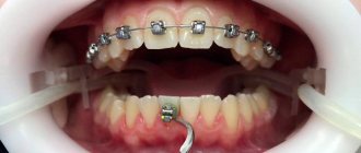
To correct the bite, you need to wear orthodontic elastics - elastic rings of different lengths, which are attached to special hooks on braces or arches. The child must secure the oroelastics himself, changing them daily. The orthodontist shows how the elastics are secured and checks whether the patient has understood everything correctly.
It is better for parents to monitor this process, since the duration and result of orthodontic treatment depends on the correct wearing of elastics. You should always have spare elastics with you, since breaks in wearing are extremely undesirable, and if one elastic breaks, both must be changed so that the traction is symmetrical.
After 1 year and 1 month:
Patient reviews
Alla, 30 years old
I agreed to install braces after the doctor said that if the bite is not corrected, I could subsequently lose all my teeth. The structures had to be worn for about two years. The result was truly a perfect smile. But after a while, the distances between the teeth became noticeable. It turned out that the dentist forgot to prescribe mouth guards. Now I wear them all the time, and my smile is gradually returning to normal.
Alexander, 41 years old
My son wore braces for almost 2 years. There were unpleasant sensations after installation and tightening. Could only eat liquid food. With the appearance of wisdom teeth, the teeth began to bunch up again. So, the effect of braces is there, but it is short-lived.
Olga, 26 years old
I had braces for over a year. It took me a long time to choose a clinic and a specialist who would install them for me. After installation, the unpleasant time of correction began. She devoted a huge amount of time to oral hygiene. I used all kinds of cleaning methods.
At the beginning of treatment, I experienced severe pain and was on painkillers. But now I’m just happy and very grateful to my doctor. The teeth are straight, the smile is almost perfect. The only thing is that I still wear mouth guards at night.