How does dental sinusitis develop?
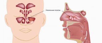
The paranasal sinuses (sinus paranasales) consist of a system of several cavities in the near-nasal space.
In the case of a cold, less ventilated sinuses are especially susceptible to developing sinusitis. The maxillary sinuses (sinus maxillaris, maxillary) are relatively well ventilated. However, the floor of the maxillary sinus is separated only by a narrow bony plate from the molars of the upper jaw. Due to this anatomy, the development of dental (odontogenic) sinusitis is quite common. The main cause of odontogenic sinusitis may be inflammation that forms in the area of the roots of the teeth and easily spreads to the mucous membrane of the maxillary sinus. Common pathogens include bacteria such as:
- Streptococcus pneumonia - streptococcus;
- Haemophilus influenza – hemophilus influenzae;
- Moraxella catarrhalis is a protobacterium of Moraxella.
Odontogenic maxillary sinusitis can also form due to tooth extraction (extraction). If one of the maxillary molars is removed, with damage to the bone grafting of the upper jaw, bacteria from the oral cavity can enter the sinus. In this case, they talk about the formation of an unnatural connection between the oral cavity and the paranasal sinus - an oroantral fistula. This trigger factor is considered one of the common causes of “dental” sinusitis.
A third dental cause of sinusitis involves inflamed roots that go unnoticed. As a result, cysts are formed that “grow” into the sinus cavity.
Lay down straws
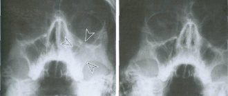
Today it is not difficult to assess the risk of developing odontogenic sinusitis. Formally, this is the result of the close contact of the maxillary sinus with the apices - the tips of the roots of the upper teeth of the chewing group. Therefore, a simple and effective diagnostic tool is a panoramic photograph of the jaw. The doctor will receive up to 90% of the information about her condition. The rest, if necessary, will be given individual photographs of each tooth. Are your teeth in satisfactory condition? Then you can repeat the examination every 2-3 years!
The development of odontogenic sinusitis is always facilitated by weakened immunity. Moreover, the state of the immune system is directly related to dental health. This means that treatment should always begin with sanitation of the oral cavity.
Acute and chronic odontogenic sinusitis
This form of sinusitis is quite painful. This occurs due to the connection between the acute form and inflammation in the area of the tooth root. However, if there are always persistent dental problems, acute sinusitis can develop into chronic inflammation of the antral sinus. The two forms of sinusitis differ in their symptoms.
Acute dental sinusitis manifests itself:
- Severe throbbing pain;
- Swelling around the cheek (can reach up to the eyelid);
- Redness of the nasal wall and turbinates;
- Secretion from the nose is mucopurulent in nature.
In addition, pressing on the affected area may cause pain. Acute dental sinusitis is usually accompanied by fever.
Signs of the chronic form of odontogenic sinusitis are often much less pronounced. Some patients experience symptoms only occasionally, such as occasional headaches.
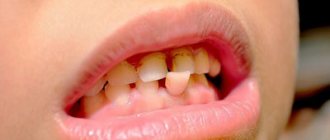
I needed to take care of my teeth!
The cause of the disease can be not only a poorly placed filling. Odontogenic sinusitis can also occur due to individual structural features of the upper jaw. But in the vast majority of cases, the disease is caused by improper dental care. For example, a patient starts caries, and necrotic decay of the pulp (dental nerve) begins. The products of necrosis cause inflammation in the perihilar tissues, which then develops through the membrane of the maxillary sinus.
Article on the topic
Expert advice: How to prevent sinusitis?
If, due to a long-term inflammatory process of the upper “sixes”, the tooth has to be removed, an additional problem may arise – a fistula. Then the only solution is surgery: a flap is cut out from the side of the oral cavity, which is used to cover the tooth socket. If the infection has managed to lead to the development of sinusitis, then it is necessary to operate on the maxillary sinus.
However, sometimes the tooth can still be saved. In such cases, the doctor carefully moves the gum with special instruments, cuts out a piece of the root, filling the void with artificial bone tissue and treating the surgical site with medications. Naturally, therapy is simultaneously prescribed for the mucous membrane of the maxillary sinus - this is the prerogative of the otolaryngologist. But it should be noted that healing is a fairly long process, at least a month to a month and a half.
Diagnosis of “dental” sinusitis
Inflammation of the antrum can have several causes and does not necessarily have to come from the teeth. Since treatment should always be cause-and-effect, the doctor must make an accurate diagnosis. In the context of odontogenic sinusitis, unilateral onset of symptoms is typical. Other complaints, such as pain, which is usually worse when bending over, are additional symptoms.
Further investigations include rhinoscopy (nasal endoscopy) and imaging techniques:
- X-ray examinations;
- CT (computed tomography);
- DVT (digital volumetric tomography);
How does an infection in a tooth cause the lining of the maxillary sinus to thicken?
The apices of the roots of the teeth are located under the Schneiderian membrane, that is, under the mucous membrane that lines the bottom of the maxillary sinus.
This is the normal anatomical structure of the sinus - when the tops of the teeth are located in it. Some people have the tops of the fourth, fifth, sixth, seventh there. Some people only have the sixth. Some have sixth and fifth. But, one way or another, they are almost always there.
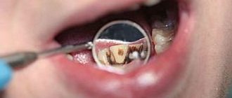
When a tooth is affected by caries, the infection penetrates through the dentinal tubules into the pulp chamber. Through it, the infection slowly progresses further through the blood and lymphatic vessels into the periodontium of the tooth.
Pulp microvessels emerge from the periodontium directly under the mucous membrane of the sinus, and over time it begins to react with inflammation.
Immune system cells responsible for protecting against infection initiate chronic inflammation. And the mucous membrane gradually thickens. This irritation happens all the time.
Infection from the carious process, which is thus restrained by immunocytes, causes a gradual degeneration of the structure of the sinus lining in the form of polyps and cysts.
Implant into a modified sinus - is it possible?
Some time after tooth extraction, the patient may decide to install an implant. If sinus treatment was not carried out in a timely manner after tooth extraction, it remains to treat sinusitis before sinus lift surgery and implant installation.
It is advisable to do this together with an otolaryngologist.
But at the same time, the patient must understand that during a sinus lift, the altered sinus may rupture because it has an altered structure. And if this does happen, the sinus lift may not take place. Therefore, odontogenic sinusitis still needs to be treated during tooth extraction. And prepare the sinus together with the otolaryngologist.
Treatment of pain in the head and nose
Treatment tactics are selected individually, depending on the exact diagnosis and the nature of the disease. Most diseases can be treated with medication, but in some cases surgery is necessary. The course may include the following stages:
- taking antibiotics is a mandatory measure for bacterial diseases, purulent inflammation, injuries, sinusitis;
- supportive treatment, which includes anti-inflammatory and vasoconstrictor drugs, antipyretics and analgesics;
- antiviral drugs;
- surgical treatment - consists of opening and washing abscesses, boils, as well as sanitizing the paranasal sinuses for sinusitis.
Doctors at the Clinical Institute of the Brain will select the most effective treatment regimen, which will include only the necessary steps. The drugs can be taken on schedule at home, only in some cases treatment in a hospital is indicated. The patient remains to follow all recommendations and regularly return for re-examination.
Prevention methods
Doctors have several recommendations on how to prevent the manifestation of the disease, which causes pain in the nose and head. They include the following activities:
- support for general immunity, including proper nutrition, sufficient vitamins, regular physical activity;
- tests for suspected allergies to identify the irritant and stop contact with it;
- timely diagnosis to identify sinusitis, neoplasms and other dangerous diseases that may progress over time.
The Clinical Brain Institute specializes in the diagnosis and treatment of all types of headaches, including those associated with nasal diseases. To receive recommendations for treatment and prevention, all you need to do is make an appointment and undergo a full examination. Our center is equipped with modern tools for diagnostics and treatment procedures, and our doctors are experienced specialists in a narrow and broad profile.
Clinical Brain Institute Rating: 5/5 — 2 votes
Share article on social networks
The relationship between nose and head pain
The nose is one of the sections of the respiratory system. It consists of several departments interconnected. If diseases occur on the visible part of the nose, they are easy to diagnose. However, nasal pain can be caused by damage to the internal parts that are located inside the skull. To understand why the nose hurts and radiates to the head, it is worth familiarizing yourself with its structure.
- The outer nose is the visible part. Its shape is formed by cartilage and several bones, as well as facial muscles. This is the first section into which the inhaled air penetrates, so it is initially purified from small particles and microorganisms.
- The nasal cavity is an expansion formed by bones and cartilage. Its posterior part narrows and passes into the nasopharynx.
- Paranasal sinuses (paranasal sinuses) are additional cavities that are located on opposite sides of the nasal cavity. There are anterior and posterior sinuses.
All parts of the nose are covered with mucous membrane. It is continuous, contains nerves and vessels, and connects to the epithelial layer of the oropharynx. The nasal mucosa has a distinctive feature - it contains olfactory receptors, with the help of which the nervous system is able to distinguish odors. In addition, it is the first barrier against viruses and bacteria that enter the body with inhaled air and can cause infectious diseases of the respiratory system.
Diseases that cause nasal congestion and discomfort reduce the supply of oxygen to the lower respiratory system. This causes insufficient nutrition of tissues, especially brain cells. Most colds are accompanied by a headache, which occurs due to respiratory failure. In addition, colds are accompanied by swelling of the mucous membrane, inflammatory processes, and increased formation of exudate.
How to treat?
If nothing serious was found in the images, sinusitis can be overcome conservatively, that is, with the help of medications and a number of physical procedures. Treatment must begin immediately, otherwise the implant may be rejected due to the inflammatory process.
If during diagnostic procedures damage to the maxillary sinuses was discovered, doctors will have to perform surgery. It looks like this:
- Administration of an anesthetic.
- Creating access to the damaged sinus.
- Drain accumulated liquid out.
- If necessary, the artificial structure is removed.
- Antiseptic treatment is carried out.
- Stitching.
In the future, the situation is resolved with the help of medications. A course of antibiotics is indicated, preferably in the form of injections. The course must last at least 14 days. Nasal drops are also prescribed to create free breathing.