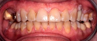
There are several types of malocclusion. Rare teeth are the most common. It is characterized by the presence of very wide spaces between dental units. At the same time, the chewing structures are externally similar to a comb with sparse teeth. This pathological condition cannot be ignored, as it can lead to the development of dangerous complications. Currently, there are several methods for treating rare teeth (a photo of the defect is presented below). The feasibility of using a particular method is assessed by the orthodontist.
Causes and consequences
The “culprit” of the pathology is a genetic factor. Large distances between chewing units are noticeable already at the stage of eruption of milk teeth. In extremely rare cases, pathology develops against the background of progression of dental diseases.
Rare teeth in humans are not only unsightly, but also dangerous to health. Initially, psycho-emotional instability appears. It is caused by complexes that have arisen and low self-esteem.
In addition, sparse teeth cause the following pathological conditions:
- Disorders of the gastrointestinal tract. Wide spaces between dental units do not allow for proper chewing of food. As a result, food enters the stomach in an insufficiently processed form. The natural consequence is indigestion.
- Tooth loss. Chewing units located far from each other are characterized by increased fragility. In addition, they are extremely vulnerable to any external influences.
- Inflammatory processes in the oral cavity. When there are large gaps between teeth, soft tissues also become more vulnerable to external influences. They are easily injured, and when pathogenic microorganisms penetrate the oral cavity, an inflammatory process develops.
To prevent the development of complications and make your smile perfect, you need to contact an orthodontist. Only a doctor can provide information on how to correct rare teeth in each specific case.
Do you want beautiful dental restoration?
8
or order a free call
Request a call
How the shades of cement were selected during fixation
The work on dental restoration was varied, and we worked with both crowns and dental modules:
- the upper anterior teeth, which were restored using “crown + tooth root” modules, were fixed with double-curing cement,
- and those teeth on which I placed veneers and crowns were fixed with light-curing cement so that in bright daylight the glue would not be noticeable
Light reflecting off the crown or veneers can make the luting cement visible. In addition, due to the fact that the working surfaces of the teeth were of a non-uniform color, with the presence of a yellow tint, it was necessary to work with different colors of cement. That is, I used more yellow cement where the roots of the teeth were and used light-neutral-colored cement on the veneers and crowns to achieve a uniform color scheme. It's very difficult to get the color perfect
.
But we managed to do
:
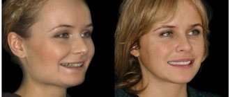
Naturally, Aryuna is now the most super beautiful:
Aryuna, as I said earlier, works on television, she is a fairly public person - she interviews TV and show business stars. She appears on camera very often, and for her such a smile is not only a guarantee of her good mood, but also great confidence in herself and her abilities. After all, an open and cool smile is the key to female success
! This job, according to her, is the ideal solution for her. Made your smile easy, quick and beautiful!
Aryuna left a video review, short but very charming
:
And in conclusion of my story about this case of restoration of the front teeth, let’s once again go through all the stages
of transforming Aryuna’s smile:
- Photo BEFORE the start of
dental restoration. Aryuna came to me with such a smile; she has old temporary structures. - View of temporary structures on the 6 anterior teeth of the upper jaw.
- Temporary structures were removed and teeth and tooth roots were prepared for prosthetics.
- Selecting the color of the veneers.
- New temporary structures were installed. Your smile transformation has begun!
- Aryuna's smile with new temporary structures.
- In the meantime, we prepared the permanent teeth. This is how they “sit” on the model.
- And this is what all the elements of the teeth that we will install for our patient look like
- The teeth have been installed. Close-up - looks great!
- View of a smile on the right
- View of a smile from the left
- FINISH!
ARYUNA'S SMILE WITH NEW TEETH!
Author:
Sergey Samsakov, orthopedic dentist was born on 02/02/1989.
Education:
2011 — Graduated from the Moscow State Medical and Dental University named after. A.I.Evdokimova
2012 — Internship in the specialty “Orthopedic Dentistry”, Moscow State Medical University named after. A.I.Evdokimova
2014 - Residency in Orthopedic Dentistry, Moscow State Medical University named after. A.I.Evdokimova
Diagnostics
After the examination, the orthodontist will refer the patient for a comprehensive examination. Based on its results, the doctor will be able to make a decision on what to do with the rare teeth of the person who contacts him.
Diagnosis may include the following studies:
- Orthopantomogram. This is a three-dimensional shot of the jaws.
- X-ray examination.
- Computer diagnostics. The method is considered the most accurate and informative. It allows you to evaluate both the structure of the jaw and the location of the teeth.
Based on the diagnostic results, the orthodontist will select the most effective method of treating rare teeth. It is important to understand that it is impossible to correct the genetic factor, but doctors are able to disguise the defect so that the patient’s smile is perfect. Contrary to popular belief, if adults have sparse teeth, braces are powerless. This method of correcting the bite is not used if a person has large distances between the teeth.
Preparing for treatment
Elimination of the defect requires a comprehensive examination of the oral cavity and selection of optimal treatment. In particular, the doctor assesses the size of the pathological holes. An x-ray examination is also prescribed, which allows you to identify the presence of hidden injuries. Regardless of the chosen scheme, before starting treatment of rare teeth, therapy for caries and other pathologies of the oral cavity is prescribed.
Self-medication is dangerous with complications!
Attention
Despite the fact that our articles are based on trusted sources and have been tested by practicing doctors, the same symptoms can be signs of different diseases, and the disease may not proceed according to the textbook.
Pros of seeing a doctor:
- Only a specialist will prescribe suitable medications.
- Recovery will be easier and faster.
- The doctor will monitor the course of the disease and help avoid complications.
find a doctor
Do not try to treat yourself - consult a specialist.
The most difficult operation to correct a malocclusion is implantation. Before it is performed, a panoramic photograph of the jaw is taken, which allows you to select the appropriate pin size.
Implantation is contraindicated in many cases. It cannot be used in the presence of oncological pathologies, tuberculosis, a number of chronic diseases and allergies to anesthesia. Implantation takes several hours. During the procedure, tooth extension is carried out in three projections: width, height and length.
After the operation, the patient should follow certain procedures for several months to prevent the development of complications.
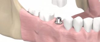
Dental implantation
The technique is the most radical. The essence of the procedure is to replace rare dental units with prostheses. The latter are screwed into the jaw using a titanium pin that imitates a tooth root.
The main advantage of implants is that they are as similar as possible to natural chewing units and fully perform the functions of the latter. As a result, food is chewed efficiently and the load between the teeth is distributed evenly. As a result, the process of forming a correct bite begins.
In addition, implanted teeth are permanent. In other words, they do not need to be removed and cared for separately.
Dental implantation is contraindicated if the patient has type II diabetes mellitus, HIV, hepatitis and severe heart pathologies.
How was the restoration of the front teeth carried out?
So, Aryuna had problem 4 front teeth; crowns and missing modules needed to be made on them:
- two missing modules with tooth roots,
- two crowns.
And we also decided to use the two upper canines
— put two Luxury veneers on them to make Aryuna’s smile more complete. Why did you decide to use fangs? – The canines had to be included in the group for restoring the aesthetics of a smile, because the 4 restored teeth will eventually, after 5 years, be noticeable in color - their own teeth will darken, but the veneers will not.
We ended up working with six teeth
.
Stages of smile restoration:
My first task
temporary crowns were removed and four teeth were prepared: two for a crown and two for monolithic crown + tooth root structures:
Second task
I had to minimally prepare the two upper canines for veneers, which I did.
I treated the fangs with micro-grinding. I also made six temporary structures in the first appointment. smile
immediately :
We removed the old temporary crowns, ground the teeth and made new temporary orthopedic structures, and after 4 days we received laboratory veneers: the color of the neck is 1M2, and the body of the tooth is 1M1:
At the second appointment, I removed the temporary structures, installed all six new teeth for fitting, and the patient really liked everything. And after that I recorded them permanently.
Installation of a bridge
The use of fixed orthodontic structures is a modern way to fill wide gaps in the dentition.
In order for the prosthesis to be securely fixed, it is necessary to have supporting dental units. It is on them that the entire structure is attached.
The best option is to make a bridge from metal ceramics. The service life of this design is 7 years or more. At the same time, the bridge retains its original shade and does not differ from adjacent natural teeth.
Contraindications to installing a prosthesis on rare teeth:
- Acute periodontitis.
- Parafunctions of the masticatory muscles.
- Insufficient height of supporting teeth.
If there are absolute contraindications, the doctor makes a decision regarding the advisability of using a different treatment method.
Why does bone tissue decrease when teeth are missing?
Very often, when planning dental restoration using implants, we are faced with a deficiency of bone tissue, which is accurately detected during computed tomography:
Let's figure out what it is and why it happens
Let's start with the fact that bone is a living tissue, with its own laws of life. In the case of implantation, we are talking about the alveolar bone, that is, the bone of the alveolar process - the bone where the roots of the teeth are located. And when we talk about bone deficiency, we are talking about a deficiency of the alveolar bone. This bone provides support and stability to teeth. Its entire structure is aimed at stabilization.
And when teeth are lost, the need for this bone disappears and its resorption or resorption occurs. Loss of alveolar bone can also occur with preserved teeth due to periodontitis. The loss of bone tissue increases many times over when using removable dentures, since the denture exerts unusual pressure on the gum and bone, pressure for which the bone was not “designed” to bear. As a result, we are faced with a situation where the bone - where the implants can be installed - is missing or insufficient.
An example of what can happen to the bone tissue of a patient who had a tooth removed more than 15 years ago and did not go to the doctor only because he was afraid of the sinus lift procedure is shown in detail in my article “He was afraid of the sinus lift and waited... 15 (!) years"
We will talk about methods of treating adentia in the next article.
Sealing
Currently, it is one of the most affordable ways to treat rare teeth. Its wide distribution is due to its low cost and ease of implementation.
The essence of the method is as follows: fillings of the same color as the natural chewing units are applied to the teeth. The procedure is considered a jewelry procedure, since the doctor must literally mold new bone structures. As a result, the teeth become wider and the gaps between them disappear. Only a specialist can visually distinguish fillings from natural dental units.
During the procedure, orthodontists use only high-quality materials. These are ceramics and porcelain.
After filling rare teeth, the functioning of the gastrointestinal tract is normalized and a correct bite begins to form.
Note: computer necrosis of teeth
Experts started talking about this non-carious lesion of teeth relatively recently; it definitely did not exist in the 20th century, however, like people who spent most of their working and free time at the computer. The death of dental tissue threatens patients who are ready to sit at a computer for more than 8 hours a day, without days off and a full night’s rest. After 3–5 years of living in this mode, a person (and these are mainly young people under 35 years old) are guaranteed to have problems with the front teeth located in the path of the electrostatic radiation of the monitor.
Symptoms of computer necrosis of teeth
Experts, comparing computer and post-radiation lesions, find much in common between them: dying areas in both cases spread over the entire surface of the supragingival part of the teeth, which acquires a dark, almost black color. The focus of necrosis is filled with softened brown tooth tissues; the dentist easily removes them with special instruments, and the patient does not feel pain. As for the teeth of the chewing group, which are exposed to relatively less computer radiation, they also do not look the best: the enamel loses its shine and acquires a gray tint. Studies have shown that not only hard tissues undergo negative changes, but also the pulp. Thus, during electroodontometry, the neurovascular bundle does not respond to an electric current of 25-30 μA. In addition, changes in the functioning of the salivary glands are observed.
Treatment of computer necrosis of teeth
The “newness” of the disease complicates diagnosis, since the dentist may never have encountered it. An X-ray examination is required (sight image, orthopantomogram, computed tomogram), in the images the teeth look translucent and have unclear contours, which indicates a deficiency of mineral substances.
Treatment of computer necrosis is complicated by the fact that during the removal of damaged tissue with a bur, the tooth falls apart; in such conditions, it is impossible to preserve the vitality of the pulp, and often the tooth itself. If recovery is possible, it will take a lot of time, as it will be carried out in several stages, including local and general therapy, preceding filling or prosthetics. At the first stage of treatment, dead tissue is removed and healthy tissue is remineralized, and the patient is recommended to take mineral-vitamin complexes containing calcium, antioxidants, and vitamin A. After the condition improves, therapeutic linings are placed in the dental cavity and a temporary filling is performed. The result of this stage of treatment, which takes up to two months, is the restoration of dentin, after which the tooth is finally filled.
Prevention of computer necrosis of teeth
Computer necrosis of teeth can be prevented by a rational approach to working at the computer. Firstly, comply with sanitary standards, which require organizing a work area of at least 6 sq.m., located in a room with an area of 20 - 24 sq.m. The monitor should be located no closer than 60 - 70 cm from the face; if there are several computers installed in a room, the distance between them should be at least 2 m. Every two hours you should take a break of 15–20 minutes, during which the room is ventilated. Secondly, monitor your diet; if you don’t get enough antioxidants, vitamins and microelements with food, then take vitamin-mineral complexes. Thirdly, use new models of monitors that have minimal electrostatic effect on the body.
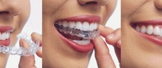
Installation of veneers
This is a highly effective correction method. Veneers are thin plates made of ceramic or porcelain. They are placed on top of natural teeth.
The main advantage of the method is that the plates cover and protect the enamel coating of the dental units. In other words, there is no need to grind or file down adjacent teeth.
Contraindications for installing veneers:
- Severe destruction of dental units from the tongue.
- The presence of large fillings.
- Grinding of teeth.
- Having a bad habit of opening bottles with your teeth, cracking nuts with them, etc.
In addition, veneers cannot be installed if six or more chewing units are missing from the mouth.
Prevention of edentia
The best prevention is competent, timely oral care. Regular brushing of teeth and gums, preventive examinations at the dentist twice a year, giving up bad habits, switching to a balanced diet - all this will help keep your teeth healthy and strong for as long as possible.
If you or your child is experiencing this disease, do not delay visiting the dentist. Experienced therapists, orthopedists and implantologists at STOMA clinics will help solve the problem of partial or complete absence of teeth: they will select a comfortable and aesthetic prosthesis that will significantly improve the quality of life, make you more confident and beautiful.
Call and make an appointment by phone: +7 (812) 416-94-37. Our clinics are located in different areas of St. Petersburg and are open from Monday to Saturday, from 9 to 21 hours.
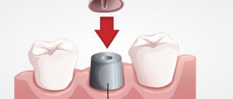
Installation of crowns
These are non-removable structures that can be used to eliminate dental defects. The installation of crowns is also indicated in case of severe destruction of dental units. The essence of the method is to insert an artificial tooth into the gum using a pin made of metal.
Installing crowns requires some preparation. The patient's natural teeth are prepared and, if necessary, pulp removal is performed. Crowns take several days to make. During this period, the patient is provided with temporary structures. The finished crowns are completely identical to the patient’s teeth. They differ only in width, which makes it possible to hide large gaps between chewing units.
The following conditions are contraindications: loose teeth, allergic reaction to the components used, periodontal pathologies. In addition, crowns are not placed on anyone under 16 years of age.
Advantages of the GrandMed clinic
- Dentistry has created an excellent diagnostic base that allows identifying non-carious dental pathologies in the early stages. Doctors have modern equipment at their disposal (radiovisiograph, orthopantomograph, computed tomograph), and all necessary clarifying tests can be done in the clinic’s biochemical laboratory.
- We employ professionals of the highest level, whose experience and qualifications allow us to treat complex non-carious pathologies, which often require the use of special techniques and non-standard approaches. Clients are provided with high-quality therapeutic and orthopedic care, including prosthetics on implants. So even in the most advanced cases, our patients can count on restoration of the beauty and functionality of their dentition.
- This disease is treated using local anesthesia. Patients of our clinic do not experience pain and feel comfortable during the procedure after injection of one of the modern anesthetic drugs that do not have side effects (Ultracaine, Ubestezin, Scandonest). You should not be afraid of the injection itself - it is performed quickly and accurately, and therefore is absolutely painless.
- Our clinic adheres to the highest sanitary standards, which is confirmed by the use of the latest sterilization equipment, which completely eliminates infection during treatment; Dentists have all the necessary consumables and disposable dental instruments at their disposal.
Rare teeth in a child
Radical treatment methods are not used for children. In addition, a child under 5 years old just needs to be monitored. This is due to the fact that after teething, the jaw continues to grow, so there must be gaps between the dental units. This is a normal physiological process. It is necessary to consult a doctor only if the space between the teeth remains very wide after the eruption of all milk teeth.
Children may have braces. In most cases, after such an adjustment, the teeth will take the required position, and the distance between them will decrease.
If the cosmetic defect remains pronounced even after removing the braces, the doctor may recommend filling or installation of veneers. The advantage of these methods is that there is no need to prepare natural teeth. It is recommended to give preference to the installation of veneers. Thin plates can not only hide empty areas, but also protect the enamel from the harmful effects of external factors.
Do you want beautiful dental restoration?
Always at your service - Sergey Samsakov, orthopedic dentist with more than 10 years of successful experience, Moscow. Expert in 3D digital smile modeling, dental inlays and dental modules, veneers and digital CEREC technology.
Patients come to me from all over Russia:
Consequences of adentia or tooth loss
How are complete dentition and facial appearance related?
The loss of even one tooth already affects the patient’s appearance, let alone when teeth are completely missing. A person ages sharply, and the effect of the so-called “senile face” appears, when the distance from the tip of the nose to the top of the chin decreases greatly. This occurs due to a decrease in the height of the lower third of the face. Since there are no teeth, the support of the lower jaw by the teeth is lost, and the jaw moves backward in the joint area, and forward and upward in the chin area.
This displacement of the jaw leads to a decrease in the tone of the facial muscles, as a result of which the cheeks and chin sag, the corners of the lips go down, even the lips become smaller, as if falling inward. The face becomes more mature.
Dental restoration has a rejuvenating effect, as it allows you to return the lower jaw to its normal position, which provides the necessary support to the soft tissues and restores the smile. After treatment, patients regain confidence and quality of life improves.
How to improve your facial contour without going to a cosmetologist
The “senile face” effect can also occur with partial loss of teeth, especially the chewing group. Sometimes a woman goes to a cosmetologist to tighten her facial contour, and he refers her to a dentist to restore missing teeth. Good, responsible cosmetologists understand that any rejuvenation procedures will not give the expected effect if there are no teeth in place.
Symptoms
The diagnosis of “uneven teeth” includes many different defects. They can hardly be called diseases; rather, they are cosmetic problems. Only in rare cases is there a serious impairment of chewing function. Usually the teeth “grind” against each other, and contact is established between them, sufficient for confident chewing.
There may be violations:
- location of teeth;
- growth directions;
- tooth sizes;
- dental crown shapes.
Humans are mammals, and their teeth are very different. But for each tooth there is a fairly accurate version of the norm, and small deviations make the dentition unsightly. Large or small teeth, protruding forward or backward, displaced to the side, not corresponding to their intended purpose (incisors, canines, molars) - patients turn to the orthodontist with such a wide range of defects.
Normally, crowns fit tightly to adjacent ones, or the gaps are minimal. Expanded ones, which are popularly called “birdhouse”, look unimportant and can cause damage to the gums when chewing.
The symptoms of malocclusion are usually obvious; an experienced orthodontist does not require additional research to clarify the diagnosis. In some cases, radiography is useful. Teeth (usually fangs) can shift due to the growth of excess teeth nearby, which normally should not be there. On an x-ray, the location and angle of inclination of the roots of teeth “number 1” and “number 2” are determined, and how healthy they are is assessed. Then they decide to keep or delete some of them.