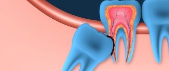
TREATMENT OF PERIODONTITIS
Price from 4000 rubles, anesthesia is included in the price.
Page Topics
- Signs of exacerbation of periodontitis (flux)
- Dental anesthesia is included in the price
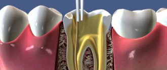
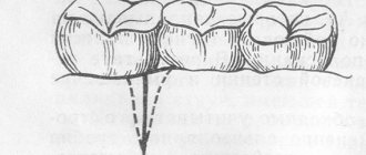
- Treatment of periodontitis - canal therapy
- Filling tooth canals with gutta-percha and sealer
- Tooth filling (filling or inlay)
- The price of periodontitis treatment is 4000 rubles
- 24/7 inexpensive
- inexpensive dentistry 24 hours
- It hurts to bite on a tooth
“Currently, the vast majority of clinicians attribute chronic periodontitis to infectious foci.” With this phrase, allow me to quote Professor Evgeniy Vlasovich Borovsky, who made a huge contribution to world and Russian dentistry. Samara dentists agree with their teacher on everything. Elimination of infectious foci in the oral cavity is the main task of Samara dentists.
It is difficult for the patient to determine the cause of the disease. He cannot correctly formulate a diagnosis, which is natural. It so happened historically and linguistically that in Russian most exacerbations of diseases in the oral cavity are called gumboil or swelling of the cheek . Well, okay, what’s wrong with this, if a person called “Exacerbation of chronic periodontitis” with the short and clear word flux, everything is clear to everyone, until the fifties this name was officially used in textbooks on dentistry and in scientific works. Now we are more inclined to Latin transcription.
What is baby tooth flux?
Periostitis is a purulent lesion of the periosteum. It contributes to the formation of an abscess and the appearance of severe toothache. It can be acute or chronic.
The acute form of the disease is difficult to miss - a lump appears on the child’s gum, filled with purulent masses. As a result, the cheek may “swell” and the cervical lymph nodes often become enlarged.
Chronic flux on a baby tooth often does not manifest itself at all. But at the same time, it has a destructive effect on the child’s jaw and causes negative changes in the functioning of his immune system. It is a consequence of incompletely cured acute periostitis, pulpitis, periodontitis or trauma.
Why is it important to treat flux
Dentists warn that advanced periostitis is very dangerous. If you ignore its occurrence and do not carry out the necessary treatment, the following consequences may occur:
- accumulation of a large amount of pus, loosening of the tooth;
- destruction of the rudiments of permanent teeth (then after the loss of milk units, the permanent ones will not erupt - the child will face complete or partial adentia);
- abscess (in place of the abscess, the tissues seem to melt, forming a huge cavity filled with pus);
- phlegmon (can lead to blood poisoning and death).
How to recognize flux in baby teeth in children
Among the main symptoms of the disease are:
- the appearance of an abscess on the gum at the base of the tooth crown;
- the occurrence of aching pain from the inflammatory reaction;
- pronounced swelling of the cheek;
- increased body temperature;
- general weakness, lethargy;
- the appearance of bad breath;
- enlargement of the lymph node located on the side of inflammation.
The flux makes the child feel unwell. He becomes capricious, whiny, and refuses to eat.
Causes of periostitis
In addition to advanced caries, swelling of the cheek due to a tooth can be caused by other factors.
Inflammation of the gums
Periodontal disease can serve as a starting point for infection to penetrate the periodontal tissue. This diagnosis requires a thorough examination and often long-term treatment.Reaction to poor-quality canal filling
If depulpation is carried out without modern diagnostic equipment, the doctor may not completely clean the root canals. As a result, the remaining nerve tissue becomes inflamed and leads to such unpleasant complications as periostitis.
Inflammatory reaction to tooth extraction
Any tooth extraction is considered a minor surgical operation. It leaves an open wound that can become infected. In this case, the inflammatory process in the tissues occurs quickly and with complications in the periodontal tissue and periosteum.
Cyst formation in periodontal tissues
A tooth cyst can develop in a person over several years, and then at one point this process can spread to the periosteum tissue. If the spread of infection has gone so far, then the most likely option is to remove the diseased tooth, cleanse the affected tissues, and thoroughly treat all tissues with antiseptic drugs.
Infection due to injury
As a result of an open fracture of the jaw, pathogens can penetrate the tissues near the tooth, both soft and bone.
Infection through lymph nodes
In rare cases, the infection enters the periosteum through blood vessels or through inflamed tissue in the lymph nodes, such as with tonsillitis.
An indirect factor in the occurrence of periostitis may be concomitant diseases, decreased immunity, sore throat, acute respiratory infections, stress, etc. By the time a tooth hurts and a cheek swells, there is probably an accumulation of pus in the periodontal tissues. This process is quite dangerous for the body, as it can be accompanied by symptoms of acute inflammation:
- increased body temperature;
- general weakness, body fatigue;
- headaches, local pain in the area of inflamed tissue;
- formation of a purulent fistula with access to the oral cavity or through the soft tissue of the cheek;
- increased pain symptoms at night or when trying to chew.
If caries can be ignored, then with periostitis, patients simply “fly” to the dentist for help. The course of the disease can be so acute that there is no way to tolerate it. The specialists of the LeaderStom network of clinics have extensive experience in treating periostitis. With such a diagnosis, therapy is required not only for the tooth itself, but also for the entire body. Therefore, be prepared to follow all doctor’s orders, take antibacterial medications and regularly rinse your mouth every half hour.
If you have a toothache or a swollen cheek, it is recommended to immediately seek help from a dentist, but if this is temporarily unavailable, the following recommendations will help you cope with the pain.
- If your cheek is swollen and your tooth hurts, you should not apply warm compresses or bandages to your face, as heat promotes even greater inflammation of the dental tissues and can cause complications.
- A cold compress should be applied to the affected side of the face.
- Be sure to take a pain reliever.
- Rinse your mouth several times with infusion of chamomile, sage, and St. John's wort.
- It is strictly prohibited to take anti-inflammatory drugs and antibiotics without a diagnosis of “periostitis” and a doctor’s prescription.
Why does flux develop?
Various factors can lead to the disease. Among the most common:
- Trivial lack of oral hygiene. Many children refuse to brush their teeth and do not want to open their mouths at the dentist. If the health of their teeth is not carefully monitored, tooth decay will occur. As it progresses, the periosteum may become inflamed, leading to periostitis.
- Untreated caries. The enamel of baby teeth is destroyed very quickly, so the infection easily penetrates into deeper hard tissues.
- Gum injuries. If a child puts foreign objects in his mouth or chews something, he can damage the mucous membrane. The abrasion will become an entry point for infection.
- Pathologies of ENT organs. Sore throat, pharyngitis, adenoids, inflammation of the tonsils - all these are factors that can cause infection and provoke deepening of the gum pockets. This eventually leads to flux.
It is very important to understand why periostitis developed. Then it will be possible to eliminate the provoking factor, and therefore the disease will not return again.
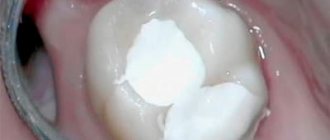
What to do with flux - treatment features
It is important for parents to remember: if a child’s illness appears, they must be shown to an experienced dentist. Attempts to open an abscess on your own can only aggravate the situation and lead to blood poisoning. The only thing is, if you can’t consult a doctor right away today, you can rinse the affected tooth with water and soda or salt. Also suitable for these purposes are pharmaceutical antiseptics designed specifically for treating the mucous membranes of the oral cavity.
The dentist always decides how best to deal with gumboil, taking into account the condition of the gums and the age of the patient. If the affected tooth is about to fall out, it is removed immediately. There is no point in stressing out a child by trying to save a unit that is about to leave the dentition anyway.
If we are talking about a tooth that the patient must live with for several more years, then conservative treatment is most often carried out. It is preferred to avoid crooked teeth in the future. Indeed, often when a tooth is pulled out prematurely, “neighbors” take its place. Because of this, the permanent unit then begins to erupt to the side, and it has to be put in place with the help of complex orthodontic treatment.
Conservative therapy involves opening a purulent neoplasm. The formed cavity is drained and washed with an antiseptic composition. If necessary, a drainage tube is installed in the gum for a while to avoid the re-formation of a purulent “bump”.
After such a procedure, special attention is paid to therapeutic rinses. The baby may even be prescribed antibiotics or anti-inflammatory drugs.
Sinusitis and complications of endodontic treatment
The maxillary sinus is the largest paranasal sinus, which is located directly above the alveolar process of the maxilla. Considering the topographic-anatomical connection between the roots of the distal teeth and the paranasal sinus, endodontic treatment of molars and premolars of the upper jaw requires special attention. Previously, it was suggested that the prevalence of maxillary sinusitis of odontogenic etiology is increasing. This trend may be caused not only by the pathology itself, but also by the higher availability of diagnostic methods such as cone-beam computed tomography.
During endodontic treatment, many complications can arise that can provoke the development of sinusitis. Thus, a regular size 10 K-file (Kerr Dental) can simply pass through the apex and perforate the Schneiderian membrane, after which all solutions used for disinfection can enter the sinus space without obstruction. Extrusion of sodium hypochlorite into the sinus area can cause burning sensations, nosebleeds, and breathing problems. The use of the EndoVac system with the principle of negative pressure (Kerr Dental) allows for safe irrigation of the root canal system, regardless of the location of adjacent anatomical structures.
During the obturation phase, endodontic sealers containing zinc oxide can provide a medium for the growth of the Aspergillus fungus. The possibility of contamination of the sealer with fungal spores, according to the authors, should be seriously taken into account when choosing an agent for obturation.
Clinical case 1
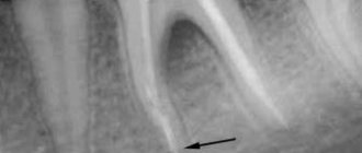
The patient was referred to the dental clinic due to broken endodontic files in the canal. The patient was a pilot by profession and during the flight he noted the discharge of pus from the nasal area. After performing radiography (photo 1), it was discovered that there were two broken fragments of the file in the mesial canals and gutta-percha was removed beyond the apex area in the palatal canal.
Photo 1
According to the CBCT scan, the left maxillary sinus was completely filled with inflammatory exudate (Figure 2), and the gutta-percha pin from the distal canal completely penetrated into the sinus space. It was also confirmed that there were two fragments of files in the mesial system (photos 3-4), with one of them fully penetrating the maxillary sinus.
Photo 2
Photo 3
Photo 4
Better visualization of files can be achieved through the use of various image filters (photo 6-9).
Photo 5
Photo 6
Photo 7
Photo 8
Photo 9
Thanks to them, it was also possible to better objectify the position of the removed gutta-percha into the lumen of the sinus. Using H-files, it was possible to remove the gutta-percha pin from the palatal canal, but the broken fragments of the files in the medial system could not be bypassed. Considering the complexity of this manipulation, the canals were sealed (photo 10), and the doctor began the surgical stage of the intervention. Periapical surgery involved resection of 3 mm of the mesial root using Impact Air (SybronEndo), after which it was possible to more extensively visualize the Schneiderian membrane, which contained one of the file fragments (Figure 11-12). The file was captured and deleted without any problems (photo 13). Photo 14 visualizes the situation after performing the retrograde filling procedure of the medial canal system.
Photo 10
Photo 11
Photo 12
Photo 13
Photo 14
Clinical case 2
The patient was referred to the clinic with complaints of dull pain in the area of the maxillary molar. The radiograph revealed the presence of various filling materials in the endospace of the tooth, some of which were fragmented, and some were brought outside the tooth. By analyzing the radiograph, it was found that the Schneiderian membrane was perforated, and an inflammatory process developed in the sinus area (photo 15).
Photo 15
Under microscope control, using ultrasound and K3XF files (Kerr Dental), it was possible to clean the canals from all materials present in them, and then close the endospace with temporary cement (photo 16). Photo 17 shows that a small portion of the previously used silver pin has protruded beyond the apex of the medial canal. Using a macrocannula from EndoVac, the author was able to extract this fragment and complete the medicinal treatment of the endospace. Photo 18 shows the situation immediately after treatment and obturation of the mesial canals. Photo 19 shows that the area of the membrane lesion has almost completely healed, and the bony floor of the sinus is noticeably restored.
Photo 16
Photo 17
Photo 18
Photo 19
Discussion
The maxillary sinus is located at a lower level than the floor of the nasal cavity, and at the same time it is closely connected with the roots of the teeth of the upper jaw. Some studies indicate that the palatal root of the maxillary molar very often protrudes into the lumen of the sinus, although other authors argue that the mesiobuccal root is characterized by the highest level of protrusion into the sinus region. Diagnosis of maxillary sinusitis of odontogenic etiology requires special attention and evaluation. The increase in cases of diagnosis of sinusitis of odontogenic etiology may be associated with the greater availability of diagnostic imaging methods such as CBCT. CBCT makes it possible to objectify the quality and quantity of the existing bone tissue around the apexes of the distal teeth, as well as to more accurately assess their position relative to the bottom of the sinus. If the thickness of the Schneiderian membrane is 0.5 mm or more, regardless of the presence or absence of radiological signs of inflammation, the doctor can assume the possible presence of an inflammatory process in the area of the apex of the tooth. Most often, perforation of the mucous membrane of the floor of the maxillary sinus is characterized by bleeding from the nose, the development of sinus obstruction and acute or chronic infection.
Studies have shown that in conditions of periapical lesions due to pulp necrosis, powerful virulence factors of bacterial agents are formed such as collagenase enzymes and lysosomal aggressive substances, which provoke the destruction of periapical tissue and can reach the maxillary sinus. Bacteria and toxins from the apical lesion may also enter the sinus area through direct diffusion through the porous maxillary bone or through the blood and lymphatic vessels, causing thickening of the sinus floor mucosa. The risk of extrusion of endodontic obturation materials into the sinus area depends on the skill of the practitioner and the obturation system used. If the root is over-treated, it is difficult to form an apical plug, resulting in filler extrusion. After extrusion, the surface of the material removed beyond the apex is at risk of infection. Thus, in cases of extrusion of zinc oxide endodontic agents, they are most often contaminated with the Aspergillus fungus. Such contamination of the material can cause fungal spores to enter directly into the sinus.
The authors recommend considering the following aspects to avoid iatrogenic errors during endodontic treatment:
- Maintain the working length during the initial formation of the canal;
- Maintain stable working length indicators during tooling using rubber stoppers;
- Use negative pressure techniques to prevent cases of extrusion of chemical agents into the postapical space;
- Pay special attention to the obturation procedure to prevent extrusion of material into the postapical space and sinus space;
- Take into account the parameters of mixing and dosage of the sealer; paste-like types should not be introduced into the channel under too much pressure.
Author: Dr. Philippe Sleiman (Beirut, Lebanon)
Prevention measures
It's no secret that it is easier to prevent any disease than to treat it later. The same is true with periostitis. To reduce the risk of developing the disease described, it is very important:
- control how thoroughly the child brushes his teeth;
- take him to the pediatric dentist every six months for a preventive examination;
- competently treat all emerging infectious pathologies;
- follow the recommendations given by the doctor after tooth extraction.
These simple rules allow you to keep baby teeth healthy until permanent teeth begin to emerge in their place.
100% Recommendations on how to relieve pain, why the cheek is swollen, medications
Thanks to anesthesia, removal of the dental nerve is painless. But postoperative pain is a common occurrence. Why does a tooth hurt after nerve removal and filling, how to deal with it, and in what cases should you consult a doctor?
Here are the answers to all these questions. Most patients experience unpleasant painful sensations during the first time after depulpation. They can last from several hours to a week, and should become less noticeable over time. If the pain increases or suddenly appears a few days after the operation, this is a reason to contact the dentist again.