Periostitis of the jaw (flux) refers to dental diseases. The localization of the source of inflammation occurs in the periosteum. A subperiosteal abscess is formed, and the peri-maxillary soft tissues swell. A person experiences painful sensations radiating to the temporal region, ear, eye; his health worsens: weakness, sleep disturbances, fever, and headaches occur.
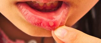
Periostitis, aka gumboil
The disease is diagnosed by inspection and palpation; an x-ray is required for confirmation. The doctor opens and drains the subperiosteal abscess and removes the tooth that is the source of infection. After this, the patient must undergo a course of physiotherapy and antibiotic treatment. It is also necessary to rinse your mouth regularly.
What kind of diagnosis is this? According to the international classification ICD-10, accepted throughout the world, periostitis of the lower jaw is assigned code K10.2.
Reasons for the development of periostitis
The causes of periostitis of the jaw are usually infection or injury. According to statistics, in practice this disease is observed in 5.4% of patients with inflammation in the face and jaw. Moreover, 95% of patients have an acute form, 5% have a chronic form. Localization of periostitis is observed one and a half to two times more often in the lower jaw. The clinical picture, both local and general, is peculiar. If the treatment process is started on time, it will end successfully, otherwise the disease progresses, which carries a high probability of severe purulent complications.
Why does this disease occur? There are several reasons. And the main one is complicated caries, in which pathogenic microflora penetrate through microchannels into the tooth cavity and then spread to the tissue surrounding the root.
Often periostitis is caused by chronic apical periodontitis. Diseased microorganisms move further and further through the channels over time. If the disease is not treated, the soft tissues are first affected, then the periosteum.
Staphylococcus always lives in the oral cavity. If the immune system is weakened, the population begins to grow and the periosteum becomes inflamed.
The hematogenous and lymphogenous form is usually caused by ENT diseases, ARVI, and infectious diseases such as measles and scarlet fever. Traumatic periostitis may occur after tooth extraction or trauma, surgical intervention, jaw fractures, infected wounds on the face, etc.
In many patients, periostitis is caused by previous hypothermia or overheating, as well as emotional or physical overload.
There are other reasons, less common: tuberculosis and other diseases that are fraught with complications spreading to the jaw periosteum.
The etiology and pathogenesis of periostitis in modern dentistry has been studied quite well. Treatment usually does not cause problems.
Treatment of periostitis in dentistry in Moscow
Periostitis is a concomitant phenomenon of dental pathologies. But sometimes patients are not even aware of a dangerous disease in the oral cavity. For example, the need for serious dental treatment for periostitis of the upper jaw often turns out to be an unpleasant surprise due to the hidden development of the pathological process.
Modern dentistry in Moscow is guaranteed to prevent such phenomena. Highly qualified specialists have unique diagnostic and treatment equipment at their disposal. If treatment of periostitis is necessary, all possible methods are used to achieve one hundred percent recovery.
Types and stages of disease development
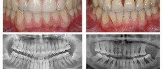
The classification of odontogenic periostitis depends on the phase of the disease and the nature of its course. In addition to examination and palpation, the dentist prescribes an X-ray examination. The resulting image allows you to assess the condition of the roots and periapical areas. The image does not show thickening of the periosteum in the first three days from the onset of the disease.
- Various types of periostitis are classified according to the international classification ICD 10 depending on the route of infection into the periosteal tissue:
- acute odontogenic periostitis of the jaws (cause - diseased tooth);
- hematogenous (infection enters through the blood);
- lymphogenous (spread through the lymph system);
- traumatic (for injuries of the periosteum).
According to the course and pathomorphology, the disease is divided into two forms: acute and chronic.
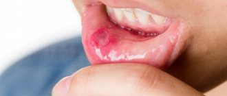
In the acute form, the symptoms are pronounced. There is swelling of half the face, he is tormented by severe throbbing pain, and pus forms. The acute serous form is characterized by such clinical manifestations as infiltration in the periosteum, serous exudate is formed in the lesion. In acute purulent periostitis of the jaw (popularly called gumboil), a limited subperiosteal focus of inflammation is formed, fistulous tracts are formed, and pus comes out through them. The course of purulent periostitis is much more severe, with bursting pain that increases when drinking hot foods. If a fistula tract does not form, the dentist cuts the periosteum to allow the pus to escape.
The course of the chronic process is sluggish, stages of exacerbation occur periodically. Young bone tissue begins to grow on the surface of the jaw. With the development of the ossifying form, ossification and hyperostosis occur quite quickly. There are two degrees of spread of this process: limited (covering one to three teeth) and diffuse purulent (almost the entire jaw is covered).
If the wisdom tooth erupts with difficulty, retromolar periostitis may develop in the lower jaw. The pus does not come out on its own due to anatomical features, which requires dissection.
Forms
- Diffuse
. Accompanied by attacks of severe toothache and signs of general intoxication: high fever, lethargy, lack of appetite. Fibrous thickening of the periosteum is observed on the lower and upper jaw. - Purulent
. The most common form of periostitis. Accompanied by high temperature (up to 39°C), the appearance of an abscess and the formation of fistula tracts. In the absence of therapy, it forms severe swelling, phlegmon, and acute osteomyelitis is possible. - Ossifying
. A common form of chronic periostitis, which is characterized by the formation of new bone and hyperostosis. Progresses very quickly. - Chronic
. It proceeds slowly without pronounced symptoms, sometimes worsening, sometimes subsiding over several months and even years. At the site of inflammation, thickening of the bone is possible. As a rule, the lymph nodes become enlarged.
Characteristic symptoms
Symptoms of acute periostitis of the jaw are determined by the form of the disease. The state of the patient’s immune system and the diseases he has are also influenced. But there are also characteristic signs that make it possible to make an accurate diagnosis.
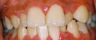
The disease develops gradually. At first, the gums swell a little, and pressing on the tooth causes pain. Having discovered such signs, the patient should visit a doctor on the same day, otherwise the cheek may become swollen by the morning.
Signs of the form of the disease with serous infiltrate are:
- red color of the mucous membrane:
- the appearance of painful swelling in the fold between the gum and cheek;
- tolerable pain;
- temperature may rise to 37°C;
- facial asymmetry;
- submandibular and postauricular lymph nodes enlarge (lymphadenitis).
With a purulent infection, the condition becomes much worse:
- signs of intoxication and pain are observed;
- temperature (up to 38°C);
- the cheek becomes swollen;
- pain radiates along the trigeminal nerve;
- pulsation is felt in the swollen part;
- a fistula may occur;
- When you press in the area between the cheek and gum, you can feel the vibrations of the fluid.
Symptoms
Inflammation of the periosteum of the tooth may be indicated by such signs as:
- formation of an abscess (abscess);
- swelling of the soft tissues of the cheek;
- enlargement and redness of the gums;
- painful, limited mouth opening;
- pathological mobility of the diseased tooth;
- unbearable pain when exposed to the source of inflammation;
- high temperature (37-38 °C);
- facial asymmetry;
- chills and lethargy due to general intoxication of the body;
- inflammation of the lymph nodes.
Symptoms of acute inflammation may subside for a while, but if left untreated, the disease progresses to the chronic stage, which is dangerous with serious complications.
| Have you noticed signs of gumboil? Contact our dental clinic as soon as possible to immediately undergo an examination and begin treatment for periostitis! |
How is periostitis diagnosed?
For an accurate diagnosis, the doctor collects anamnesis, conducts an external and internal examination, prescribes an x-ray and examines the x-ray. Since the symptoms of some dental problems are identical, the surgeon must have a good understanding of their symptoms.
When conducting diagnostics, doctors look for similarities and differences between chronic periostitis of the jaw or acute and other diseases of the oral cavity. An x-ray helps differentiate the disease from others (apical periodontitis, phlegmon, abscess, inflammation of the salivary gland, osteomyelitis).
Apical periodontitis is characterized by the presence of a purulent focus located at the top of the root.
Cellulitis and abscess are manifested by external changes on the skin. An infiltrate appears on the affected area, the skin over it turns red and becomes shiny.
With sialadenitis, it is necessary to palpate the salivary gland to determine its density. Osteomyelitis is diagnosed by X-ray - in the early stage of the disease, it shows decaying bones, in the late stage - formed sequesters.
Clinical manifestations
Periostitis begins with pain and local swelling. These phenomena usually develop in the area of the tooth, which is the cause of the disease. The disease can begin with a serous form, but in many cases a purulent focus forms initially.
The general condition worsens. The patient experiences severe pain, which makes it difficult to eat, talk, and open the mouth. Swelling forms, which spreads to the soft tissues of the face, and flux forms.
When examining the oral cavity, redness of the mucous membrane is noted. At the site of localization of the pathological process there is a clearly visible infiltrate. Over time, pus accumulates and exits into the oral cavity through the resulting hole - a fistula.
The chronic form proceeds smoothly. Pain occurs periodically, swelling is mild. There may be enlargement of regional lymph nodes and changes in facial contours.
For differential diagnosis, determining the condition of the jaw bone tissue and identifying affected teeth, an X-ray examination is performed. In acute periostitis, the bone tissue is not changed; in the chronic form, changes are observed in the form of areas of regeneration. X-rays provide diagnostically valuable information, so one cannot do without an image.
Periostitis can occur in isolation or be combined with other inflammatory diseases: periodontitis, osteomyelitis, cysts. It is also important to identify them, since a complicated process requires different treatment tactics.
Stages of the operation
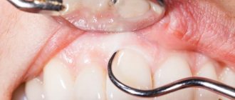
When treating periostitis of the jaw, the surgeon must cleanse the cavity of pus so that the process does not spread deeper. To do this, he cuts the periosteum. The operation consists of several stages:
- Anesthesia . The affected area is numbed using modern drugs.
- Periostotomy . The surgeon makes a soft tissue incision along the fold between the gum and cheek, capturing the periosteum. This ensures the release of purulent exudate to improve the patient’s condition.
- Wound drainage . A rubber or gauze graduate is installed into the incision. This ensures the drainage of pus.
After opening the purulent lesion, the surgeon asks the patient to rinse his mouth with a disinfectant solution.
After this, chlorhexidine and similar solutions are used to wash the wound. Measures such as irrigation with dimexide with oxacillin and 15-minute applications of dimexide liniment are also effective.
After examining an x-ray of the tooth that caused periostitis, the doctor decides whether it can be preserved and treated in the future to eliminate the source of infection, or whether tooth extraction is necessary.
Acute purulent periostitis of the jaw is an acute purulent inflammation of the periosteum, the alveolar process of the upper jaw or the alveolar part of the lower jaw, less commonly the alveolar part and the body of the jaw. The process can primarily affect the periosteum from the vestibular surface, less often from the palatine (palatal abscess) or lingual surface.The cause of the disease is a mixed infection: streptococci and staphylococci, various types of gram-positive and gram-negative bacilli and often putrefactive bacteria.
Acute purulent periostitis is a complication of acute or chronic periodontitis in the acute stage. The inflammatory process can develop with difficulty in teething, suppuration of radicular cysts, and periodontal diseases. Sometimes the disease occurs after conservative dental treatment, during traumatic tooth extraction.
In acute and aggravated chronic periodontitis, the outflow of pus through the root canal or gum pocket is sometimes insufficient or impossible. Exudate spreads from the periodontium towards the periosteum. Often, an ulcer forms in the wall of the dental alveolus, through which purulent exudate penetrates into the periosteum. Sometimes microorganisms spread into the periosteum through the lymphatic vessels.
Sensitization and general factors play a certain role in the development of the disease: cold, overwork, stressful situations that reduce the body’s defense reactions.
Clinical picture. Characteristic complaints are pain, swelling of the soft tissues of the face, poor general health, and increased body temperature. At first, the pain is insignificant, then it intensifies and spreads to the entire jaw, radiating along the branches of the trigeminal nerve: to the ear, temple, eye. A swelling of the soft tissues appears, which progressively increases, while the pain in the tooth subsides. Complaints of headache, malaise, and poor sleep are possible. Body temperature is elevated - 37.5-38 °C, sometimes up to 38.5-39 °C; In some patients, general weakness, fatigue, loss of appetite, and insomnia due to pain are possible.
In case of acute purulent periostitis of the upper jaw, on the side of the vestibule of the mouth, upon external examination, diffuse soft swelling is visible due to collateral edema. Its localization and distribution depend on which tooth caused the disease. Periostitis of the upper jaw, associated with damage to the incisors, is characterized by significant swelling of the upper lip, spreading to the wings and bottom of the nose. The spread of the purulent process from the canine and premolars is characterized by the localization of collateral edema in the middle and lower thirds of the face, significant swelling of the eyelids, as a result of which the palpebral fissure is sharply narrowed. If the cause of the disease is premolars, then swelling of the buccal, zygomatic, and parotid areas occurs; from the third molar of the upper jaw, in addition to the listed areas, swelling spreads to the temporal region.
If the inflammatory process is associated with the incisors of the lower jaw, then swelling of the lower lip, chin and often submental areas appears, if with the canine and premolars, then there is collateral swelling of the lower cheek, the area of the corner of the mouth, descending into the submandibular region.
With inflammation from the molars of the lower jaw, collateral swelling of the lower part of the buccal, submandibular and parotid-masticatory areas is observed. The spread of the process to the periosteum of the lower jaw branch involves the masticatory and medial pterygoid muscles in the inflammatory process, which leads to their inflammatory contracture of the I-II degree. With the development of a purulent process from the third molar of the lower jaw, the swelling can spread down to the lateral parts of the neck. The patient experiences pain when swallowing and speaking.
Regional lymph nodes are enlarged, painful, and often fused. The inflammatory reaction of the lymph nodes is especially pronounced in children.
In acute purulent periostitis of the jaw, hyperemia of the mucous membrane over the affected area is noted on the vestibular side of the oral cavity. The vault of the vestibule of the mouth is smoothed due to inflammatory infiltration; palpation of this area is sharply painful. After a few days, the periosteum breaks through and pus penetrates under the mucous membrane of the alveolar process. Along the arch of the vestibule of the mouth, a roller-like bulge appears, covered with a thin mucous membrane; upon palpation, fluctuation is clearly visible. Percussion of the affected tooth may be painless, the tooth is sometimes mobile. Then the abscess can spontaneously open into the oral cavity. In such cases, the pain in the tooth subsides and the inflammation begins to subside.
With the development of the process in the lower jaw on the lingual side, collateral edema with a package of enlarged lymph nodes in the submandibular triangle is possible. Due to infiltration of the medial pterygoid muscle, mouth opening is painful and limited. In the oral cavity, from the alveolar part of the body of the lower jaw, swelling and infiltration of the periosteum are detected; palpation of this area is painful. Swelling and hyperemia of the mucous membrane can spread to the sublingual region, sometimes the anterior velopharyngeal arch and the pterygomandibular fold. Movement of the tongue becomes difficult.
A palatal abscess develops with chronic or acute periodontitis of the lateral incisor, the root of the first premolar and the palatal roots of the maxillary molars. An external examination reveals enlarged, painful lymph nodes on the affected side, and a limited hemispherical or oval infiltrate on the hard palate. The enlargement of the abscess leads to smoothing of the transverse palatal folds. With the development of a palatal abscess associated with molars, inflammatory swelling from the hard palate spreads to the mucous membrane of the soft palate, palatoglossus arch, and pterygomandibular fold, causing pain when swallowing.
The accumulation of purulent exudate under the periosteum of the hard palate causes exfoliation of soft tissue from the bone. This is accompanied by pain, often pulsating in nature, aggravated by talking and eating. A week or more after the onset of the disease, the abscess breaks out and pus pours into the oral cavity.
Treatment. Complex treatment is carried out: surgical opening of the abscess, conservative drug therapy, etc. In the initial stage of acute periostitis of the jaw (acute serous periostitis), treatment can begin with opening the tooth cavity, removing decay from the canals and creating conditions for outflow, in other cases - with removing the damaged tooth that is the source of infection. All manipulations are performed under infiltration or conduction anesthesia. These therapeutic measures, together with lidocaine or trimecaine blockade with antibiotics, proteolytic enzymes along the transitional fold to the bone, and drug therapy can help to subside the inflammatory phenomena.
In case of acute purulent periostitis of the jaw, emergency surgical intervention is performed - opening the purulent subperiosteal focus and creating an outflow of exudate (primary surgical treatment of a purulent wound). This operation is usually performed on an outpatient basis, in some cases in a hospital.
Surgical treatment for acute purulent periostitis is performed under local anesthesia - conduction or infiltration anesthesia. For infiltration anesthesia, a thin needle is used, through which an anesthetic solution is slowly injected under the mucous membrane and the tissue is infiltrated along the intended incision line. The needle should not be inserted into the abscess cavity. Medicinal preparation of patients has a good effect. Sometimes the operation is performed under anesthesia.
If the subperiosteal abscess is located in the area of the vestibule of the mouth, then it is better to make the incision with a beak-shaped scalpel parallel to the transitional fold through the entire infiltration area; dissect the mucous membrane, submucosal tissue and periosteum to the bone, respectively, of 3-5 teeth. To prevent the edges of the wound from sticking together and to ensure the drainage of pus, a narrow strip of thin (glove) rubber is loosely inserted into the wound.
When the abscess is localized under the periosteum in the area of the tubercle of the upper jaw, the incision is made along the transitional fold in the area of the molars of the upper jaw, but to open the inflammatory focus, a rasp or a grooved probe should be passed from the incision along the bone in the direction of the tubercle of the upper jaw (back and inward). In the same way, a purulent focus is opened in case of periostitis of the upper jaw, which has spread to the canine fossa.
It is recommended to open the inflammatory focus during periostitis from the lingual surface of the lower jaw by making an incision in the mucous membrane of the alveolar part to the bone at the site of the greatest protrusion of the infiltrate. A grooved probe is passed down the surface of the bone and, pushing back the periosteum, allows the pus to drain out.
For palatal abscess, the incision is made in the area of greatest tissue bulging, slightly away from the base of the alveolar process, or at the midline of the palate, parallel to it. Then a wide strip of thin (glove) rubber is inserted into the surgical wound, which avoids sticking of the edges of the wound and creates conditions for good outflow of pus. The best results are obtained by excision of the wall of the abscess - a small section of the mucous membrane of a triangular shape, which ensures a freer outflow of pus.
To open the inflammatory focus in the area of the periosteum of the jaw branch, on its outer and inner surfaces, special techniques are used. In case of periostitis on the inner surface of the jaw branch, an incision is made with a sickle-shaped scalpel with a limiter or a regular scalpel to the bone, tissue is dissected in the retromolar region (at the base of the palatoglossal arch), and a rasp is passed to the inner surface of the jaw branch, creating an outflow of exudate from the source of inflammation.
The subperiosteal abscess along the outer surface of the lower jaw branch should be opened with an incision made from the vestibular side at the level of the second and third large molars along an oblique line to the bone, then with a rasp they pass subperiosteally in the direction of the angle of the lower jaw, retracting the masticatory muscle outward. After opening the lesion, a rubber strip must be inserted deeply into the wound for drainage. The lack of effect from such an intervention the next day is the basis for hospitalization and surgical intervention through external access. In children, indications for hospitalization should be expanded; children under 10 years of age are treated in a hospital.
The opening of periosteal lesions in children should be gentle. The periosteum must be peeled off well, but not to injure the bone with the instrument to avoid injury to the tooth buds.
After opening the purulent focus, it is advisable to let the patient rinse his mouth with a weak solution of potassium permanganate or 1-2% sodium bicarbonate solution, and it is also necessary to rinse the wound with a solution of chlorhexidine. A good effect is achieved by irrigating the abscess cavity with a solution of dimexide with oxacillin and applying a 40% solution of dimexide liniment to the wound for 15 minutes.
If the tooth that was the source of infection is destroyed and does not represent functional or aesthetic value, then it should be removed simultaneously with the opening of the subperiosteal abscess. This will improve the emptying of the purulent focus and will contribute to a faster elimination of inflammatory phenomena. Tooth extraction is sometimes postponed due to perceived technical difficulties or the unsatisfactory condition of the patient. In other cases, the tooth is preserved: its cavity is opened, the root canal is freed from decay products, and then conservative treatment of chronic periodontitis is carried out. It is especially important to remove the sources of infection in children - baby teeth, and if conservative treatment is impossible, permanent teeth.
Drug treatment of acute purulent periostitis consists of prescribing sulfonamide (sulfadimethoxine, sulfadimezin, etc.) and nitrofuran (furazolidone, furadonin) drugs, pyrazolone derivatives (analgin, amidopyrine, phenacetin, etc., as well as their combinations), antihistamines (diphenhydramine, suprastin , diazolin, etc.), calcium supplements, vitamins (multivitamins, vitamins C 2-3 g per day).
The patient is invited to an appointment on the 2nd day after surgery. During examination and questioning, the degree of subsidence of inflammatory phenomena is determined and, depending on this, additional treatment is prescribed. During dressing, local treatment of the wound is carried out, following the recommendations given earlier.
In case of acute purulent periostitis of the jaw, for a more rapid cessation of inflammatory phenomena on the 2nd day after opening the abscess, physical treatment methods should be prescribed: light and heat therapy (sollux lamp), warm baths of antiseptic or deodorizing solutions, ointment dressings (levomikol, levamisole, petroleum jelly, 20 % camphor oil, sea buckthorn oil, rose hips), UHF, microwave therapy, fluctuarization, laser therapy with helium-neon and infrared rays. Exercise therapy is recommended.
In most cases, inflammatory phenomena quickly (after 2-3 days) subside. If the subsidence of inflammation is delayed, then a blockade is carried out 2-3 times: intra-or extraoral infiltration with 0.25-0.5% trimecaine solution, 40-50 ml of lidocaine with enzymes, antibiotics (lincomycin). Weakened patients, patients with concomitant diseases, children and patients at risk, as well as persons with increasing inflammatory phenomena are prescribed antibiotics. A prerequisite for the effectiveness of antibiotic therapy is opening the abscess (primary surgical treatment). In a clinic setting, it is advisable to prescribe broad-spectrum antibiotics (semi-synthetic penicillins, tetracycline, oletethrin, oxacillin, macrolides). It is mandatory to prescribe metronidazole derivatives 200,000 units 3 times a day for 5-6 days, in the hospital - injections of these drugs 3-4 times for 6-7 days.
"Surgical Dentistry" edited by Robustova T.G.
Fourth edition. Moscow "Medicine" 2010
Surgical intervention
With serous inflammation, as a rule, conservative therapy is limited. Pulpitis or periodontitis is treated, the patient is prescribed a course of physiotherapeutic sessions (UHF), rinsing with disinfectants. This leads to resorption of the infiltrate.
The acute purulent form requires surgical intervention to open the subperiosteal or submucosal abscess.
The milk tooth that caused periostitis must be removed. Severely damaged permanent teeth are also removed. Teeth that are capable of performing their functions need to be treated.
Differential diagnosis
These diagnostic methods are used if a person has symptoms of several diseases that are similar in course. They help make an accurate diagnosis. For example, to distinguish acute and purulent periostitis from abscess, phlegmon, lymphadenitis and other ailments. Chronic, aseptic and specific periostitis are detected using x-ray examination; they reveal thickenings, growths on the bones, necrotic changes in tissue, etc.
If x-rays do not allow an accurate diagnosis, then doctors resort to a biopsy.
Drug treatment
The second stage of treatment after surgery is drug therapy. The patient is washed with modern antiseptic compounds (Chlorhexidine, Ethacridine, Dimexide with Oxacillin), and sterile bandages are applied. To impregnate them, use a disinfectant solution or ointment. Antibiotics (Oletetrin, Oxacillin, Lincomycin) must be prescribed to prevent complications and relapses. Nitrofurans (Furadonin), sulfonamides, painkillers, antihistamines (Suprastin) may be prescribed; vitamins. In the period after the operation, the patient must follow a gentle diet and rinse with antiseptics.
Historical reference
Tooth abscess, “tooth fire” - these are just some of the “folk” names for this disease that have come down to us from antiquity. In those days, a patient who came to a doctor with a gumboil, in the overwhelming majority of cases, left him... without a tooth, because the removal of the causative tooth seemed to be the only reliable “remedy” for all dental diseases. However, it is impossible not to mention the existing artifacts - the found skulls dating back to the time of the Old Kingdom, with traces of drilling in the jaw, in all likelihood to release pus - proving that even in those days attempts were made not to get rid of diseased teeth, but to treat them1.
But since medicine in those days was an “expensive pleasure,” people looked for ways to help themselves on their own, and traditional medicine always came to their aid. For example, in the treatment of gumboil, ground garlic was used as the most powerful natural antibiotic. Clove oil, peppermint oil and oregano oil, which have analgesic, bactericidal, and antiseptic properties, were widely used, as were warm salt water, apple cider vinegar, sesame seeds, herbal rinses, and even raw potatoes. Among the more “modern” folk remedies, rinsing with hydrogen peroxide and baking soda is known. Of course, all these “medicines” had extremely low effectiveness, in many cases they were simply useless, and in some cases they were dangerous, since they took up precious time, during which the active inflammatory process led to sepsis. And only at the end of the 19th and beginning of the 20th centuries, when medical science began to develop steadily and rapidly, did doctors have truly effective means of combating periostitis.
Physiotherapy
After surgery, the patient is recommended to undergo a course of physiotherapy to stop the inflammatory process, solux lamp, ultrasound. These are procedures such as: UHF, fluctuarization, microwave, laser, electrophoresis. Usually 5 to 7 sessions are prescribed.
Is it possible to do without it? Such a serious disease as periostitis of the jaw cannot be cured at home; qualified professional help is needed from a professional who will eliminate the source of infection. You can use folk remedies only in cases where the pain comes on suddenly, and you need to somehow improve the patient’s condition. But you need to visit a doctor as soon as possible. Before this, you can rinse your mouth with soda, salt, a decoction of herbs that have an anti-inflammatory effect, apply cold compresses to reduce swelling and relieve pain.
Indications and contraindications
Periostitis therapy is carried out when the following signs appear:
- swelling of soft facial tissues;
- redness and swelling of the gums;
- constant toothache, which intensifies when the tongue touches the tooth.
Surgical intervention is indicated for inflammatory processes and gumboil formation. Swollen gums hold pus and need to be removed. Indications for periostotomy:
- abscess (the gum is cut to remove the pus);
- serous periostitis (removal of suppuration);
- inflammation after canal filling (an incision is made to eliminate swelling).
The operation is not performed with the following contraindications:
- malignancies and chemotherapy;
- mental disorders;
- blood diseases (hemophilia, leukemia);
- heart and vascular diseases;
- acute respiratory infections;
- diabetes.
What to do for prevention
The main preventive measure is to constantly monitor the condition of the oral cavity. Preventive measures include:
- daily hygiene of teeth and gums;
- visiting the dentist twice a year;
- use of professional hygiene procedures, removal of tartar;
- timely treatment of caries and other pathologies;
- If you have problem gums, you need to be systematically treated by a periodontist.
To prevent periosteal disease, doctors recommend having an x-ray every year.
Periostitis of the jaw often develops even in children.
If you notice swelling on the face of a child complaining of pain, immediately take him to the dental clinic. Self-medication is unacceptable! Taking some medications may disrupt the picture of the disease, which will complicate the diagnosis and may result in serious complications. Moscow metro station Zvezdnaya, Danube Avenue, 23
What causes it and how is it treated?
Periostitis is caused by various microorganisms, including:
- sticks;
- streptococci;
- staphylococci.
They enter the periosteum from incisors affected by various ailments. Under the influence of these aspects, against the background of stress, hypothermia and weakened immunity, pus accumulates in the inflammatory focus. As a result, an abscess appears, then fistulas, through which the purulent substance penetrates out.
Periostitis occurs as a result of:
- presence of infection in the gum or tooth;
- advanced dental diseases (periodontitis or caries);
- with certain viral diseases, microorganisms penetrate the lymph from the blood;
- difficult teething;
- mechanical damage to teeth;
- ulcers on the face.
Flux is treated with conservative and surgical methods. The use of one or the other treatment method depends on the severity of the disease and its progression. In case of serious inflammation of the periosteum, surgical intervention is preferably performed.