Dentist-therapist
Spivak
(Therapist) Evgenia Ivanovna
Experience 28 years
Dentist
Make an appointment
Stomatitis is a characteristic inflammation of the oral mucosa. More common in children, this is due to the habit of not washing their hands and “tasting” surrounding objects. It can act as an independent disease or develop against the background of mechanical damage, infections or dental diseases. Manifestations of pathology are different: from small ulcers to large inflammations over the entire surface of the mucosa. Provided that you seek medical help in a timely manner and take a course of medication under the supervision of a doctor, it can be completely cured.
Causes of stomatitis
The list of causes of stomatitis is varied. Provoking factors include:
- diseases of the gastrointestinal tract and oral cavity;
- consequences of chemotherapy;
- mechanical injuries to the gums and oral mucosa, burns from hot food;
- unbalanced diet, hypovitaminosis;
- side effect in the development of cancer;
- consequences of hormonal disorders;
- addiction to nicotine and alcohol;
- infection of the oral mucosa;
- chronic diseases;
- lack of proper oral hygiene;
- long-term use of medications.
Knowing and eliminating the causes of stomatitis will allow you to successfully cope with the disease and reduce the risk of its recurrence.
List of categories of patients at risk
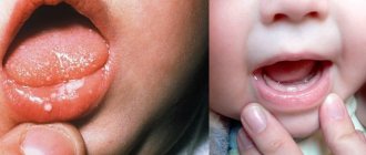
- Children under 7 years old. More than half of cases of stomatitis occur in preschool children due to the lack of the habit of regularly washing their hands.
- Elderly people with diseases of internal organs and loss of several teeth.
- Persons living in unsanitary conditions, as well as those who refuse to observe the rules of personal hygiene.
- Smokers, regardless of length of service.
- Persons with reduced immunity after severe illness, chemotherapy or organ transplantation.
- Patients who have undergone a long course of antibiotic treatment.
- Persons with asthma who use inhalers without following hygienic rules for their use.
The frequency of cases of stomatitis is from 5 to 20%, depending on living conditions: less in large cities, more in rural areas where there is no centralized water supply or the water quality is considered unsatisfactory.
Candidal stomatitis
In 80% of healthy children, Candida fungi can be found in the oral cavity. They get there during childbirth, from nipples and pacifiers, from care products, in contact with the mother's skin, with food during eating and usually do not cause any problems. Candidal stomatitis, or thrush, occurs when immunity decreases1.
Predispose to the disease:
- prematurity and postmaturity;
- developmental defects and concomitant diseases;
- treatment with antibiotics and hormones;
- artificial feeding;
- poor care and poor feeding hygiene;
- Using the wrong nipples1.
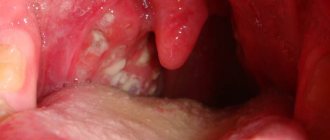
What does candidal stomatitis look like? In children's mouths, whitish or whitish-gray dotted formations appear on the reddened mucous membrane. They merge into films of a cheesy nature; when the films are rejected, bright red painful erosions are formed. Because of the pain, the baby becomes restless, cries often, sleeps poorly, and refuses to eat1.
In severe cases of candidiasis, a cheesy coating may appear on the palate and on the lateral surfaces of the tongue, on the tonsils and the back wall of the pharynx - candidal tonsillitis and pharyngitis develop1.
If your child has a fungal infection, you should always consult a doctor; he will tell you how and what to treat the mucous membrane with candidal stomatitis, so that the child does not have complications and recovers faster. Treatment, as a rule, involves treating the oral cavity with drugs with antifungal activity (chlorhexidine, hexethidine). In severe cases of the disease, antifungal agents, probiotics and immunomodulators are prescribed1.
Up to contents
Types of stomatitis
Depending on the reasons that caused the inflammation, infectious and non-infectious diseases are distinguished:
- viral stomatitis - caused by the consequences of an infectious disease or the effects of pathogenic microorganisms on the damaged oral mucosa. It looks like bubbles with transparent contents that gradually become cloudy. Ulcers form at the site of burst blisters;
- bacterial – consequences of damage to the body by staphylococci or streptococci. Under favorable conditions, they quickly spread throughout the mucous membrane, on which purulent foci form;
- fungal - characterized by the formation of numerous ulcers with a white coating;
- radiation – the consequences of ionizing radiation in the form of painful erosions and hardening of the mucous membrane;
- chemical – associated with burns of the mucous membrane, after healing of which dense scars form;
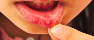
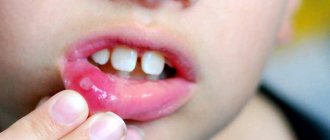
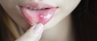
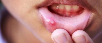
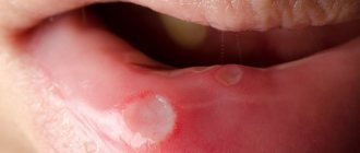
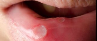
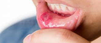
- aphthous - one of the popular types of stomatitis, is associated with reduced immunity and the consequences of gastrointestinal diseases. Small ulcers with a diameter of up to 5 mm are painful and quickly grow in size;
- allergic – caused by exposure to external irritating factors, goes away completely without treatment when the allergen is eliminated: foods, medications, incorrectly selected dentures, etc.
Acute herpetic stomatitis
Herpes
is the most common viral infection.
About 90% of people on earth are infected with herpes. Herpesvirus infections became one of the significant medical and social problems in the USA and Western European countries 20 years ago. However, in our country this group of infectious diseases remains little known. This is due to the fact that patients are observed by different specialists - dermatologists, dentists, gynecologists, ophthalmologists, therapists, etc. Currently, one of the most common diseases in childhood is herpetic infection, which is explained not only by the widespread prevalence of the herpes simplex virus (HSV) , but also by the peculiarities of the formation of the immune system in the developing child’s body. Herpetic infections in general are among the most common and poorly controlled. According to WHO, among viral infections, diseases caused by HSV rank second after influenza. The incidence of HSV infection in children aged 6 months to 5 years is 60%, and by the age of 15 it is already 90%. The incidence of acute (primary) herpetic stomatitis in children increases every year. N.F. was the first to point out the role of HSV in diseases of the oral mucosa at the beginning of the 20th century. Filatov (1902). He suggested the possible herpetic nature of the most common acute aphthous stomatitis among children. This evidence was obtained later, when HSV antigens began to be detected in the epithelial cells of the affected areas of the oral mucosa. According to the international classification of diseases and health problems, latest (tenth) revision (ICD 10, Geneva, 1995), this disease is registered as acute herpetic stomatitis (AHS). OGS not only ranks first among all lesions of the oral mucosa, but is also included in the leading group among all infectious diseases of childhood. Moreover, in every 7-10th child, AGS early becomes chronic with periodic relapses. The widespread occurrence of the disease in children from 6 months to 3 years is explained by the fact that at this age the antibodies received from the mother through the placenta disappear in children and the lack of mature specific immune systems. Among older children, the incidence is significantly lower due to acquired immunity after herpetic infection in its various clinical forms. In the development of herpes infection, which manifests itself mainly in the oral cavity, the structure of the oral mucosa in children at different ages and the activity of local tissue immunity are of great importance. The highest prevalence of OGS under the age of 3 years may be due to age-morphological characteristics, manifested by high permeability of histohematic barriers during this period and low level of cellular immune reactions due to the thinness of the epithelial cover with low levels of glycogen and nucleic acids, friability and poor differentiation of the basal membrane and fibrous tissues. connective tissue structures (abundant vascularization, high content of mast cells with their low functional activity, etc.). The appearance of lesions on the oral mucosa is preceded by lymphadenitis of varying severity. In moderate and severe clinical forms, bilateral inflammation of the submandibular lymph nodes often develops, but all groups of cervical lymph nodes can also be involved in the process. Lymphadenitis in OGS not only precedes the rash of lesions in the oral cavity, but accompanies the entire course of the disease and persists for 7-10 days after complete epithelialization of the elements. Both specific and nonspecific immune factors play a role in the body’s resistance to disease and its protective reactions. Studies of nonspecific immunological reactivity in AGS have revealed violations of the body's protective barriers, reflecting the severity of the disease and periods of its development. Moderate and severe forms of stomatitis led to a sharp suppression of natural immunity, which was restored 7-14 days after the child’s clinical recovery.
Clinical picture
AHS, like many other infectious diseases, occurs in mild, moderate and severe forms. During the development of the disease, two phases are distinguished - catarrhal and rash of lesions.
During this period, symptoms of damage to the oral mucosa appear.
Initially, intense hyperemia of the entire mucous membrane occurs, and after a day, less often two, elements of the lesion are usually found in the oral cavity. The severity of AGS is assessed by the severity of toxicosis manifestations and the nature of mucosal lesions. The mild form of AGS
is characterized by the external absence of symptoms of intoxication of the body; the prodromal period is not clinically manifested.
The disease begins suddenly with an increase in temperature to 37-37.5°C. The general condition of the child is quite satisfactory. A child sometimes exhibits minor inflammation of the mucous membrane of the nasal cavity and upper respiratory tract. Sometimes hyperemia and slight swelling occur in the oral cavity, mainly in the area of the gingival margin (catarrhal gingivitis). The duration of this phase is 1-2 days. The vesicle stage is usually monitored by parents and the doctor, as the vesicle quickly bursts and turns into aphtha. Afta is a round or oval-shaped erosion with smooth edges and a smooth bottom with a rim of hyperemia around it. In most cases, against the background of increased hyperemia, single or grouped lesions appear in the oral cavity, the number of which usually does not exceed 6. Single rashes. The duration of the disease development is 1-2 days. The period of extinction of the disease is longer. Within 1-2 days, the elements acquire a marble-like color, their edges and center are blurred. They are already less painful. After epithelialization of the elements, the phenomena of catarrhal gingivitis persist for 2-3 days, especially in the area of the anterior teeth of the upper and lower jaw. In children with this form of the disease, as a rule, there are no changes in the blood; sometimes only towards the end of the disease does slight lymphocytosis appear. Natural immunity in a mild form of stomatitis suffers slightly, and during the period of clinical recovery, the child’s body’s defenses are almost at the level of those in healthy children, that is, in a mild form of stomatitis, clinical recovery means the complete restoration of the impaired defenses of the body. The moderate form of OGS
is characterized by quite clearly defined toxicosis and damage to the oral mucosa during all periods of the disease.
Already in the prodromal period, the child’s well-being worsens, weakness, moodiness, loss of appetite appear, and possible catarrhal sore throat or symptoms of acute respiratory disease. The submandibular lymph nodes enlarge and become painful. The temperature rises to 37-37.5°C. As the disease progresses during the development of the disease (catarrhal phase), the temperature reaches 38-39°C, headache, nausea, and pale skin appear. At the peak of the rise in temperature, increased hyperemia and severe swelling of the mucous membrane, elements of the lesion appear both in the oral cavity and often on the skin of the perioral area. In the oral cavity there are usually from 10 to 20-25 lesions. During this period, salivation increases, saliva becomes viscous and viscous. There is pronounced inflammation and bleeding of the gums. Rashes often recur, which is why when examining the oral cavity one can see elements of the lesion that are at different stages of clinical and cytological development. After the first eruption of lesions, body temperature usually drops to 37-37.5°C. However, subsequent rashes are usually accompanied by a rise in temperature to the previous level. The child does not eat, sleeps poorly, and symptoms of secondary toxicosis increase. ESR rises to 20 mm/h, leukopenia, sometimes slight leukocytosis, is more common. The level of band neutrophils and monocytes is usually within the upper limits of normal, lymphocytosis and plasmacytosis are detected. An increase in the titer of herpetic complement-fixing antibodies is detected more often than after a mild form of stomatitis. The duration of the period of extinction of the disease depends on the degree of resistance of the child’s body, the presence of carious and damaged teeth, and the rationality of therapy. Under unfavorable conditions, the elements of the lesion merge, their subsequent ulceration occurs, and ulcerative gingivitis develops. Epithelization of the lesion elements takes up to 4-5 days. Gingivitis, severe bleeding gums and lymphadenitis last the longest. The severe form of AGS
is much less common than the moderate and mild forms. During the prodromal period, the child has all the signs of an incipient acute infectious disease: apathy, adynamia, headache, musculocutaneous hyperesthesia, arthralgia, etc. Symptoms of damage to the cardiovascular system are common: brady- or tachycardia, muffled heart sounds, arterial hypotension. Some children experience nosebleeds, nausea, vomiting, and pronounced lymphadenitis of not only the submandibular, but also the cervical lymph nodes. During the development of the disease, the temperature rises to 39-40°C. The child has a mournful expression on his lips, and his suffering, sunken eyes attract attention. There may be a mild runny nose, coughing, and the conjunctivae of the eyes are somewhat swollen and hyperemic. Lips are dry, bright, parched. The mucous membrane of the oral cavity is swollen, clearly hyperemic, and gingivitis is pronounced. After 1-2 days, lesions begin to appear in the oral cavity - up to 20-25. Often, rashes in the form of typical herpetic blisters appear on the skin of the perioral area, eyelids, conjunctiva of the eyes, earlobes, and on the fingers in a felon type. Rashes in the oral cavity recur and therefore at the height of the disease in a seriously ill child there are about 100 of them. The elements merge, forming large areas of necrosis of the mucous membrane. Not only the lips, cheeks, tongue, soft and hard palate are affected, but also the gingival margin. Catarrhal gingivitis turns into ulcerative-necrotic. There is a sharp putrid odor from the mouth, profuse salivation mixed with blood. Inflammatory phenomena on the mucous membrane of the nasal cavity, respiratory tract, and eyes worsen. Streaks of blood are also found in secretions from the nose and larynx, and sometimes nosebleeds are observed. In this condition, children need active treatment from a pediatrician and dentist, and therefore it is advisable to hospitalize the child in the isolation ward of a pediatric or infectious diseases hospital. The period of extinction of the disease depends on the timeliness and correctness of treatment and on the child’s history of concomitant diseases. Despite the clinical recovery of a patient with a severe form of AHS, during the period of convalescence there are profound changes in homeostasis.
Diagnostics
To diagnose HSV, the following research methods are used:
- virological methods for detection and identification of herpes simplex viruses
- polymerase chain reaction (PCR)
- detection of HSV antigens
- registration of immune response to HSV
- cytomorphological methods
- assessment of immune status
The presence of HSV antigens in biological material is determined using serological methods:
- neutralization reactions (RN)
- enzyme immunoassay (ELISA)
- radioimmunoassay (RIA)
- Complement fixation reactions (CFR)
- passive hemagglucination reactions (RPHA)
The diagnosis of AGS is made based on the clinical picture of the disease. The use of virological and serological diagnostic methods in practical healthcare is difficult. This is primarily due to the complexity of special research methods. In addition, with these methods, results can be obtained, at best, towards the end of the disease or some time after recovery. Such a retrospective diagnosis cannot satisfy the clinician.
Symptoms of stomatitis
The type of stomatitis can often be determined by characteristic signs of the disease associated with the cause of its occurrence. All of the above types of pathology are united by:
- the appearance of characteristic ulcers and areas of inflammation on the oral mucosa;
- the formation of white, yellow or green plaque, under which traces of mucosal erosion are visible;
- redness on the gums;
- enlarged lymph nodes;
- unpleasant bloody or putrid taste in the mouth;
- soreness of the affected areas;
- increased salivation.
Signs of stomatitis in adults and children can be more or less pronounced depending on the state of the immune system, compliance with personal hygiene rules and living conditions.
Are you experiencing symptoms of stomatitis?
Only a doctor can accurately diagnose the disease. Don't delay your consultation - call
Symptoms
Common symptoms of stomatitis are pain, burning, swelling of the mucous membrane and redness, and bad breath. Salivation also changes - it can be excessive or insufficient, with dry mouth. In some cases, there may be an increase in temperature and enlargement of the submandibular lymph nodes.
The types of stomatitis in children and treatment methods differ, as do the specific manifestations. For example, with aphthous stomatitis, only one large ulceration may appear, but with viral stomatitis, inflammation almost always takes the form of numerous elements or a rash merging into one focus.
Candidal stomatitis is characterized by the formation of a white or yellowish cheesy coating. It is easier to recognize traumatic stomatitis because it is preceded by damage or the source of injury is nearby - a chipped tooth or a rough filling.
Possible complications of stomatitis
The requirement to immediately consult a doctor at the first signs of stomatitis is dictated by concern for the health and well-being of the patient. Ignoring this rule may cause:
- damage to the mucous membrane of the entire oral cavity;
- development of infectious diseases of internal organs;
- inflammation of the gums and loss of some teeth;
- digestive problems;
- infection of loved ones through kissing, sharing utensils, etc.;
- diseases of the heart, lungs and blood.
Self-medication can lead to similar consequences. Therefore, if you suspect stomatitis, you should immediately seek help from a therapist or pediatrician.
Diagnosis of the disease
In most cases, an external examination of the patient is sufficient to make a diagnosis. If teeth and gums are affected, a dentist is involved in consultation. If a secondary nature of the disease is suspected, causing inflammation of the mucous membrane, highly specialized doctors can begin working with the patient.
Diagnosis of infectious stomatitis has its own characteristics. Special laboratory tests allow you to determine the type of pathogenic organism that caused the inflammatory process:
- oral swab for testing for fungi and bacteria;
- blood test for the presence of antibodies to pathogens of infectious diseases of internal organs;
- smear or PCR (polymerase chain reaction) blood test to detect and analyze the DNA of viruses.
If the disease is accompanied by an increase in body temperature and a serious deterioration in health, the patient is referred for blood and urine tests. This will identify possible complications or infectious diseases that cause inflammation of the oral mucosa.
Treatment options
When visiting a doctor in the first stages of the disease, treatment of stomatitis at home is symptomatic and lasts about a week. When an infectious cause of the disease is established, the action plan changes:
- a course is prescribed that relieves inflammation and other main symptoms;
- local gels and ointments are prescribed;
- drugs are selected that act on the causative agent of the disease to completely destroy it.
The complex of drugs for the treatment of infectious stomatitis includes antibiotic compounds, painkillers, antiseptic and antihistamine medications. The duration of this course is about 10 days. If the diagnosis establishes the secondary nature of the disease and serious pathologies are identified in the patient’s body, the treatment process may be delayed until the possible provoking factors are completely eliminated.
Additional methods to speed up the recovery process and increase the effectiveness of medications for stomatitis include:
- avoidance of foods that cause irritation to the mucous membranes;
- increasing the volume of fermented milk products in the diet;
- rinsing the mouth with calendula decoctions;
- lubricating ulcers with sea buckthorn oil, aloe juice or petroleum jelly.
Treatment of stomatitis in adults should be carried out under the supervision of a physician. This approach will eliminate possible side effects and promptly adjust the course to increase its effectiveness.
Prevention of stomatitis
You can eliminate the risk of inflammation of the mucous membrane and the development of stomatitis by following the following recommendations:
- stop smoking and drinking alcohol;
- use antibiotics only as prescribed by a doctor, strictly in the indicated doses;
- maintain oral hygiene;
- promptly treat diseases of teeth and gums;
- reduce the amount of sweet foods and yeast baked goods;
- choose toothpastes and rinses from leading manufacturers;
- Avoid eating too hot, cold or spicy foods;
- take vitamin complexes and strengthen your immune system.
Additional recommendations based on the patient’s health condition will be provided by the attending physician.
Allergic stomatitis
Allergic damage to the mucous membrane most often occurs in the form of contact stomatitis and chronic aphthous form of the disease1.
Allergens can include medications, food products, varnishes and paints that coat toys, toothpastes, mouth rinses, chewing gum, and dental metals included in braces1,2.
With allergic stomatitis in children, erosions and ulcers may appear in the mouth, but more often the matter is limited to redness and swelling of the mucous membrane1.
Treatment is avoiding contact with the allergen, rinsing or irrigating the mouth with antiseptic solutions to prevent infection. If necessary, doctors recommend taking antihistamines1,2.
Chronic recurrent aphthous stomatitis often occurs in schoolchildren and adolescents due to allergies2. In addition, its development can be provoked by diseases of the gastrointestinal tract, upper respiratory tract infections, disorders of the nervous system, and hypovitaminosis1,2.
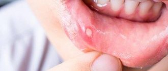
With aphthous stomatitis in children, itching and burning first appear in the oral cavity. The mucous membrane at the site of the lesion becomes red and swollen, then an aphtha forms on it - a round or oval erosion 0.5-1 cm in diameter rising above the surrounding tissues with a red rim along the periphery and a bottom covered with a grayish-white coating. Aphthae are extremely painful, and if many of them form, they cause significant distress to the child1,2.
How many days does aphthous stomatitis last in children? With a mild course of the disease, the elements of inflammation persist for up to 5-7 days, then they heal without scar formation2 and do not appear for quite a long time. However, in severe cases, aphthae can occur constantly, and then many elements of inflammation can be found on the mucosa at different stages of development1,2.
To treat aphthous stomatitis, doctors use:
- antihistamines;
- immunomodulatory drugs;
- vitamins;
- probiotics2.
As a local therapy, it is recommended to treat the mucous membrane with drugs with analgesic, antiseptic, proteolytic (protein-breaking), anti-inflammatory and regenerating effects2.
Up to contents
Questions and answers
How to treat stomatitis at home?
Prescribing medications for stomatitis is possible only after examination by a specialist and establishing the causes of the disease. All types of stomatitis are treated at home according to the prescribed course of medications for internal use and local healing agents. The patient is admitted to the hospital only in an emergency, if the examination reveals a serious pathology of the internal organs.
Is stomatitis contagious or not?
The fungal or infectious nature of stomatitis makes the patient a source of spreading the disease to others. If the disease is caused by an allergic reaction, gastrointestinal pathology or mechanical damage to the gums, there is no danger of infecting loved ones. It is possible to indicate the exact cause of the development of the inflammatory process only after examining the patient and studying the test results. Before visiting a doctor, you should limit contact with family and strictly observe personal hygiene rules.
How long does it take to treat stomatitis?
If you consult a doctor early, you can completely cope with the disease within a week. If the inflammation is caused by a problem in the internal organs, stomatitis takes longer to treat, depending on the degree of development of the underlying pathology. The sooner the risk factors are eliminated, the faster the symptoms will be eliminated.