A tumor in the ear can be benign or malignant. The Yusupov Hospital is equipped with modern diagnostic equipment from leading European and American manufacturers. This allows otolaryngologists to establish an accurate diagnosis in the shortest possible time. Oncologists take an individual approach to choosing a treatment method for each patient. All complex cases are discussed at a meeting of the expert council with the participation of professors and doctors of the highest category. Ear cancer is treated using the latest techniques.
Ear cancer is diagnosed in 2% of all malignant tumors and in 12% of tumors of the ENT organs. Tumors of the external ear account for up to 95% of all ear tumors. In 85% of cases of malignant neoplasms, a tumor occurs on the earlobe and auricle, and in 10% - in the external auditory canal.
Polyp in the ear
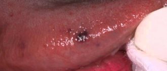
Benign tumors of the ear include polyps. They arise as a result of the proliferation of granulation tissue. The polyp may be located in the external auditory canal or middle ear. Neoplasms localized in the ears can spread to other parts of the skull.
Most often, a polyp is a complication of a chronic inflammatory process in the ear. At the site of chronic inflammation of the mucous membrane, gradual tissue proliferation occurs, replacing normal connective tissue. When the pathological process is localized in the middle ear, the formation may remain invisible to conventional otoscopy for a long time. As the polyp grows, it “falls out” into the external auditory canal through the perforation of the eardrum.
A polyp in the ear is manifested by the following symptoms:
- Suppuration, sometimes mixed with blood (stopping the flow of pus can be caused by blockage of the ear canal by a polyp);
- Itching, noise and pain in the ear;
- A feeling of constriction, the presence of a foreign body in the ear cavity;
- Decreased or loss of hearing;
- Headaches.
In the absence of adequate treatment, a polyp caused by an infection in the ear often becomes the cause of chronic otitis media, supports the inflammatory process and prevents the penetration of drugs to the site of infection. The growth of the polyp leads to blockage of the ear canal and deafness. Under certain conditions, there is a risk of its degeneration into a malignant tumor.
For polyps of small size, in some cases, otolaryngologists carry out conservative treatment with creams containing glucocorticoids and antibacterial drops. If the disease is fungal in nature, antifungal drugs are used. However, the main treatment for a polyp in the ear is surgery.
The polyp is cut off on an outpatient basis with a special loop or using another instrument: a curette, an ear conchotome. Radical surgery is performed in a hospital. The operation is performed if the fistula is localized in the semicircular canal. An alternative treatment option is laser removal of polyps. A modern method of treating a polyp in the ear is OTO NUS therapy in combination with endoural LILI (exposure to the pathological focus with low-frequency ultrasound through various medicinal solutions).
Make an appointment
Lump on the jaw joint near the ear
Head of the Institute for the Treatment of Joints: “You will be amazed at how easy it is to cure your joints by taking the product every day for 147 rubles .
Dental diseases
There are the following types of dental diseases, in which pain in the jaw can radiate to the ear:
- caries;
- periodontitis;
- gingivitis;
- alveolitis;
- periodontal abscess.
- dry mouth;
- hoarseness;
- pain when swallowing;
- fever;
- sore throat.
Lymphadenitis
When the lymph nodes are involved in the inflammatory process, swelling also forms. Most often this occurs in the corner of the lower jaw, in front of or behind the ear. Lymph nodes react to any inflammatory process in the area of their functional activity: otitis and mastoiditis, tonsillitis, periodontitis, sinusitis, skin infections, etc.
Signs of lymphadenitis are mainly local in nature. These include the usual signs of inflammation such as swelling, redness and tenderness. The affected lymph nodes increase in size, and the skin over them becomes hotter. During an acute purulent process, an abscess forms in the thickness of the tissue, which leads to an increase in local symptoms and a deterioration in the general condition.
Signs of lymphadenitis are mainly local in nature. These include the usual signs of inflammation such as swelling, redness and tenderness. The affected lymph nodes increase in size, and the skin over them becomes hotter. During an acute purulent process, an abscess forms in the thickness of the tissue, which leads to an increase in local symptoms and a deterioration in the general condition.
Glomus tumor of the middle ear
Tympanic paraganglioma (glomus tumor of the middle ear) develops from glomus bodies, which are located on the medial wall or roof of the tympanic cavity, jugular - on the bulb of the jugular vein. Paraganglioma is a benign neoplasm, but mature forms of the tumor have infiltrating and locally destructive growth.
Due to the impossibility of total removal, a glomus tumor of the middle ear can pathologically spread to vital structures of the body (brain stem, internal carotid artery). It can destroy the walls of the temporal bone pyramid, penetrate the posterior cranial fossa and cause compression of the medulla oblongata. Glomus cells often affect vessels over a considerable distance, leading to various complications with a fatal outcome. Patients complain of a “pulsating” noise in the ear. During an objective examination behind the eardrum, the doctor sees a pulsating red mass. As the tumor grows, the following symptoms occur:
- Hearing impairment;
- Facial asymmetry;
- Dysphonia (speech disorder);
- Dysphagia (swallowing disorder).
The Yusupov Hospital has accumulated extensive experience in diagnosing and treating patients with glomus tumor of the middle ear. Otolaryngologists determine the degree of invasion of the glomus tumor of the middle ear into adjacent structures using computed and magnetic resonance imaging of the temporal bones with contrast, angiography and retrograde jugulography. Doctors make the final diagnosis based on the results of histological examination.
If the glomus tumor of the middle ear is widespread, angiography is mandatory. The study is necessary to confirm the vascular nature of the neoplasm, determine its size, location and sources of blood supply. This plays a role in the possibility of embolization, a minimally invasive procedure that is an alternative to surgery.
The procedure is aimed at preventing blood supply to the damaged area, which helps to reduce the size of the tumor and achieve a good effect with further surgical removal of the identified tumor. Total surgery is performed in the presence of a glomus tumor that does not spread beyond the middle ear. For subtotal (incomplete) removal of the tumor, and depending on the patient’s age, radiation therapy or stereotactic radiotherapy (gamma knife) is used.
Preventing ear inflammation on vacation
In addition to the basic advice to avoid sudden changes in temperature, hypothermia, swimming in dirty ponds, people who have problems with nasal breathing are advised to cure it in time, since swelling and difficulty in nasal breathing contribute to the development of inflammation in the ear.
If you or a member of your family has otitis media or its symptoms, you should consult with an ENT doctor before going on vacation. The doctor will provide the necessary instructions, prescribe ear drops depending on the form of the disease, recommend antibacterial drugs, as well as vasoconstrictor nasal drops to relieve swelling from the mouth of the auditory tube.
Even with a slight runny nose while traveling by plane, before takeoff and landing, it is recommended to use vasoconstrictor drops in the nose, which will avoid the development of complications from the ears.
Middle ear hemangioma
Benign tumors of the middle ear also include hemangioma and various neurogenic neoplasms. Hemangiomas of the middle ear are manifested by the following symptoms:
- Decreased hearing;
- Ear congestion;
- Feeling of noise.
Often the first symptom of the disease is a slowly occurring paralysis of the facial muscles on the side where the hemangioma is located. For middle ear hemangioma, otolaryngologists usually perform abdominal surgery or widely remove the mastoid process.
Chemodectoma
Chemodectoma of the middle ear develops from glomus bodies, which are normally located at the bottom of the tympanic cavity, on the dome of the bulb of the internal jugular vein and in the temporal bone. They differ in structure from glomus bodies, which are located in other areas. Depending on the histological structure and the ratio of cell accumulations, there are 3 types of glomus tumors: adenoid-like, alveolar and angioma-like. According to the clinical course, limited and widespread forms of chemodectoma are distinguished.
Chemodectomas are observed at different ages and can be multiple in both ears. Sometimes neoplasms have a malignant course from the very beginning, despite the benign structure of the chemodectoma.
Chemodectomas, which are located in the tympanic cavity, in the initial period of the disease cause hearing loss and pulsating noise in the ear. At this time, the neoplasm appears through the eardrum. Then the tumor protrudes it and causes hyperemia (redness). Gradually, the neoplasm penetrates the external auditory canal and looks like a polyp. When you try to remove it, bleeding occurs. Sometimes patients report ear pain.
Chemodectomas that originate in the jugular vein bulb first destroy the dome of the jugular fossa and spread into the tympanic cavity. As the tumor increases and bone destruction occurs, symptoms of damage to the VII-XII pairs of cranial nerves develop. Patients are bothered by noise in the ear, and otoscopic changes occur. Chemodectomas can grow into the cranial cavity.
Diagnosis of chemodectoma is carried out using radiography of the jugular fossa, pyramid of the temporal bone, attic-antral region, mastoid process. X-ray examination includes radiography of the temporal bone in three main projections and tomography in direct and lateral projections.
Chemodect treatment is surgical. Small tumors that do not destroy the eardrum are removed or exposed to ultra-low temperatures. Tumors that have spread to the external auditory canal, mastoid process, or antrum are also subject to surgical treatment. Otolaryngologists perform operations of varying scope - from tympanotomy to extended radical surgical interventions on the ear. Sometimes cryotherapy is used. For tumors that destroy the pyramid and spread into the cranial cavity, external gamma irradiation is performed, which often causes growth arrest or a decrease in chemodectoma.
Diagnostics
To find out the reason for the appearance of a lump on the jaw, a comprehensive examination is prescribed. First, the doctor examines and collects the patient’s complaints. If a patient has lymphadenitis, at the initial stage of the disease the following symptoms appear:
- slight enlargement of lymph nodes, their mobility;
- pain of the nodes on palpation, turning the neck (pain can radiate to the ear or other parts of the body nearby);
- general weakness;
- low-grade temperature ;
- decreased sleep quality;
With inflammation of the lymph nodes, accompanied by purulent processes, the patient has complaints of:
- increased pain when moving the jaw;
- redness of the skin in the area where pus accumulates;
- severe swelling of the lymph nodes, their pain even without pressure;
- temperature rise to 38 ºC or more.
In the last stages of lymphadenitis, the patient’s well-being deteriorates significantly: inflammatory processes spread to the neck and shoulders, fever appears, and bluish skin is observed at the site of inflammation.
Since, in addition to inflammation of the lymph nodes, there are other reasons for the appearance of lumps on the jaw, it is impossible to make a correct diagnosis based on the clinical manifestation of the pathology. Therefore, laboratory and instrumental diagnostic methods are used.
The patient is prescribed:
- blood test (general, advanced);
- bacteriological examination of mucus from the tonsils and upper respiratory tract;
- radiography of the jaw (in different projections);
- ultrasound examination of pathological formation;
- CT or MRI of the head;
- biopsy (if necessary).
Regardless of the mechanism of occurrence of a lump on the jaw, it is important to begin taking therapeutic measures in a timely manner. The earlier treatment is started, the lower the risk of complications.
Osteoma
Osteoma in the ear (exostosis, osteophyte) develops mainly from the compact layer of the posterior wall of the bony part of the external auditory canal. Much less often, neoplasms are found on the lower and upper walls of this section. Endophytic osteomas penetrate into the mastoid process. Osteoma is a benign tumor that grows rather slowly.
Osteoma has the appearance of a round formation, which is covered with a skin layer, very dense when palpated with a Vojacek probe. It is treated surgically. The operation is performed after the tumor has grown to medium size. In this case, removing the tumor is technically as convenient as possible. If the tumor is small, there is a risk of not completely removing the pathological tissue. If the osteoma is large, it is possible to capture a significant part of the healthy bone tissue during surgery. This will cause a large bone defect.
Mumps
Mumps can also cause a tumor. It is the salivary glands located next to the ears that have become inflamed, which means they have noticeably increased in size. At the same time, the patient, and these are mainly children, experience an inflammatory process of the throat mucosa, an increase in temperature and a general weak condition. This disease, which has a popular name - “mumps”, is accompanied by painful manifestations in the affected areas. The danger of mumps is that it is transmitted by airborne droplets, and the infection easily spreads to a healthy person. Therefore, complete isolation of the patient is required.
Lipoma and atheroma
The area of skin around the ear contains a huge number of sebaceous glands. For this reason, lipomas and atheromas often form behind the ear. Lipomas that form behind the ear grow slowly and are often not cancerous. They are a soft-elastic formation with a smooth surface, surrounded by a capsule. Lipoma has the appearance of a wen.
Atheroma is a cavity formation filled with sebum. Formed due to blockage of the sebaceous glands. Atheromas occur for the following reasons:
- Disorders of fat or carbohydrate metabolism;
- Genetic predisposition to increased oily skin;
- Hormonal imbalances and diseases of the endocrine system;
- Hyperhidrosis is a disease associated with excessive sweating;
- Failure to comply with personal hygiene rules.
Atheroma is a rounded formation protruding above the surface of the skin, which can reach up to 4.5 cm in diameter. When the tumor becomes infected or inflammatory reactions occur, the following symptoms occur:
- Pain behind the ear;
- Redness of the skin;
- Burning and itching;
- Fluctuation is a symptom that indicates the presence of fluid in a cavity formation.
When pressure is applied to the walls of the atheroma or they are damaged, the viscous mass contained inside comes out to the surface of the skin. It has a white color and an unpleasant odor. When atheroma suppurates, the contents have a green-yellow tint. Lipomas and atheromas behind the ear are removed surgically. Modern treatment methods are used - laser or radio wave removal.
Effective medical assistance
Seeking help from a doctor if you have a lump on your head near your ear is the best decision. First of all, he finds the starting point and finds out whether the patient has any diseases, since the formation of a lump can be affected by:
- infection getting under the skin. Most often this happens due to an unprofessional puncture of the earlobe or ear cartilage from behind;
- development of a chronic disease, such as diabetes, etc.;
- increased production of sebaceous glands;
- weakened immune system;
- increased sweating.
Adenoma behind the ear
A benign tumor, a parotid adenoma, often develops in the parotid region. The structure of the neoplasm resembles the salivary gland itself. The cause of the development of benign tumors of the salivary glands is the formation of altered glandular epithelium.
The neoplasm is enclosed in a capsule, has a soft-elastic consistency, and is not fused to the skin and surrounding tissues. The skin above the adenoma behind the ear is not changed. It is treated surgically. To undergo examination and treatment of benign tumors of the ear and parotid region, call the contact center of the Yusupov Hospital.
What is the main reason?
Most often, help is required due to the appearance of a hard lump near the earlobe due to inflammation of the lymph nodes. Moreover, several small bumps may appear. They roll under the skin and hurt. The high temperature begins to worry, the general condition deteriorates sharply. Itching and redness appear. In this case, the doctor diagnoses lymphadenitis. This disease is not considered particularly dangerous, but the source of inflammation must be removed as quickly as possible, since this disease has the ability to take an acute or chronic form, as well as with the formation of pus. If no treatment is given, sepsis may occur. It differs from other similar seals in that in this case it is not sebaceous deposits. The swollen parotid lymph nodes are to blame for this due to inflammation and the entry of pathogenic microorganisms into the body.
Causes and types of ear cancer
Malignant neoplasms in the ear area occur against the background of pathological processes called precancerous. The morphological structure of malignant tumors of the external ear is diverse. In 61% of cases, morphologists identify epithelial cancer, 38% - glandular cancer.
In the external auditory canal, basal cell carcinoma is 2-3 times less common than squamous cell carcinoma. Squamous cell carcinoma with varying degrees of keratinization predominates. The initial localization of malignant neoplasms of the auricle is the posterior surface, the area of the postauricular fold and the earlobe.
The primary tumor of the external auditory canal occurs on one of its walls. The pathological process predominantly develops in the area of the lower and posterior walls of the ear canal. Ear cancer can also develop on scars. A tumor behind the ear can also be cancerous.
Make an appointment
Tumors of the external ear
Malignant neoplasms of the external ear are represented by the following tumors:
- Squamous cell carcinoma;
- Basal cell carcinoma;
- Adenocarcinoma from ceruminoma;
- Adenoid cystic carcinoma.
Oncologists detect the following soft tissue tumors in the ear and jaw area:
- Hemangioma;
- Neurofibroma;
- Neurilemmoma (schwannoma).
Fibrosarcoma or rhabdomyosarcoma can develop in and around the ear. Papillomas most often occur on the skin of the auricle. If the neoplasms are located in the external auditory canal, they often fill its lumen, resembling polyps that come from the middle ear. Ceruminoma of the external auditory canal is a very rare and long-growing tumor arising from the sebaceous (sulfur) glands.
Ceruminoma is usually observed in people over 20 years of age. With this type of ear cancer, the symptoms are as follows:
- Ear congestion;
- Hearing loss;
- Pain and discharge from the ear.
In the initial stage, the tumor is located on the wall of the external auditory canal. The tumor is pink. As it grows, it fills the ear canal and looks like a polyp. Radiologically, good pneumatization of the mastoid process is determined. Gradually, the tumor spreads to the middle ear and its walls, destroying them. These changes are determined on radiographs.
Mixed tumors of the external auditory canal are secondary. They most often originate from the parotid gland and penetrate the external auditory canal.
Nevi (benign pigmented tumors of the auricle and external auditory canal) do not differ in clinical course from nevi that are located on other areas of the skin. Soft tissue tumors (fibroma, hemangioma) arise from fibrous, fatty, muscle, vascular and other tissues.
Fibroma is most often located on the earlobe, at the sites of puncture with a needle for wearing earrings. Dimensions vary from 5 mm to 4 cm. Less commonly, the tumor is localized at the ascending branch of the helix of the auricle and at the entrance to the external auditory canal.
Hemangiomas develop in all parts of the ear. Capillary and cavernous forms of vascular tumors are more often observed. The first ones very often disappear in childhood. Cavernous hemangiomas are located in the thickness of the auricle. They occur in the form of individual or multiple neoplasms. They have a soft consistency and a bluish tint. Hemangiomas of the auricle can affect its edge and other parts. They often spread towards the external auditory canal, closing its lumen, and bleed when injured.
Malignant ear tumors
Locally destructive tumors of the ear include basal cell carcinoma, and malignant tumors include cancer, melanoma, and sarcoma. The most commonly observed are basal cell carcinomas and cancer. The listed tumors of the outer ear, affecting the skin and spreading to the cartilaginous and bone parts of the ear, grow into the scalp, bones of the face and skull, and the parotid salivary gland. They grow either slowly or very quickly.
Cancer of the outer ear often occurs at the site of injury, long-term inflammatory processes, or age-related skin changes. Develops under the influence of the following predisposing factors:
- Burns;
- Frostbite;
- Household and professional hazards.
Cancer of the outer ear can be endophytic (flat ulcer with infiltrated edges) or exophytic (warty nodules with a wide base). A cancerous tumor, having arisen in one or another part of the auricle, infiltrates and gradually destroys the entire shell, then spreads to adjacent tissues and organs. Cancer of the external auditory canal can grow into the auricle, mastoid process, middle ear, parotid salivary gland, skull bones and cause paralysis of the facial muscles on the side of the tumor.
Cancer of the external auditory canal in the early stages of development is usually impossible to recognize, since patients do not show any complaints, and the externally pathological process resembles weeping eczema or a chronic inflammatory process, which is manifested by the formation of pale granulations. During this period, the first signs of ear cancer are often observed: itching and pain in the external auditory canal.
Cancerous tumors of the external auditory canal grow faster than tumors of the auricle and are characterized by severe bleeding. Further growth occurs towards the auricle or towards the middle ear, or in all directions at the same time. As the cancerous tumor spreads to the middle ear, surrounding tissues and bones, excruciating pain occurs and the following symptoms develop:
- Paralysis of facial muscles;
- Deafness;
- Metastases in regional lymph nodes.
When will you need specialist help?
But it happens that the health condition may worsen, and the culprit in this case is a small lump on the jaw near or behind the ear.
Doctors advise not to hesitate and visit a medical facility if:
- periodically arising pain bothers you, over time it becomes more and more disturbing;
- nearby lymph nodes enlarge;
- the color of the damaged area is significantly different from the healthy one;
- the general condition is increasingly deteriorating, weakness, a feeling of fatigue after little physical activity, and an increase in body temperature may appear.
To avoid visiting a doctor, some people begin to use medical and folk remedies, trying to squeeze out the contents that have accumulated in the lump near the ear in children and adults. Most often, this leads to sad consequences: the inflammatory process begins to progress, the tumor increases, and the general state of health deteriorates sharply.
What is required of the patient during this period is to prevent infection from entering the affected area, ensuring its sterility.
Treatment
For benign tumors, surgical treatment is performed. The following methods are used:
- Local excision with a scalpel;
- Electroexcision and coagulation;
- Cryogenic exposure.
To prevent recurrence of the tumor, the operation is performed radically, taking into account possible erosion of cartilage and bone. The postoperative period after ear tumor removal proceeds in most cases without complications. Sometimes, with a localized tumor, it becomes necessary to resect the auricle.
For stage I cancer and basal cell carcinoma of the auricle, surgical and radiation methods are used. If, after close-focus radiation therapy, remnants of the tumor remain, then after the radiation epidermitis subsides, electroexcision of the tumor is performed. Cryogenic therapy is also effective.
Cancerous tumors of the auricle of stage II and basal cell carcinomas of the same size are subjected to surgical and combined treatment. First, preoperative radiation therapy is performed. After 2 weeks, electrosurgical resection of the affected area of the auricle with the underlying cartilage is performed. Elderly patients obtain good results after using cryosurgery.
For stage III ear cancer and basal cell carcinoma, combined treatment is carried out. 2 weeks after the end of preoperative radiation therapy, wide radical surgery is performed. Surgeons often have to excise the external auditory canal. The resulting defect is closed with free skin grafting or local tissue.
Make an appointment
Treatment of benign middle ear tumors
Treatment of fibroids, endotheliomas, osteomas is carried out surgically. Electrocoagulation and radiotherapy are used to treat angiomas. For advanced osteoblastomas, radiotherapy is used.
In recent years, otolaryngologists have often observed glomus tumor. It ranks first in frequency among benign tumors of the middle ear. It develops from glomus (glomeruli), which are often found along the tympanic nerve, the auricular branch of the vagus nerve, and, less commonly, the superior petrosal nerve. They can be located in the mucous membrane of the tympanic cavity, adventitia of the jugular vein bulb. Glomus have a size from 0.5 mm to 2.5 mm, surrounded by a capsule. They consist of numerous intertwined capillaries and precapillaries, as well as special epithelioid or glomus cells. Innervated by the glossopharyngeal and vagus nerves.
The tumor, which originates from the tympanic cavity, gradually protrudes the eardrum and grows into the external auditory canal. It causes destruction in the tympanic cavity, which leads to hearing loss and facial paralysis. The neoplasm can grow into the jugular fossa, causing its destruction and paralysis of the IX, X and XI cranial nerves. Sometimes it grows into the posterior fossa, causing the corresponding symptoms. The tumor grows slowly, but has infiltrative growth and often bleeds. As a result, it is classified as a clinically low-quality neoplasm.
The diagnosis is based on the results of the examination:
- Clinical picture;
- Otoscopy;
- X-rays;
- Histological examination.
Sometimes the presence of Brown's symptom helps to make an accurate diagnosis - the cessation of pulsation of the tumor when the pressure in the external auditory canal increases using a Siegle funnel. Since radical surgery often cannot be performed due to severe bleeding, oncologists use radiation therapy or perform surgery followed by radiation. Positive dynamics are observed after 20-25 injections into the tumor of a 10% solution of quinine hydrochloride, 0.5 ml.
If the tumor spreads to the carotid artery canal, freezing is performed using a Cooper cryosurgical probe. At a temperature of -180°C, the neoplasm tissue is frozen and completely removed. The artery wall is protected from freezing by the blood in the artery.
Treatment of malignant ear tumors
Sarcoma is a malignant tumor of the middle ear. It can be round cell, spindle-shaped, or myxosarcoma. It occurs very rarely, more often in children. In the presence of sarcoma, oncologists perform radiation therapy or electrocoagulation of the tumor followed by irradiation.
Much more often, doctors at the Yusupov Hospital diagnose basal cell and squamous cell carcinoma of the middle ear. The tumor predominantly develops due to chronic suppurative otitis media, and is therefore diagnosed late. The occurrence of precancerous changes (papillomatous growths) is promoted by caries of the bone walls of the tympanic cavity in chronic purulent otitis media. It supports chronic inflammation of the metaplastic epithelium of the mucous membrane of the tympanic cavity, which is constantly irritated by purulent secretions.
Most often, the cancerous tumor comes from the attic-antrum region or the tympanic ring. Middle ear cancer is characterized by rapid infiltrative growth, especially in young people, spreading to the parotid gland, inner ear, mandibular joint, and cranial cavity. This makes radical removal of the tumor much more difficult. Cancer cells metastasize early to regional lymph nodes.
A malignant tumor of the middle ear is characterized by the following symptoms:
- Ear pain;
- Headache;
- Discharge of foul-smelling pus, often mixed with blood;
- Bleeding dense granulations that quickly recur after removal;
- Early peripheral facial palsy;
- Sudden hearing loss.
In later stages, cochlear and vestibular function declines. Intracranial complications (meningitis) develop quite quickly. Oncologists provide combined treatment for middle ear cancer. In case of stage III cancer (damage to the cartilaginous and bone parts of the ear with metastases to regional lymph nodes), extensive surgery with diathermocoagulation is performed. The outer ear, temporal bone (subtotal resection is performed), parotid gland, and articular process of the lower jaw are removed as a single block. In stage IV, radiation and chemotherapy are performed.
Diagnosis and treatment
If the tumor is accompanied by severe pain, increases in size and grows, then it is important to visit a doctor as soon as possible. To make an initial diagnosis, you need to see a therapist or dentist. If the lymph nodes are inflamed, drug therapy will be prescribed; if cancer is suspected, the treatment will be carried out by an oncologist.
Self-medication is dangerous with complications!
Attention
Despite the fact that our articles are based on trusted sources and have been tested by practicing doctors, the same symptoms can be signs of different diseases, and the disease may not proceed according to the textbook.
Pros of seeing a doctor:
- Only a specialist will prescribe suitable medications.
- Recovery will be easier and faster.
- The doctor will monitor the course of the disease and help avoid complications.
find a doctor
Do not try to treat yourself - consult a specialist.
Treatment methods for a tumor under the ear are varied and will depend on the diagnosis and severity of the disease. For inflammation and viral diseases, treatment will be with special pharmaceutical drugs; for cysts and malignant tumors, surgery will be necessary to remove the tumor.
Why can our articles be trusted?
We make health information clear, accessible and relevant.
- All articles are checked by practicing doctors.
- We take scientific literature and the latest research as a basis.
- We publish detailed articles that answer all questions.
The following factors will be the reason to carry out an early diagnosis of the compaction under care:
- greatly enlarged lymph ;
- throbbing acute pain in the area of the tumor;
- the tumor festers , the skin changes color.
Only an experienced specialist will be able to accurately diagnose the cause of the lump under the ear. For example, a dense formation upon palpation may indicate a benign tumor; the size of the compaction will indicate the likelihood of a malignant process. With severe throbbing pain, we can talk about severe purulent inflammation or the beginning of the development of complications.
To make an accurate diagnosis, you will also need to undergo an ultrasound and x-ray. Only on the basis of their testimony will the doctor be able to accurately determine the nature of the tumor and formulate the correct treatment program.
If the cause of the compaction is lymphadenitis, then first it will be necessary to overcome the infectious focus or virus that caused the inflammation. The patient is prescribed antiviral drugs, as well as drugs to strengthen the immune system.
Antibiotics and antifungal drugs may also be prescribed to eliminate the infection and stop the purulent process. In these cases, external hygiene, including the use of antiseptics, is also important. To remove the wen, the patient is given a cortisol injection and the operation is performed under local anesthesia.
If there is severe pain and purulent foci, you should under no circumstances begin self-treatment; the choice of the right medications and treatment methods is chosen only by the attending physician. It is important to remember that any pharmaceutical drugs and traditional methods can only worsen the patient’s condition.
In general, any tumor formations under the ear are removed surgically or with a laser. It is important to consult a specialist as early as possible; if the neoplasm feels elastic and mobile to the touch, then serious complications are possible.
Tumor of the inner ear
Inner ear symptoms occur in patients with tumors located outside this anatomical structure. Otolaryngologists do not know any neoplasms that would arise in the ear labyrinth. True cholesteatomas, which are rarely found in the inner ear, refer only to tumor-like formations, the structure of which is fundamentally different from the structure of known “tissue” tumors.
Neuroma of the vestibular-cochlear nerve is a benign encapsulated tumor that primarily develops in the internal auditory canal from the neurolemma of the vestibular nerve and subsequently grows in the direction of the cerebellopontine angle. As the tumor grows, it fills the entire space of the lateral cistern of the brain, significantly stretching and thinning the sections of the cranial nerve of the cerebellopontine angle that appear on its surface (facial, vestibulocochlear, intermediate and trigeminal). This leads to trophic disorders and morphological changes in these nerves, which disrupt their conductivity and distort the function of the organs they innervate. Filling the entire internal auditory canal, the tumor compresses the internal auditory artery, which nourishes the structures of the inner ear. Exiting into the area of the cerebellopontine angle, the tumor puts pressure on the arteries that provide nutrition to the cerebellum and brain stem.