From this article you will learn:
- what is dental pulp and its functions,
- structure and architectonics of pulp layers,
- histological preparations.
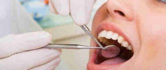
Dental pulp (pulpa dentis) is a specialized type of loose fibrous connective tissue rich in blood vessels and nerve fibers that fills the cavity of the tooth, i.e. pulp chamber and root canals. Due to the presence of a large number of receptor nerve endings in it, the pulp provides very high pain sensitivity of tooth tissue to the effects of mechanical, physical, and chemical stimuli.
Inflammation of the pulp (pulpitis) most often develops as a result of pathogenic bacteria entering it. This occurs when the carious process spreads to the deep layers of dentin surrounding the pulp chamber, but in some cases the cause of inflammation can be physical trauma to the tooth or a thermal burn (for example, due to errors when preparing a tooth for a crown). Most often, pulp inflammation occurs acutely, with severe pain, but in some cases the course can be asymptomatic - with a predominance of hyperplastic processes.
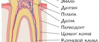
Structure of the dental pulp (diagram) –
Due to the abundance of blood vessels in the pulp, as well as due to the presence of narrow apical openings at the apexes of the roots, with the development of acute pulpitis, inflammatory edema always increases rapidly. Since the volume of the pulp is limited by the walls of the pulp chamber, inflammatory edema leads to compression of the veins and lymphatic vessels, thereby disrupting the outflow of fluid. This leads to the development of necrosis and death of the pulp. In addition, swelling also leads to compression of nerve endings, which actually causes the development of acute pain.
It should be noted that the dental pulp contains entire nerve plexuses, including a large number of receptors that are capable of perceiving stimuli of any kind - pressure, temperature, chemical and physical influences. But at the same time, effector nerve endings are also described in the pulp. Part of the nerve fibers of the pulp (together with the processes of odontoblasts) exits it into the predentin and the inner zone of the peripulpal dentin, located in the dentinal tubules.
What does dental pulp consist of?
Anatomical structure of the dental pulp
From an anatomical point of view, the tissue is divided into two zones. The coronal dental pulp has a loose structure and is involved in dentinogenesis; all layers of the dental pulp in this part are penetrated by an extensive network of capillaries and nerve cells. The root pulp of a tooth is denser because it does not contain a large number of cellular elements, but is saturated with collagen fibers. Through the apical foramen, the canals communicate with the periodontal tissues and allow minerals and nutrients to reach the tooth walls.
The pulp and dentin of the tooth form a strong complex - the hard tissue protects the tooth pulp from external irritants, and it, in turn, helps the formation of dentin.
The pulp of the front tooth smoothly passes from the crown to the root part, the dental pulp of the molars has clear boundaries - the mouths of the dental canals.
Histological structure of dental pulp
The pulp contains a large number of different elements:
- Elastin and collagen fibers supply the organ with hyaluronic acid, reducing susceptibility to toxins and bacteria.
- Odontoblasts and stellate cells are responsible for the regeneration of dental pulp.
- Leukocytes, lymphocytes and fibroblasts support the vital activity of the epithelium and organize communication between cells.
- A branched network of nerve processes forms Rashkov's plexus and provokes the occurrence of pain sensitivity when exposed to stimuli - the innervation of the dental pulp occurs due to the trigeminal nerve.
- Vessels and capillaries provide the blood supply to the dental pulp, which is necessary to nourish the tissues.
Composition of dental pulp
The fabric is 74% water, the remainder being organic and inorganic layers. Pulp cells include protein compounds, acids, lipids, glucose and various enzymes, which allows the epithelium to actively consume and process oxygen.
Many people mistakenly believe that the pulp is a nerve. The opinion is incorrect, since in addition to nerve plexuses, the tissue contains blood vessels and collagen fibers. |
What is the purpose of depulpation?
The goal of the procedure is to achieve several important results that will allow the patient to wear the prosthesis for a long time. Moreover, during the entire period of wearing the person will not be bothered by pain and the need to remove, for example, a crown. Let's take a closer look at what effect should be achieved:
- the pulp will not overheat during grinding of the stump and, accordingly, will not become inflamed (i.e., pulpitis will not appear under the crown),
- cariogenic bacteria will not penetrate into the pulp (i.e., pulpitis is again excluded),
- It will be possible to place pins or a core inlay into the canals: this will strengthen the restoration, i.e. it will last longer.
Don't know what type of prosthetics to choose?
We will help in the selection, advise where to read more information and compare types of prosthetics.
Consultation with an orthopedic doctor in Moscow clinics is free! Call now or request a call
Working hours: from 9:00 to 21:00 - seven days a week
Age-related changes in dental pulp
The pulp of temporary and permanent teeth has a similar structure and only becomes thinner over time. Before the roots are formed, the pulp of a baby tooth is concentrated in the coronal part. Later, the tissue begins to spread into the dental canals through the apical foramen and grow into a wide network. The dental pulp in a child has a massive and dense structure, as well as a large fiber size.
The development of dental pulp continues throughout life, but with age, regeneration processes slow down: the number of active cells decreases, which leads to vascular fragility, insufficient tissue nutrition, the tooth suffers from odontoblast atrophy, that is, the impossibility of dentin formation. The described changes apply to older people.
How much does depulpation cost?
How much does tooth removal cost? The cost of the procedure ranges from 3000-4000 rubles per 1 tooth in small cities, and in large cities, when treated under a microscope, from 3000-4000 rubles per 1 root canal (there can be 3-4 of them in one tooth). It is more profitable if the clinic offers turnkey installation of the prosthesis - absolutely all manipulations, examinations and materials are included in the total price.
[1] Shashmurina V.R. Biomechanical features of dental depulpation, 2021.
Author: Dulgarov Zh. G. (Thank you for your help in writing the article and the information provided)
Functions of the dental pulp
The main role of the pulp is to perform several tasks to support the vital functions of the tooth.
- Plastic function.
Formation of basic dentin, as well as the formation of hard tissue in case of damage. - Protective function.
Preventing infections from entering the periodontium through canals, removing dead cells, maintaining regeneration processes. - Sensory function.
A signal about the presence of an external or internal irritant to preserve tooth health. - Trophic function.
Supply of nutrients to dentin and tooth enamel.
Features of care after removal
Oral care after dental nerve removal and prosthetics must be regular and thorough. Twice a day you need to use a brush and paste, follow the correct cleaning technique (only sweeping movements). It is recommended to rinse your mouth after each meal, and also to exclude toffees, chewing gum, hard pieces of food, and not to chew nut shells or seeds. In order to promptly identify and eliminate dental problems, you should visit an orthopedic dentist at least 1-2 times a year.
“I had a crown placed on a post about 7 years ago. They did it very well and treated it under a microscope. But after the prosthetics, the dentist told me not to forget to come to them every year or every six months for an examination. At the same time, professional hygiene was carried out. I’ve never had any problems in all this time, I eat whatever I want.”
Anna, review from irecommend.ru
What can cause pulpitis?
Burn of tooth pulp
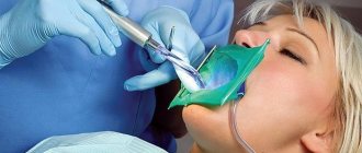
As a rule, a pulp burn during tooth grinding occurs as a result of medical error or carelessness. Before prosthetics, the crown part is treated at high temperatures. Insufficient cooling during the preparation process can lead to a burn, which will provoke subsequent inflammation of the dental pulp.
Dental pulp hematoma
After a tooth injury, there is a possibility of getting a hematoma, that is, bleeding into the dentin. The crown acquires a reddish tint and painful sensations occur when pressure is applied. However, a bruise that has not turned into pulpitis or necrosis does not need treatment and goes away on its own over time.
Caries
Through the smallest cracks in the tooth cavity, infection and pathogenic bacteria penetrate into the pulp. Advanced caries is one of the most common causes of pulpitis.
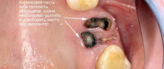
Any damage or complications of diseases can lead to disastrous consequences if left untreated. Dental pulp necrosis is the process of tissue death due to the spread of infected cells. The anomaly can be recognized by the grayish color of the tooth and incessant aching pain. During treatment, the doctor will perform depulpation, clean and seal the canals. |
Complications
After tooth depulpation, pain may continue for some time. To alleviate the patient's condition, doctors prescribe analgesics to relieve pain. If suddenly unpleasant sensations persist for more than two weeks and signs of inflammation appear, then you should be wary. It is necessary to urgently seek medical help to prevent the development of complications (for example, periodontal abscess, cystic formation, granuloma).
They develop due to the following reasons:
- the affected nerves were not completely removed;
- areas left affected by caries;
- dental canals are cleaned incorrectly or incompletely;
- open channels are incorrectly or incompletely sealed;
- After the manipulation, the patient did not follow the recommendations for oral care.
Therapeutic methods for treating dental pulp
Therapeutic or conservative are methods of treatment that make it possible to do without removing tissue. These methods of preserving dental pulp are not available to all patients; there are certain indications:
- age not older than 40 years;
- deep caries;
- acute serous-purulent or fibrous pulpitis;
- opening of the dental pulp due to trauma;
- lack of medical intervention in the inflammation process to date.
As a rule, the dentist treats the surgical field with antiseptic solutions, removes the affected tissue and places the drug into the cavity. There are two methods for performing this procedure.
- Indirect covering of the dental pulp.
An antibacterial agent is placed at the bottom of the cavity to stimulate the production of bone substance. Regeneration occurs naturally. - Direct covering of the dental pulp.
The medication is applied directly to the soft tissue to preserve the viability of healthy cells, then isolated with a pad.
Next, the tooth is restored with temporary materials, and after a secondary examination, it is finally restored.
Contraindications
The nerve cannot be removed if there are contraindications. Here is their list:
- first and last trimester of pregnancy;
- the presence of foci of suppuration in the oral cavity;
- hemorrhagic diathesis;
- acute respiratory infections;
- mental disorders;
- epilepsy;
- some pathologies of the heart and blood vessels (post-infarction state, hypertensive crises);
- leukemia;
- stomatitis;
- hepatitis of an infectious nature.
In such cases, the procedure is not advisable. Doctors offer alternative treatments.
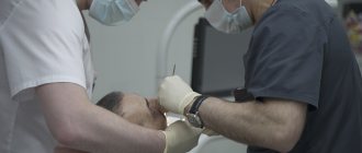
Surgical methods for treating dental pulp
In cases where therapeutic methods are powerless, the only option left is to remove the dental pulp. This option is used in cases where the patient’s immunity is weakened, the disease is at an acute stage, or the tooth will be used in the future as a support for prosthetics. Surgery involves amputation or extirpation of the dental pulp.
- Amputation of the dental pulp
- during the procedure, only the coronal part is removed. Prescribed for acute pulpitis or mechanical damage to the dental pulp. - Extirpation
is the complete removal of the pulp. Used for all forms of pulpitis.
The pulp can be removed vitally, that is, under anesthesia without prior killing. If this method is not possible, the doctor resorts to devitalization of the dental pulp - the toxic substance is left in the cavity for about a day, after which the dead tissue is painlessly removed. Means for devitalizing dental pulp include arsenic or paraformaldehyde paste.
Purpose of individual cell classes
The individual components of the pulp perform different tasks:
- fibroblasts are needed for collagen synthesis;
- lymphocytes are responsible for the production of immunoglobulins;
- macrophages are needed to organize antitoxic work and remove dead cells;
- odontobalsts perform a plastic function, that is, the production of primary dentin by the pulp, the formation of a secondary one after the process is completed (the synthesis is constant, with age this leads to a decrease in the volume of the tooth chamber).
The tooth cavity is not tightly closed, so in the absence of proper care there is a risk of infection. In addition, the condition of the tissues is negatively affected by injuries, unsuccessful filling and other factors. Therefore, inflammatory processes may begin to develop inside. At the first signs, which also include pain, you should immediately contact a dentist, who will diagnose and prescribe treatment. If addressed early, the tissue can be treated, leaving the tooth alive.
Treatment methods
Primary or acute pulpitis is eliminated using three methods. Once the tooth has been diagnosed and opened, the following treatment options may be used:
- conservative (biological);
- extirpation (the exact procedure depends on the condition of the tissue);
- amputation, that is, removal of the affected pulp.
Biological treatment is aimed at restoring the condition and health of tissues and shows effectiveness only at the initial stage. For therapy, treatment is carried out with special drugs that have an anti-inflammatory effect. To do this, the cavity is opened and a tampon soaked in antimicrobial compounds or antibiotic-based solutions is placed inside. After eliminating the inflammation process, the cavity is sealed with a temporary filling. The therapy period takes about five to seven days, after which the cavity is opened, washed with a disinfectant solution and sealed.
With the extirpation method, the condition of the tissue and the viability of the organ are first determined. For the chronic stage with signs of development of the gangrenous process, vital methods of treatment are recommended; for the chronic form, preliminary devitalization is recommended. If the condition is acute diffuse, treatment is selected by the doctor based on other indicators. The main technique is to completely remove the affected pulp, which can no longer be restored. Next, the cavity is processed and sterilized, closed with a temporary filling mass for the period of elimination of inflammatory processes.
The third treatment option is to remove the pulp mass, that is, amputate it. Depending on the working conditions, two methods are distinguished - vital and devital elimination of affected tissues. The first case is usually observed in pediatric dentistry, when the mass is removed at the canal level, after which a filling is installed for a period of up to six months. After six months, the temporary polymer plug is removed and a permanent filling is placed. The second method involves the use of means to kill the pulp, which is then left in the canal for further observation.
The doctor chooses a treatment method based on the diagnosis performed and taking into account the possibility of maintaining the functionality and aesthetics of the series. If the disease is detected at an early stage, removal can be avoided, but the Patient is required to contact the dentist in a timely manner and strictly follow the rules of care. This is a mandatory condition for preserving tissue, eliminating the development of complications, inflammatory and other lesions of the pulp. Regular scheduled examinations are also needed, during which problems can be identified in a timely manner and treatment can begin.
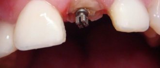
Depulpation during prosthetics
If you were going to do dental prosthetics, you must first prepare the tooth so that the crown is not larger than the usual size.
Before covering the tooth with a crown, it was filed down, removing 3 mm of tissue. In the case of a large pulp chamber, the doctor definitely depulps the tooth. Otherwise, the tooth will hurt under the crown.
If the patient has undergone depulpation and the person begins to experience pain, then it is not necessary to undergo prosthetics immediately. You need to wait for the pain to go away. If there is no pain for a long time, then it is already possible to create and install crowns.