Cheek cancer is a malignant neoplasm of the oral cavity.
Examination of patients with symptoms of cheek cancer at the Yusupov Hospital is carried out using modern diagnostic equipment from leading companies in the USA and European countries. Oncologists provide comprehensive treatment for cancer of the buccal mucosa. The medical staff is attentive to all the wishes of the patient. Chefs provide special dietary meals. Professors and doctors of the highest category develop tactics for managing patients with cheek cancer at a meeting of the expert council. Early diagnosis of the disease and multidisciplinary therapy improve the prognosis and five-year survival of patients after treatment.
Causes of cheek cancer
Cancer of the buccal mucosa develops under the influence of the following provoking factors:
- Use of tobacco in any form (cigarettes, cigars, pipes, chewing tobacco);
- Alcohol abuse (the risk of developing cancer increases when the use of alcohol and tobacco is combined);
- Infection with carcinogenic forms of human papillomavirus.
One risk factor is exposure to sunlight. Both family history and genetic predisposition, as well as exposure to mutagenic environmental factors, play a role in the development of cheek cancer. The formation of a malignant tumor occurs in several stages. The most important is the disruption in the functioning of oncogenes and genes that inhibit tumor growth. The development of malignant neoplasms of the cheek is associated with inactivation of the p16 gene, mutations in the p53 gene, and the introduction of the human papillomavirus.
The mechanism of development of cheek cancer
Malignant tumors of the cheek occur against the background of precancerous changes in the epithelial and subepithelial layers (leukoplakia or erythroplakia). The risk of leukoplakia degenerating into invasive cheek cancer is about 4-6%, with erythroplakia it reaches 30%. Subsequently, dysplasia transforms into “cancer in situ”, which penetrates into surrounding tissues and metastasizes to local and regional lymph nodes.
In some patients, even those cells that do not initially raise suspicion of dysplasia upon initial microscopy may gradually become malignant. As the tumor process progresses, distant metastases occur in the bones, lungs, and liver. Buccal cancer can grow through the skin.
Symptoms of cheek cancer
Cheek cancer in the initial stages of the tumor process is asymptomatic. Any ulcer that is not prone to rapid healing and any area of hyperkeratosis should be regarded as an early stage of cancer. In the early stages of cheek malignancy, there is little or no pain. As the size of the cancer tumor increases, the following symptoms appear:
- Pain;
- Induration and infiltration of underlying tissues;
- Enlargement of regional lymph nodes, including damage to the lymph nodes of the submandibular triangle, jugular-digastric group and deep cervical lymph nodes.
What does cheek cancer look like? Any non-healing oral mucosal ulcer should be assessed as a potentially malignant tumor and consultation with an oncologist should be sought.
Clinical picture
The specific type of growth occurring on the inside of the cheek can be determined by evaluating symptoms.
Papillomas are often multiple in nature. The clinical picture of such growths depends on their location. Common symptoms characteristic of papillomas include:
- no pain ;
- rough surface of the neoplasm;
- presence of a leg;
- light pink shade.
Some papillomas reach large sizes, thereby interfering with the process of chewing food. With each bite, the patient damages the tumor, which leads to bleeding in the oral cavity. Regular injuries contribute to the growth of the growth.
Why can our articles be trusted?
We make health information clear, accessible and relevant.
- All articles are checked by practicing doctors.
- We take scientific literature and the latest research as a basis.
- We publish detailed articles that answer all questions.
When papilloma simultaneously affects the mucous membranes of the cheeks and larynx, the patient experiences:
- problems ;
- change in voice (wheezing appears);
- difficulties with diction;
- attacks of suffocation if papillomas reach large sizes and block the airways.
The following symptoms indicate damage to the tonsils:
- edema;
- sensation of a foreign body in the throat;
- hoarse voice;
- problems ;
- difficulty eating .
The development of papillomas on the tongue is accompanied by the appearance of:
- ulcers;
- bleeding.
It is important to note that papillomas very rarely degenerate into malignant tumors. This transformation is possible due to the use of chemical drugs in the treatment of neoplasms.
The surface of the lump caused by injury has a red or purple tint. In addition, such neoplasms are distinguished by their convex shape. This lump is characterized by severe pain. It often bleeds.
The lipoma is small in size. It rarely reaches three centimeters in diameter. Its development is not accompanied by the appearance of unpleasant symptoms. The only sign indicating the presence of a lipoma in the oral cavity is a characteristic spherical neoplasm of a dense structure.
Atheroma has a rounded shape and a colorless shell. As it grows, it reaches 10 cm in diameter. When palpating the atheroma, no pain occurs, but the neoplasm begins to move under pressure. In advanced cases, the patient experiences a number of side effects:
- pain syndrome;
- suppuration in the affected area;
- formation of a focus of inflammation;
- increase .
You can distinguish a mucocele from other seals on the mucous membrane by the appearance of this neoplasm: it has a red or blue tint. This growth does not cause pain. The mucocele is soft to the touch, and its interior is filled with fluid. The tumor can open on its own, which can lead to the spread of infection throughout the body.
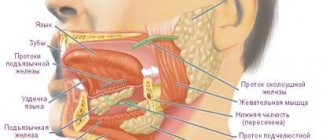
The cancer most often occurs under the jaw or near the ear. The pathology of the salivary gland is characterized by the appearance of numbness, which occurs when pressure is applied to the facial part of the head in the affected area.
The development of a cancerous tumor is not always accompanied by pain. It occurs when a tumor compresses the trigeminal nerve. In such circumstances, the patient experiences severe pain that radiates to the tonsils.
The clinical picture of a cancer tumor depends on the stage of development of the latter. At the initial stages, due to the presence of a malignant tumor, the functioning of the facial muscles worsens.
Diagnosis of cheek cancer
If a tumor of the mucous membrane of the cheek is suspected, oncologists at the Yusupov Hospital conduct an examination using a mirror, palpation of the tumor and lymph nodes. If a long-term non-healing ulcer is detected, a biopsy is performed. If a negative result of histological examination of the material obtained during the biopsy is obtained, the suspicion of the malignant nature of the tumor remains, the biopsy is performed again.
Oncologists clarify the stage of the tumor process, assess the extent of spread of the malignant tumor from the buccal mucosa to adjacent tissues, and find out whether there are metastases to regional lymph nodes and distant organs. If the presence of metastases is suspected, computer and magnetic resonance imaging, ultrasound, and scintigraphy are used. Distant metastases are found in 20% of patients at the time of diagnosis.
Computed tomography and magnetic resonance imaging make it possible to assess the condition of the deeper anatomical structures of the oropharynx and surrounding tissues. If there is a suspicion of metastases to the lymph nodes or tumor infiltration of the floor of the mouth, a cytological examination of the aspirate obtained under ultrasound guidance is performed. To exclude distant metastases, a chest x-ray in two projections and an ultrasound examination of the abdominal organs are done.
Considering that the prognosis for cheek cancer is serious, a tomography of the neck, chest and upper abdominal cavity is performed. Using bone scintigraphy, I exclude bone metastases. Positron emission tomography allows one to identify the source of metastases in cases of undetected primary tumors.
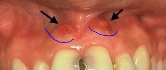
Diseases in which a lump on the gum is accompanied by pain
But the situation is much more complicated when the appearance of periodontal compaction is accompanied by severe pain. In such cases, we are talking about more serious diseases. If a tooth hurts and a lump appears on the gum: what could be the reason?
Periostitis
If periodontitis is not treated on time, the pus, instead of coming out through the fistulous tract, can go into the bone, causing its inflammation - periostitis (popularly known as gumboil). This disease has a more severe course: the temperature rises, swelling of the oral mucosa spreads to the soft tissues of the cheeks and lips, the lymph nodes become inflamed, and the person experiences acute, twitching pain. Such a lump can also form as a result of tooth extraction.
Attention!
Flux is a dangerous purulent disease, which, if delayed, can lead to inflammation of the jaw bone, phlegmon and sepsis. At the first signs characteristic of periostitis, you need to urgently contact a dental clinic!
Periodontitis
Periodontitis is a rapidly developing gum disease that leads to the destruction of the tissues that support the tooth. The reasons are the formation of tartar, lack of vitamins in the food consumed, and improper load on the teeth when chewing. The gums begin to bleed, itching appears, the teeth become loose, and gaps form between them. Then, in advanced cases, periodontal pockets appear between the tooth and the tissue, bad breath due to the release of purulent exudate, the necks of the teeth become exposed, and pain occurs when eating cold, hot, sour and salty foods. Due to the accumulation of pus in the pockets, the gums become covered with light-colored small bumps - a breeding ground for pathogenic bacteria. The appearance of such symptoms is a good reason to urgently contact a dental clinic: delay will lead to tooth loss.
Gingivitis
If bright red bumps appear on the gums, small in diameter, the gums bleed and are slightly hyperemic, we may be talking about hypertrophic gingivitis - the initial form of the inflammatory process of periodontal tissues. The disease occurs due to poor quality oral care and, in the absence of timely treatment, leads to serious complications, including periodontitis.
Important! Any pathology associated with the oral cavity can lead to serious health consequences. You should visit a dentist not only for treatment, but also for disease prevention!
Treatment for cheek cancer
Malignant tumors are successfully cured using radical radiation therapy while preserving the function of the oral cavity. Radiologists implant radiation sources because it is possible to irradiate a small volume of tissue at a high dose. Radioactive isotopes of cesium, gold, radium, iridium, tantalum, which have the same efficiency, are used as sources.
For small tumors, the size of which does not exceed 1 cm, they are limited to implantation of a radiation source, without resorting to additional external influence. In cases of slightly larger tumors that are not suitable in size for implantation, external irradiation is used along with source implantation.
Traditionally, large cheek tumors are treated with external beam radiation. Recently, oncologists have been using a combination of radiation and chemotherapy. Mobile lymph nodes are radically excised. During prophylactic removal of lymph nodes without signs of damage, in a significant number of cases, foci of micrometastasis are found in them. For this reason, radiologists prefer to perform prophylactic irradiation of the neck in patients without evidence of lymph node involvement by the external beam, sometimes in combination with surgical treatment.
After surgery for cheek cancer, a cosmetic defect is formed. The results of surgical intervention are improved using the technique of microvascular free skin grafting. If a patient develops dry mouth after radiation therapy, he is prescribed oral pilocarpine. The drug increases salivation, which is usually accompanied by minor side effects - sweating and increased urination. For primary or secondary treatment of tumors of the buccal mucosa, patients are prescribed chemotherapy drugs.
Early diagnosis of a cheek tumor allows for effective treatment. If the tumor process is at a late stage, the prognosis worsens. If you experience unpleasant sensations in the oral cavity or identify ulcers of the buccal mucosa, undergo an examination at the clinic by calling the Yusupov Hospital.
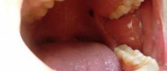
Treatment or what to do if a blood ball appears in the mouth
A blood ball in the mouth, as a reaction to external or internal irritants, goes away on its own within seven days. If this does not happen, then you should contact a specialist who will diagnose and prescribe treatment. It is important to determine its nature, fullness, size and location.
Most often, conservative therapy (less often surgical intervention) is carried out with the help of antiseptics (Chlorhexidine or Miramistin), which prevent the spread of bacteria, heal the wound and the blood ball resolves.