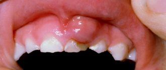
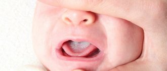
Quite often, parents may notice a soft bump that appears on their child’s gum when pressed. What could be the reasons for this phenomenon?
- inflammation of the roots (both baby teeth and molars, permanent ones);
- the beginning of tooth eruption.
Why does an abscess occur?
There can be many reasons for the formation of a lump (fistula, swelling), but the most common of them is undetected or ignored caries. Not all children maintain oral hygiene and eat properly, and not all parents carefully control this, which causes caries to appear on one or several teeth at once.
If caries is not treated, it will develop into pulpitis, extending beyond the tooth area and affecting the upper part of the root. As a rule, a lump appears just near the tooth whose root is inflamed. Typically, a lump can be noticed near a tooth with caries or with a filling that was placed long ago or poorly; Another common reason for its appearance is an injury received from a strong blow or an accidental fall, which can trigger the onset of the inflammatory process.
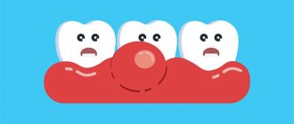
Stages of development of an abscess:
- As a result of caries, the infection penetrates into the pulp, affecting the root tip.
- Pus begins to form near the top of the root.
- Purulent formations fall under the mucous membrane of the gums.
- A cyst appears, looking like a small lump from the outside.
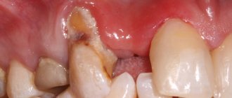
The most important symptom of an abscess is a soft and painful swelling, even with slight pressure.
Sometimes, due to an excess of pus, the lump may burst under pressure, and then a fistula appears - a small hole in the gum, which is connected with the source of inflammation located in the upper region of the root. A feature of the fistula is the constant release of purulent formations.
If inflammation decreases for some reason, the fistula may close on its own. However, when the child’s immunity decreases and the inflammatory process begins again, accompanied by pus formation, the appearance of a fistula will not be long in coming.
Possible complications
In itself, a hard or purulent ball on a child’s gum does not pose a threat. But it is represented by the diseases that caused this symptom.
An infection that affects the soft or hard tissues of the gums can quickly spread to other areas and organs, and the immune system of children is not yet able to effectively fight germs.
As a result, the pathological process can spread to the paranasal sinuses, palatine and pharyngeal tonsils, and cause orbital and meningeal complications.
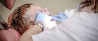
How to treat an abscess on the gum?
If an abscess or fistula appears, then you should not expect that the situation will resolve on its own: you need to make an appointment with a dentist. Treatment of an abscess on a baby tooth and a molar one will be different.
The appearance of an abscess on a baby tooth indicates periodontal inflammation. Such teeth must be removed, since the inflammatory process in the upper part of the root, accompanied by the formation of pus, may well spoil the molar, which will soon erupt in place of the milk tooth. This happens because the roots of temporary teeth are located next to the rudiments of molars. Bad bacteria and infection can also enter the lymph nodes under the jaw, causing them to become inflamed. The occurrence of a fistula means that pus will constantly seep into the mouth, which can cause the development of tonsillitis, since the tonsils will become infected. Various colds in a child are a direct consequence of an abscess on a tooth. Also, do not forget that toxins formed in the area of inflammation will definitely enter the bloodstream: this can result in allergic reactions, asthma and other serious general somatic diseases.
If we are talking about an abscess on a molar, then ordinary treatment is required for adults (provided that the tooth is not fundamentally damaged and can still be saved from removal).
Having discovered an abscess in a child, you should not try to cure it on your own. You can easily find a lot of advice on the Internet on how to relieve inflammation, but no amount of rinsing or even taking powerful antibiotics can eliminate the inflammatory focus located at the root of the tooth. Moreover, it is useless to hope that the tooth ache will stop. A bursting lump (this often happens) indicates only a short respite, during which, meanwhile, bacteria will still continue to enter the bloodstream and spread throughout the body. This situation can turn into a serious problem if the child has diseases of the respiratory system, heart and other internal organs.
Often, dentists suggest leaving teeth near which the inflammatory process is occurring, persuading the parents of a young patient to carry out the silvering procedure. The arguments for refusing removal can be very compelling: possible problems with diction and pain during the removal operation. However, such dentists neglect the information contained in every textbook on dentistry, which clearly states that a baby tooth with a purulent focus must be removed. Silvering will only complicate the situation, and an unresolved inflammatory process can lead to asthma, tonsillitis and even diabetes: in comparison with these diseases, temporary problems with diction will seem like sheer nonsense.
But the formation of an abscess can be prevented, and this is not difficult to do - just teach the child to maintain oral hygiene. At an early age, a child cannot be adequately responsible for his actions, including those related to the care of the oral cavity, teeth and gums, so responsibility for the procedure lies entirely with his parents, who must control how and when the child brushes his gums and teeth . It is the parents who should buy the most effective baby toothpaste that guarantees maximum protection against caries.
Treatment
When an abscess appears in the area of a baby tooth, the doctor will first numb the site of manipulation, then open the formation and remove pus from its cavity, after which he will remove the baby tooth, the damage to which caused the development of purulent inflammation. Next, the child will be prescribed a course of antibiotics and rinses.
If an abscess forms over a permanent tooth, the doctor, after examination and local anesthesia, will make an incision in the gum and, if the abscess is very large, install drainage. If the pulp is infected, the tooth canals are opened, depulpation is performed, and then a filling is installed.
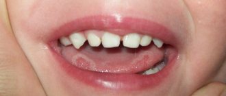
A lump on an area of the gum where there is no tooth yet: what to do in this case?
A couple of weeks before the tooth erupts, a cyst may appear on the gum, looking like a lump filled with a clear or bluish liquid. Such bumps are by no means common: dentists do not consider such formations to be a pathology requiring treatment. In addition, such formations on the gums in no way indicate an inflammatory process.
According to statistics, a small percentage of children are affected by the appearance of such a cyst. The child may not even suspect that he has a lump in his mouth, because if he touches it, there is no pain. However, a dental examination is still necessary, because the inflammatory process may still begin, in which case intervention will be required. The presence of inflammation is indicated by such signs as increased body temperature, pain when touching the lump, and swelling of the mucous membrane.
Most parents do not like the fact that their child has a lump in his mouth: in this case, you can ask the dentist to make an incision under anesthesia, as a result of which the fluid inside the cyst will come out. As a rule, when part of the cyst is removed, the crown of the tooth about to erupt is visible.
When to see a doctor?
If a child has a ball on his gum, it is necessary to see a doctor in any case. Only a qualified dentist will be able to determine the cause of such formation, identify it and provide appropriate treatment if necessary.
But in some cases it is important to visit a doctor as early as possible. Signs indicating a disease that potentially threatens complications are:
- pain in a tooth or gum, which constantly intensifies, spreads to the corresponding side of the face, radiates to the neck, temple, ear;
- when pressing on the ball, blood or pus is released from it (or from neighboring tissues);
- the child's lymph nodes under the lower jaw are enlarged, one side of the face and eye are swollen;
- there is an increase in body temperature.
The listed symptoms should be a reason for an immediate visit to the dentist, as they indicate an active infectious process and a high risk of complications.
How to help a child at home with suppuration?
If suppuration and the formation of a lump on the gum appear, it is recommended to contact a dentist as soon as possible, but, unfortunately, for various reasons this is not always possible.
If an adult can endure pain (although, of course, situations are different), then it is much more difficult for a child to endure painful sensations, and it is not easy for parents to watch their child suffer. There are ways to alleviate the baby's condition. Naturally, they will not replace full-fledged treatment, so you can resort to them only in situations where going to the doctor is impossible right now.
These include:
- take as much warm liquid as possible, which reduces intoxication of the body;
- eat liquid food (moderately warm, but not hot!), as it injures already damaged gums;
- If the pain becomes severe, you can use painkillers. For example, Nurofen or Paracetamol - the exact dosage depends on the age of the child, so before use you should carefully read the instructions for the drug;
- to reduce swelling, you can resort to cold - any frozen product from the freezer compartment of the refrigerator is wrapped in a soft cloth and applied to the cheek;
- use rinsing solutions that temporarily relieve pain - chamomile decoction or the drug chlorhexidine, which reduces irritation.
What to do if your child has a purulent lump
If a child has a lump with pus on his gum, you should not try to solve the problem yourself. Only a doctor can remove the growth after conducting diagnostic examinations. Before treatment, it is necessary to find out why the gums may become inflamed and enlarged.
How you can help your child at home
In order not to harm the baby’s health and not to aggravate the course of the disease, safe and low-toxic substances should be used in treatment. Before consulting a dentist, you can help your child in the following ways:
- Feed food at room temperature.
- Increase the amount of water consumed.
- When brushing your teeth, reduce the impact of the brush on soft tissues and teeth.
- After hygiene procedures, rinse your mouth with infusion of chamomile or oak bark.
- Every 4 hours, carry out intraoral baths with an aqueous solution of chlorhexidine.
It is difficult to carry out medical procedures on a one-year-old child without the recommendations of the attending physician, so you need to contact the dentist in any way and talk about the baby’s condition.
Treatment of an abscess
The principle of treatment depends on the reason why the abscess could appear. Lump removal is carried out by a dentist and includes several stages:
- Excision of the lump with a scalpel.
- Cleansing the wound from pus and other decomposed substances.
- Antiseptic wound treatment.
If a lump has formed near a carious or decayed tooth, the dentist performs extraction under local anesthesia. If the tooth can be saved, the surgeon cleans out the dental canals, removes the source of infection and fills the tooth with medicinal pastes and light-curing materials.
Treatment of cones in children using traditional medicine
The folk method of treating lumps in the mouth is effective in cases of complex therapy. Using folk remedies alone will not bring the desired result, but will only complicate the problem. To treat bumps on the gums, the following can be used:
- Soda-salt solution.
In 250 ml. boiled water should be diluted with 1 teaspoon of baking soda and table salt. Rinsing should be done every 4 hours for 2-3 days.
- Phytotherapy.
For therapeutic purposes the following are used: chamomile flowers, oak bark, calendula, sage, lemon balm. To prepare a medicinal infusion, you need to mix 2 tbsp. l. dry herbs in a glass of hot water. Rinsing is carried out with a warm solution 2-3 times a day.
- Honey with salt.
Liquid honey is mixed with table salt in a 2:1 ratio. The mixture is used as applications 2 times a day after brushing your teeth.
Preventive measures to prevent the formation of lumps on the gums
To reduce the likelihood of an abscess to a minimum, a child from childhood should be taught to carefully observe oral hygiene.
The list of the most effective measures includes:
- Brushing your child's teeth twice a day. First, parents should brush their child’s teeth themselves, showing how to do it correctly, gradually giving the child more independence in this matter;
- rinsing your mouth after every meal;
- minimizing sweets in the diet - it is important to ensure that the child keeps caramel and candies in his mouth as little as possible;
- Do not introduce your child to chewing gum (the product is especially harmful if it contains sugar).
What is fibroma
This is a benign neoplasm in the form of a small node on a stalk or broad base, which consists of fragments of connective tissue. In most cases, it is localized on the mucous membranes of the gums, lips, palate, on the inside of the cheeks, and a little less often on the tongue. The pathological process is more often encountered by primary schoolchildren and adolescents aged 6-15 years.
The patient is not in pain; at a very early stage he does not notice the pathology. As the thickening grows, it is easily felt and accidentally injured.
Can fibroids in the mouth resolve on their own, without medical intervention? In most cases, surgical excision of the tumor is required. However, do not worry about the complexity and duration of the operation. In less than an hour, an experienced doctor will eliminate the defect without subsequent complications and a long rehabilitation period. Currently, clinics use laser and radio wave techniques.
Despite the relative harmlessness of fibrous growths, they still need to be treated even in the absence of discomfort. If the seals increase in size and are constantly exposed to traumatic effects, it is highly likely that an infection will enter the wound. In this case, treatment will be very long, difficult and will not always lead to positive dynamics. In addition, the lack of therapy sometimes leads to the degeneration of a neoplasm into a malignant one.
Diagnosis of fibroids of the upper and lower jaw
The doctor will not prescribe treatment until he is sure that the diagnosis is correct. To do this, diagnostic procedures are carried out, the results of which will confirm or refute the fears of doctors.
First, the patient is asked to describe the symptoms. The dentist examines and palpates the tumor. However, this is not enough to develop therapeutic tactics, since it is extremely important to determine the depth of tumor growth into soft tissue. For this purpose, an ultrasound examination is performed.
In difficult cases (ulcers, development of an inflammatory process in a pathological area of the gum, etc.), a biopsy is indicated. After surgical removal of the tumor, fragments are necessarily sent for histological analysis.
The examination is necessary not only to establish a diagnosis, but also to identify the factors that provoked the disease. A full dental examination is carried out to confirm the presence of inflammation. In addition, one cannot do without radiography, orthopantomogram and other images in different projections.
If a person has dentures, a consultation with an orthopedic dentist may be necessary. This is necessary to eliminate the possible traumatic effects of artificial elements on the mucous membranes.
Differential diagnosis
Biopsy remains one of the most informative methods for distinguishing fibroids from other benign neoplasms. The study is indicated for suspected papilloma, lipoma, epulis of various structures, neurofibroma, cyst, squamous cell carcinoma, wart, etc.
If the growth is localized on the tongue or sublingual part, it is extremely important to differentiate it from all existing seals. Timely diagnostic measures make it possible to detect cancer at the earliest stages and provide high-quality therapy with a short recovery period.
Classification of fibroids
Pathology is divided into several types according to criteria. This includes the density of the benign neoplasm, the nature of its origin, as well as the severity of clinical manifestations.
Dense fibroma
Characterized by a solid consistency. This is due to the fact that the contents are quite hard fibers of connective tissue. They include a small number of cores. Formations of this type are most often found on the gingival surfaces and palate.
Soft fibroma
The fibrous structures are thin and freely located, so their clusters are characterized by a high degree of softness. This type is most often observed on the tongue and the inside of both cheeks. Benign neoplasms of a mixed type, combining the signs of all the varieties listed above, can be found on the sublingual part and on the mucous membranes of the floor of the oral cavity. For example it could be:
- Fibrolipoma. It is hard to the touch because it contains fibrous fibers. It can be eliminated surgically, with laser, or radio waves.
- Fibrohemangioma. As a rule, it is provoked by infectious processes occurring in the child’s body. It is extremely rare in adults. It never degenerates into malignant tumors. Most often it is treated surgically, in some cases it can resolve on its own.
Fibroma from irritation
This is not a tumor formation in its usual form, but the result of reactive hyperplasia. Chronic pathology is characterized by the development of focal lesions, which are caused by systematic mechanical action and subsequent injury.
The most common cause is installed crowns, fillings, and dentures. In the latter case, the disease is called prosthetic granuloma. The orthopedic structure exerts continuous pressure on the alveolar process, leads to its resorption, moves forward and contributes to the formation of compactions, which are associated with the inflammatory process.
The risk group includes not only patients who have undergone prosthetics, but also people with untimely cured caries, in adulthood, with foreign objects in the oral cavity (for example, with piercings). Studies have shown that women are more prone to such hypertrophic transformations than men.
Symmetrical fibromas
Doctors diagnose such tumors in the area of the third molars in the areas between the gums and the roof of the mouth. The tumor is hard to the touch and resembles a bean in shape.
It should be noted that this type of compaction does not apply to true fibromatosis. These are just overgrown tissues in the gingival membranes, accompanied by the process of scarring and other changes of a similar nature.
Lobular fibroma
It occurs as a result of reactive hyperplasia with systematic trauma to delicate sensory fibers with prostheses or other orthopedic structures. The main distinguishing feature of such formations is their rough, textured surface. When palpated, tubercles are felt.
Fibrous epulis
This is a dense growth of pinkish tissue that does not cause pain or other discomfort. The edges are often hyperemic, have clear boundaries and irregular shape. The base is quite wide.
The vestibular part of the gums is usually affected. There are cases of neoplasms occurring in the interdental spaces in the form of a saddle with spread to the intraoral surface.
Quite often, a dental unit located in a pathological area has a poorly fitted metal crown, extensive carious lesions, or a prosthetic clasp. It is these structures that are the provoking factor in the occurrence of a chronic inflammatory process with the formation of granules, which over time are transformed into mature connective fibrous fibers.
In dentistry, there are also angiomatous epulis. They are brighter in color, somewhat softer to the touch and bleed. In this case, blood appears not only at those moments when the surface is affected mechanically, but regardless of the presence or absence of a traumatic factor. When conducting diagnostic studies, many vascular branches are detected in the pathological area.