Author of the article:
Soldatova Lyudmila Nikolaevna
Candidate of Medical Sciences, Professor of the Department of Clinical Dentistry of the St. Petersburg Medical and Social Institute, Chief Physician of the Alfa-Dent Dental Clinic, St. Petersburg
The human oral cavity has its own microflora and its own specific diseases arise. Let's talk about one of them - fibrous epulis of the gums: what kind of disease it is, what are the causes of its occurrence, how is the treatment carried out.
What it is
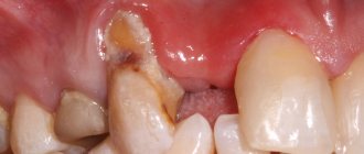
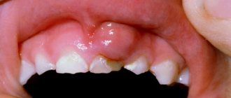
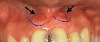
Epulis is a tumor-like neoplasm that develops on the alveolar process of the jaw. It also has other names - epulid, supragingival, giant cell granuloma. More often it is localized in the area of incisors, canines and small molars of the upper jaw, less often the tumor is detected in the lower jaw. The growth has a round or irregular shape and a wide stalk; the size of the formation varies from 3 mm to 5 cm.
Despite the fact that such a tumor looks frightening, it practically does not cause concern to the patient, with the exception of a violation of aesthetics and the sensation of a foreign body in the oral cavity. Large tumors may cause difficulty chewing and swallowing food.
If epulis is accompanied by bleeding, infection may occur, which will lead to complications.
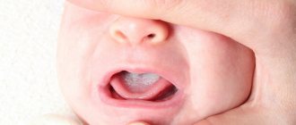
The disease mainly affects adults, but the neoplasm can also appear in children; in the latter, it occurs during teething. Studies have shown that women are more susceptible to developing epulis than men.
Forms of the disease
According to the ICD, aphthous stomatitis is assigned code K12.0. There are two forms of the disease - acute and chronic. The second is characterized by frequent relapses and may be a consequence of inadequate or untimely treatment of acute inflammation.
An acute disease is characterized by severe symptoms. It begins quickly, severe pain occurs at the site of mucosal damage, and the child may refuse to eat. In some cases, body temperature rises, weakness and lethargy occur.
The chronic form is characterized by a sluggish course; the child’s general well-being does not suffer. The disease can recur up to several times a year.
Varieties of epulis
Conventionally, there are 3 types of epulis:
- Fibrous. The neoplasm is dense, the basis is coarse fibrous tissue. Characterized by slow growth. There is no pain or bleeding.
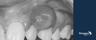
- Angiomatous. The tumor contains a large number of blood vessels and is localized mainly in the lateral parts of the jaw. With this form of the disease, bleeding is observed even with slight pressure, for example, with a toothbrush or food. When pressed, the tumor is painless and has a soft consistency.
- Giant cell. The tumor can reach large sizes, displacing teeth that are located in the epulis growth zone. The mucous membrane of the epulis is bluish-red in color. On palpation, the neoplasm is painless, but may bleed moderately if injured.
Depending on the nature of the process, epulis can be benign or malignant. In the first case, the neoplasm is characterized by slow development and, as mentioned above, in most cases does not cause pain. The tumor size in this case rarely exceeds 10–20 mm. When the process becomes malignant, rapid tissue growth is observed. In this case, the process is often accompanied by pain, bleeding gums and affects the root canals of neighboring teeth.
What reasons can lead to its appearance?
The main cause of epulis on the gums is considered to be constant mechanical impact (rubbing) of soft tissues.
Injury to one area can cause gum swelling:
- poorly installed seal;
- uneven edges of the opposite tooth, which is destroyed or chipped;
- tartar;
- poor quality or incorrectly installed prosthesis;
- agonist tooth for malocclusion;
- adjacent teeth when teeth “pile up.”
Supragingival tissue occurs and, as a consequence, injury to epithelial tissue as a result of a burn, bruise or constant ingestion of irritating foods. In pregnant women, tumors occur due to hormonal imbalances. Epulis on a child’s gums can form due to general causes or disturbances in the eruption of permanent teeth.
Diagnosis and treatment of epulis
Diagnosis of epulis comes down to collecting complaints, clinical examination, radiography and histological examination of the material.
The primary task in making such a diagnosis is to eliminate the provoking factor: professional cleaning of dental plaque, treatment of caries and its complications, replacement or correction of orthopedic structures according to indications. In fibromatous and angiomatous forms, dynamic observation is indicated, since after sanitation of the oral cavity and elimination of the causes of the disease, the tumor may decrease in size until it disappears completely.
Treatment of epulis is carried out surgically. The growth on the gum under local anesthesia or general anesthesia is excised with a scalpel or laser within the healthy tissue along with the periosteum. The second method is preferable, since the laser simultaneously coagulates the vessels and stops bleeding. With giant cell epulis, the area of bone tissue involved in the process is also removed. To do this, use a bur or cutter. The wound is closed with gauze with an iodoform mixture or a formed mucoperiosteal flap. If necessary, the material is sent for histological examination.
Intact teeth in the area of tumor localization must be removed with a high degree of mobility and severe exposure of the roots. If the bone lesion is extensive or epulis recurs, partial resection of the alveolar part along with the teeth is performed.
To speed up the healing of a fresh wound, the doctor prescribes compresses with medicinal ointments and rinses to the patient.
If the ball in your mouth does not cause discomfort
The color of the bubble will help the doctor make a diagnosis. A white lump on the gum indicates the presence of exostosis or purulent exudate. A red or bloody ball indicates the development of inflammation. If the growth matches the shade of the tissue, it is the initial stage of epulis, flux, or a malignant tumor. When the tumor does not hurt, this indicates the presence of one of the pathologies:
A fistula is a white ball on soft tissues that appears under or on a tooth. There is a hole on the surface for the release of pus. If, when pressed, suppuration flows out of the bladder, the patient does not feel pain. If the hole is closed with pus or bloody clots, the patient will experience discomfort with any impact.
A fistula is often formed due to advanced periodontitis, accompanied by periodontal hyperplasia. Overgrown tissue is good soil for the proliferation of microorganisms. In this case, the patient urgently needs treatment for periodontitis.
In the absence of therapy, the fistula enters a chronic stage, which can only be eliminated through surgical treatment. The progression of the disease must not be allowed, otherwise you may lose healthy molars.
Hematoma is a round lump on the inner surface of the cheek. Sometimes it occurs in the form of a dark bluish swelling on top of the gums. Blood accumulates in or around the root of the molar. The mucous membrane grows, the patient experiences discomfort and cannot completely close the jaw. The main reasons: consequences of filling or tooth extraction, gum damage, poor blood clotting.
Hematomas are generally not dangerous. Processes take place in the body through which soft tissues are cleared of bloody clots. After some time, the bubble disappears, but if the seal remains, you need to visit a dental clinic.
Exostosis is a hard blister that is an abnormality in which the bones protrude and protrude from the jaw. Gradually, the lump increases in volume, which causes discomfort and pain. Exostosis can be provoked by various reasons:
- jaw damage;
- heredity;
- congenital disorders;
- tissue diseases after molar removal.
An examination by a dentist or an x-ray will help detect the disease. The formation will need to be removed if the development of a malignant tumor is suspected.
Epulis is a pedunculated bubble in the form of a mushroom-shaped growth on the periodontium. The tumor may be the same color as the gums or red. The reasons causing the development of pathology include:
- improper filling of the molar or too large a filling;
- dental plaque, stone;
- jaw damage;
- malocclusion;
- hormonal imbalance;
- poor prosthetic material or incorrect prosthetics.
The symptoms of epulis resemble gingivitis; for diagnosis, the dentist prescribes radiography and histology to the patient. With their help, the degree of destruction of bone tissue at the site of the epulis lesion is determined. Mostly, pathology occurs in children during the growth of primary molars and in women.
Papilloma or fibroma is a bubble on the gum, sometimes a benign formation that does not pose a threat to the health or life of the patient. They are formed in people of different genders and ages. Predisposing factors to the appearance of a lump can be: damage to the mucosa, stress, systemic pathologies, heredity.
Papilloma is an enlargement of the papillary layer of skin. The bubble grows gradually, but with reduced immunity, systemic pathology, or stress, growth accelerates, but without turning into a malignant tumor. A papilloma neoplasm looks like a smooth, soft lump on the mucous membrane of a white or pink shade on a thin stalk.
Papilloma often does not create any discomfort. But after some time it may increase in size. You should consult a dentist and get tested.
Preventive actions
Monitoring the condition of the teeth and oral cavity will reduce the likelihood of tumors.
- Timely sanitation of the oral cavity. Visiting the dentist for a preventive examination and professional hygiene twice a year will prevent the growth of caries, the appearance of defects in fillings and the formation of tartar, leading to gum injury and the appearance of epulis.
- Prevention of gum injury. If systematic injury to soft tissue occurs as a result of poorly fitted orthopedic or orthodontic structures, consult your doctor about this problem. He will adjust the crowns or dentures.
If these preventive measures are followed, the prognosis is favorable and epulis does not recur.
We hope that our article about epulis will be for informational purposes only. And if you want to soothe your gums, make them strong and strong, try the unique two-component mouth rinse ASEPTA ACTIVE.
This is the only rinse with a combination of chlorhexidine + benzydamine for the treatment of inflammatory periodontal diseases. The product has a combined effect: antimicrobial, anti-inflammatory and analgesic. Instant anesthetic effect allows you to quickly reduce pain.
Dentists' recommendations
Epulis is a gum tumor that poses a great danger to the health of teeth and the entire body. Treatment of the pathology is complex and lengthy, but even with high-quality treatment, the risk of relapse cannot be ruled out.
It is not always possible to prevent the occurrence of epulis on the gums of a child or an adult, but with timely consultation with a specialist, complications, complex treatment, long recovery and aesthetic defects that remain after surgery can be avoided.
The best prevention is to visit the dentist every 6 months. If you are concerned about discomfort and a feeling of rubbing of the mucous membranes or gums, you should immediately consult a dentist, without waiting for a tumor or other pathologies to occur.
Clinical researches
Repeated clinical studies have proven that the two-component mouth rinse ASEPTA ACTIVE more effectively combats the causes of inflammation and bleeding compared to single-component rinses - it reduces inflammation by 41% and reduces bleeding gums by 43%.
Sources:
- Clinical and laboratory assessment of the influence of domestic therapeutic and prophylactic toothpaste based on plant extracts on the condition of the oral cavity in patients with simple marginal gingivitis. Doctor of Medical Sciences, Professor Elovikova T.M.1, Candidate of Chemical Sciences, Associate Professor Ermishina E.Yu. 2, Doctor of Technical Sciences Associate Professor Belokonova N.A. 2 Department of Therapeutic Dentistry USMU1, Department of General Chemistry USMU2
- The effectiveness of the use of Asept “adhesive balm” and Asept “gel with propolis” in the treatment of chronic generalized periodontitis and gingivitis in the acute stage (Municipal Dental Clinic No. 4, Bryansk, Kaminskaya T. M. Head of the therapeutic department Kaminskaya Tatyana Mikhailovna MUZ City Dental Clinic No. 4, Bryansk
- Study of the clinical effectiveness of treatment and prophylactic agents of the Asepta line in the treatment of inflammatory periodontal diseases (A.I. Grudyanov, I.Yu. Aleksandrovskaya, V.Yu. Korzunina) A.I. GRUDYANOV, Doctor of Medical Sciences, Prof., Head of Department I.Yu. ALEXANDROVSKAYA, Ph.D. V.Yu. KORZUNINA, asp. Department of Periodontology, Central Research Institute of Dentistry and Maxillofacial Surgery, Rosmedtekhnologii, Moscow
- The role of anti-inflammatory rinse in the treatment of periodontal diseases (L.Yu. Orekhova, A.A. Leontyev, S.B. Ulitovsky) L.Yu. OREKHOVA, Doctor of Medical Sciences, Prof., Head of Department; A.A. LEONTIEV, dentist; S.B. ULITOVSKY, Doctor of Medical Sciences, Prof. Department of Therapeutic Dentistry of St. Petersburg State Medical University named after. acad. I. P. Pavlova
If the formation on the gum causes discomfort
If a bubble near a molar causes pain, this indicates an infectious inflammatory process. Discomfort is also typical with gum injuries and hematomas. Painful formations can be caused by a cyst, cancer, fibroma, periostitis, periodontitis.
Gingivitis - the disease affects only one gum, and the periodontium near the molar remains uninjured. The main symptoms of the pathology are swelling, bleeding, and peeling of the epithelium. Gingivitis often occurs against the background of halitosis. Sometimes pathology develops as a result of endocrine and metabolic disorders.
When treating, it is necessary to eliminate the cause of gingivitis. Professional oral hygiene, diagnosis and treatment of metabolic disorders are performed. To eliminate inflammation and prevent further spread of bacteria, antibiotic therapy is administered. For pain and severe discomfort, analgesics are prescribed.
Periodontitis - purulent bumps on the gums appear as a result of degeneration of the alveolar process, through which the tooth root is held in the alveolus. Tissues can be destroyed due to poor oral hygiene and internal pathologies.
In the initial stages, periodontitis is treated by teeth cleaning at the dental clinic and further proper care. An advanced form of the disease, in which the teeth become loose, pus accumulates, requires surgical intervention - restoration of the alveolar processes or removal of molars.
Periostitis or flux is a dense formation near a problematic molar with carious lesions. Patients complain of pain radiating to the temple, chest, neck, ear. The condition gradually worsens and the temperature can rise to 38 degrees. In the oral cavity, lesions due to periostitis are hyperemic. A purulent fistula forms. When the discharge is removed, the discomfort decreases.
Causes of periostitis: trauma, periodontitis, osteomyelitis, weak immune system, infectious pathologies occurring in the body and vitamin deficiency.
Symptoms of aphthous stomatitis
The onset of acute aphthous stomatitis in children may resemble ARVI: there is malaise, increased body temperature, and more profuse salivation may appear. The main symptom of the disease is the appearance of ulcers in the mouth: first, a red dot appears on the mucous membrane, which subsequently becomes an ulcer (aphtha) within 2-3 days. Accidental touching causes pain. The child refuses food and hygiene procedures.
Single erosions occur more often, but there are cases of multiple aphthae formed in groups. They usually measure up to several millimeters, but in severe cases the diameter of the ulcer reaches one centimeter. The disease is characterized by the appearance of a bright red rim around the aphthae; a grayish or yellow film-like coating forms on it.
There are also general symptoms that accompany aphthous stomatitis. These include:
- sleep disturbances (due to pain or discomfort);
- decreased appetite;
- pain while talking;
- increased fatigue, lethargy;
- whims, irritability.
Early treatment of aphthous stomatitis in children can prevent complications, shorten recovery time, and prevent further development of the disease.
Classification
There are several types of epulis. Each of them has its own number in ICD-10 (International Classification of Diseases, 10th revision).
Fibrous (K06.82 - benign neoplasms):
Location area: vestibular side of the gum;
- Round or oval shape;
- Slow growth;
- Surface smooth/lumpy surface;
- Doesn't bleed;
- Wide base;
- Thick consistency;
- Light pink (to match the gum color).
Angiomatous:
- Location area: neck of the tooth;
Giant cell (K06.81 - other specified changes in the gums):
- Location area: alveolar part;