Caries is a common dental disease that destroys the hard tissues of teeth. According to statistics, it affects about 95-98% of the population of economically developed countries. In the treatment of caries, medicinal pads can be used. In what cases and for what they are used, we will tell you in this article.
In this article
- What is a therapeutic pad in dentistry?
- In what cases is a lining used for caries?
- Why are medicinal pads used in the treatment of deep caries?
- How is a therapeutic napkin installed for deep caries?
- Materials of therapeutic pads
- How do combined pastes work?
- Pros and cons of using medicinal pads in dentistry
In what cases is a lining used for caries?
Caries is a slowly progressive pathological process that goes through several stages. At an early stage, a carious lesion looks like a light spot on the enamel, while there is still no hole or pain in the tooth. As the disease progresses, the spot turns into a carious cavity. At the stage of superficial caries, this cavity is located within the tooth enamel; with medium and deep caries, it affects dentin tissue.
For caries in the spot stage and the superficial form of the disease, pads with a therapeutic effect are not needed; for average caries, they can be used according to indications. Most often, medicinal dental pads are used for deep caries. The peculiarity of deep caries is that the carious cavity is located as close as possible to the pulp - the neurovascular bundle, or dental nerve. At this stage, treatment is quite painful and there is a high risk of complications in the form of pulpitis and periodontitis.
Dental linings help increase the effectiveness of treatment of deep caries.
Why are medicinal pads used in the treatment of deep caries?
The purpose of a healing pad is to protect the pulp. After the dentist has removed the tissue affected by caries, cleaned and prepared the carious cavity for filling, a very thin layer of dentin, the bone tissue of the tooth, remains between it and the dental pulp. This creates a high risk of microbes entering the dental nerve and developing pulpitis. To prevent the spread of infection into the pulp, stop the inflammatory process and stimulate the production of a new layer of dentin, a special gasket is applied.
When a gasket is temporarily applied, the entire carious cavity is covered with it and a temporary filling is installed for a period of two weeks to several months. Permanent linings for deep caries are applied pointwise in the area of the tubercles of the neurovascular bundle. In the second case, along with the therapeutic one, an insulating gasket is installed.
Preparation
A qualified dentist places a Life lining only after preliminary preparation of the diseased tooth. If you begin the process of applying material without preparation, the quality of treatment will be low and the risk of negative consequences will be high.
Preparation includes:
- administration of anesthesia;
- dissection of affected tissues;
- removal of all necrotic tissue;
- drying the cavity.
The procedure is a preparatory stage before filling a tooth, but can only be carried out after high-quality treatment of the pathology and complete removal of tissue affected by caries.
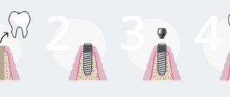
How is a therapeutic napkin installed for deep caries?
Depending on the form and complexity of the disease, as well as the chosen method of caries treatment, the dentist can install the lining by direct or indirect application. The first option assumes that it is applied directly to the pulp area, ensuring direct contact between the gasket and the dental nerve. The method is used for tooth injuries with exposure of the nerve, for fibrous pulpitis. The second method is to apply a lining using dentinal tubules without direct contact with the pulp. It is often chosen for the treatment of deep stage caries and acute pulpitis.
Materials of therapeutic pads
Several types of dental dams are used in the treatment of deep caries:
- varnishes, solutions, light-curing cements and polymers based on calcium hydroxide;
- zinc-eugenol and combined pastes.
Preparations based on calcium hydroxide have a powerful bactericidal effect. The application of these compounds to the bottom of the carious cavity prevents the penetration of microbes into the dental pulp and the development of the inflammatory process. Calcium hydroxide also promotes the production of new bone tissue in the area between the cavity and the nerve of the tooth.
Aqueous suspensions based on calcium hydroxide are usually applied in a thin layer to the bottom of the carious cavity, dried and a temporary filling is placed on top. They are replaced after 30-45 days. Also, in the treatment of dental caries, chemically cured cements are often used. They are pastes containing ether and hydroxide in a 1:1 ratio. These pastes harden directly in the patient's mouth and have excellent insulating properties in relation to fillings. Light-curing polymers must be used very carefully, because there is a risk of burning the dental nerve. At the same time, they have very high strength and, in the hands of a professional dentist, can be an effective means of treating caries. Zinc-eugenol pastes are widely used as an antiseptic for temporary fillings.
Properties of the drug Dycal (Daykal)
Dycal is a calcium hydroxide-based cushioning material. The material is used to protect the pulp. The substance promotes the formation of secondary dentin.
The material has the following properties:
- clinically effective;
- hardens quickly;
- differs in strength in the early period;
- does not dissolve in oral fluid, is resistant to saliva;
- has a natural dentin shade or ivory color (Dycal Ivory);
- easy to apply, filling the necessary areas;
- does not affect the color of the final restoration.
The base composition includes calcium phosphate and tungstate, zinc oxide, iron oxide. The catalyst contains calcium hydroxide, zinc stearate, zinc and iron oxides. The cushioning material Daikal is characterized by high strength.
Instructions for use
Dycal is used as a therapeutic lining material before using the main filling material. The base and catalyst are mixed in equal proportions. The finished mass should have a homogeneous consistency, without streaks. Mixing is carried out within 10 seconds.
Before use, the dental cavity is washed and dried. The substance is applied in a thin layer: from 0.8 to 1 mm. You should not use very thin pins in your work. Remaining material is removed. Next, an adhesive or restorative material is applied. The working time of the material is 2 minutes 20 seconds, hardening occurs within two to three minutes.
Special information
When working with the drug, avoid contact with the skin and mucous membranes of the mouth and eyes. Upon contact, irritation, an allergic reaction, and a rash may occur. Ingestion of the product is prohibited. During restoration work, you should wear gloves on your hands and protective goggles on your eyes.
Gasket material Daikal
At high temperatures and humidity, the working time is significantly reduced, since the mass begins to harden much faster. In this connection, work is carried out at an air temperature of about 21 degrees, air humidity should not exceed 50%. After each use, the tube of material should be tightly closed. It is prohibited to use products that have expired.
The price of the drug ranges from 1,700 to 2,000 rubles.
Analogs of material:
- Vitrebond
ENDOFIL (Endofill) - filling material for root canalsViedent VladMiVa: instructionsPulposeptin in dentistryZincoxide eugenol paste for filling root canalsSilidont 2: instructionsCemfil 15 Stomadent
How do combined pastes work?
The composition of combined pastes includes oils, fillers (zinc oxide or white clay), as well as medications. The therapeutic effect of a particular pad directly depends on the latter:
- Stimulating dentin growth and promoting enamel remineralization. These drugs are called odontotropic, they are based on fluorides, collagen, calcium glycerophosphate and other substances. They are used when it is necessary to increase the layer of bone tissue between the pulp and a deep carious cavity.
- Possessing anti-edematous and anti-inflammatory effects. They help stop the inflammatory process in the dental pulp; they are based on prednisolone and hydrocortisone. Typically, such preparations are temporarily placed at the bottom of the cavity and covered with a bandage on top. If the dentin layer next to the neurovascular bundle grows too slowly, then a spacer with an odontotropic effect is applied.
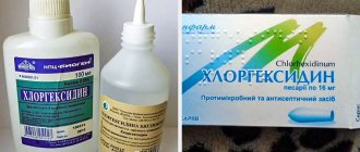
- Pads with an antiseptic effect based on metronidazole or chlorhexidine disinfect and prevent the growth of bacteria.
- Proteolytic enzymes contained in the drug are needed in the treatment of acute focal pulpitis, as well as in the treatment of deep caries.
Most combined pastes harden poorly and have low strength, so they are used as a temporary therapeutic material. After their removal, a different type of therapeutic pad is usually used.
Modern aspects of the use of gaskets and adhesive systems (Part 2)
A completely different situation arises when using composite filling materials, the high-quality use of which is almost impossible without adhesive systems, since the bonding strength of composites to tooth tissue is much lower than that of amalgams. Modern adhesive systems bind to dentin with a force exceeding 20 MPa and are capable of reliably fixing the composite in the prepared cavity. If you place the composite with a spacer made of phosphate or polycarboxylate cement, the adhesion force of which is 0.7 MPa, the spacer can easily move due to shrinkage of the filling. Liner-type glass ionomer cement gaskets are not much better. When a decision is made to combine a composite with another material, which in a given situation plays a binding role, we are usually talking about the sandwich technique. In this technique, the amount of glass ionomer cement must be greater than or at least equal to the amount of the composite, so that during the polymerization process shrinkage of the composite, the glass ionomer did not peel off from the walls of the cavity. Based on this, should a permanent filling material, which fills the cavity to the enamel-dentin border and its amount is greater than or equal to the amount of the composite covering it, be called a base lining. In addition, I believe, it is illogical to consider the sandwich technique as one of the varieties of base gaskets, since this technology does not perform a separating, but, on the contrary, a connecting function in cases where the use of adhesive systems is ineffective. Therefore, a more appropriate definition is glass ionomer filling coated with a composite . Sometimes you can come across a recommendation to place a composite filling with so-called flowable composites, to eliminate internal stress and better adapt the composite to dentin. These composites have a reduced modulus of elasticity, resulting in the creation of a “super-adaptive” layer at the bottom of the cavity, which allows the polymerization stress to be compensated by creating an elastic “cushion” under the filling. However, since we are talking about treatment, where two composite materials are combined, differing only in their viscosity and amount of filler, this technology has nothing in common with gasket technology. This method undoubtedly has its advantages, but the layer of flowable composite in this case cannot be considered a gasket. It is also difficult to agree with the opinion of the authors who consider the adhesive system to be one of the types of liner gasket. Despite the fact that it tightly seals the smallest cracks in the dentinal tubules, preventing the penetration of microbes and other irritants into the dental pulp, the primary function of the adhesive system is to bind the filling material to tooth tissues, and not their protection from the allegedly toxic effects of the restorative material. Adhesive systems and gaskets are essentially diametrically opposed means, so their combined use is often problematic. For example, it is possible that sealing dentinal tubules with an adhesive system on surfaces not covered with a gasket may negatively affect the process of formation of replacement dentin under a therapeutic dressing due to a violation of the functional state of the dentine-pulp complex. In addition, bacteria from the smear layer, which remain in the unetched area under the therapeutic and protective lining, can penetrate the dental pulp and cause inflammation. The most vulnerable place is where the insulating lining is attached to the adhesive system and the composite, since it is here that the marginal adhesion of the material can be disrupted due to polymerization shrinkage. In addition, I believe that it is inappropriate to talk about the use of spacers and adhesive systems without taking into account the condition of tissues affected by caries, protective-adaptive zones of the carious process, polymerization shrinkage of filling materials, etc. It is especially important for the clinician to take into account the presence of zones of mineralized and replacement dentin in prepared carious cavity. What is the point of using, for example, in case of deep caries a therapeutic lining, the sole purpose of which is to stimulate the formation of replacement dentin, if it has already formed naturally as a result of the protective forces of the tooth? What is the point of using a protective gasket in modern conditions if science has proven that only adhesive systems of the latest generations can create a reliable marginal fit of the filling material to the tooth tissue? It is necessary to understand that dentin is a living organic tissue, after preparation of which a so-called “dentin wound” is formed ", in the treatment of which it is necessary to take into account the physiological characteristics of the dental tissues. In particular, the dentin surface is always wet and its drying in clinical conditions is practically impossible, since the movement of fluid in the dentinal tubules leads to the renewal of moisture on the dentin surface. In addition, excision of pathologically altered tooth tissues is carried out exclusively surgically using a drill, resulting in unsealing of dentinal tubules and dentinal fluid located in them under constant pulp pressure of 20-30 mm. rt. Art. sprayed onto the dentin surface. It should also be noted that the diamond bur does not cut, but erases tooth tissue, i.e., dentin is “crushed” and an infected smear layer is formed on the surface of the prepared cavity. The contents of this layer are rubbed into the dentinal tubules with a bur, which prevents the adhesion of various filling agents. In this regard, any types of spacers and even hydrophobic enamel bonding agents and composites cannot be reliably fixed to dentin, and, as a result, debonding occurs—separation of the material from the tooth tissue. The only remedy that can be used, especially if the peripulpal tissue is softened dentin is completely removed, it is a hydrophilic bonding agent capable of wetting the wet surface of dentin and penetrating deeply into the dentinal tubules, forming a hybrid layer consisting of polymer resin and collagen fibers. This layer ensures reliable fixation of the composite to dentin, prevents postoperative sensitivity and is an effective protective a barrier against the invasion of microorganisms and chemicals into the dental cavity. This fact is confirmed by the fact that 4 main foci of infection can occur in a filled tooth:
1) microorganisms of the smear layer accumulated before and after surgical and restorative treatment measures;
2) microorganisms accumulated on the surface of the tooth in the gap formed between the wall of the cavity and the filling as a result of polymerization shrinkage of the latter; 3) microorganisms accumulated in cavities formed as a result of detachment of gaskets or adhesive systems; 4) microorganisms that directly penetrated into the dentinal tubules. It is the fourth source of infection that is especially difficult to neutralize, since the components of the treatment pads are not able to penetrate deeply into the dentinal tubules, affecting microbes accumulating in the dentinal fluid. Such an effect can only be achieved using the latest generation of adhesive systems, which are capable of not only penetrating deeply into the dentinal tubules , but also to seal them. Taking into account this fact, at present, before using therapeutic and insulating gaskets, you need to think carefully about everything and decide whether they are worth using in a particular situation and, most importantly, whether it is worth leaving softened material at the bottom of the cavity dentin, which significantly interferes with adhesive measures and at the same time is a source of infection. Based on many years of practical activity, I consider it scientifically justified to use only adhesive systems when filling shallow (superficial and medium caries), as well as fairly deep cavities with a dense layer of peripulpar dentin or mineralized walls and bottom (replacement dentin). The only indication when it is necessary to use therapeutic and insulating pads is the presence at the bottom of a deep carious cavity of a thin layer of partially demineralized, softened peripulpal dentin, the removal of which in this case is undesirable (see below). However, many clinicians in such situations, and even in cavities with exposed pulp, prefer to use adhesive systems instead of indirect or direct pulp capping. They consider conditioning the dentin and its subsequent sealing with a primer to be the best tactic (Schmidseder J., 2004). Proponents of this technique believe that glutaraldehyde contained in the adhesive has bactericidal properties, and the bonding material produces adrenaline-like substances that cause contraction of the smooth muscles of the vascular wall, resulting in vasoconstriction and cessation of bleeding from the exposed pulp during direct coating. In addition, they believe there is a very real possibility of the formation of replacement dentin as a result of exposure to the components of the adhesive system. At the same time, Professor Nakayama convincingly proved that the bond strength of the adhesive with the affected dentin is much weaker than with intact dentin, so its use in the situation discussed above is inappropriate. It is also known that even an irreversibly inflamed pulp can produce reparative dentin, therefore, in cases where, based on the current situation, removal of softened dentin does not make sense (insolvency of the patient, unformed root apex, allergic status, etc.), it is necessary to artificially stimulate the formation replacement dentin for its subsequent mineralization. At the same time, it should be noted that the existing methods of using therapeutic pads are not without drawbacks, therefore, I offer a modified method of using them to supporters of indirect pulp coverage. The first stage of treatment is to stop the inflammatory process in the pulp. This is achieved by using drugs that have a strong but short-term effect. For example, the drug “Pulpomixine” (Septodont), which is applied under a temporary bandage for 1-3 days, has proven itself well in such a situation. This drug also performs a diagnostic role, since if an exacerbation of the process occurs during this time, tooth depulpation is indicated. In the future, if the course of the pathological process is favorable, the use of drugs containing calcium hydroxide is indicated to normalize metabolism and stimulate the formation of replacement dentin. It is advisable to leave these preparations for a certain period of time under a hermetically sealed layer of aqueous dentin, on top of which a layer of glass ionomer cement is applied. The layer of aqueous dentin in this case performs a protective function, as far as possible inactivating the mixture of polycarboxylic acids released by glass ionomer cement (polyacrylic polyitaconic and polymaleic acids), which are able to neutralize the alkaline reaction of the treatment pad, thereby reducing its therapeutic effectiveness (see below). It is necessary to understand that the pulp is a fairly stable tissue and has a great potential for restoration, as a result of which it becomes necrotic only when all compensatory mechanisms in it have been exhausted. It was found that the rate of deposition of reparative dentin during the first three weeks is 3.5 microns per day, and then it decreases significantly. By the 132nd day after treatment, the formation of replacement dentin almost stops. Therefore, a period of four to six months, in contrast to the applied two-week period for applying therapeutic pads, is considered the most optimal diagnostic guideline for the possible formation of replacement dentin, during which, I believe, it is not advisable to disturb the integrity of the medical pad, as this can negatively affect the process of formation of protective and adaptive layers. In addition, such a long period allows a more reliable assessment of the quality of the formed dentin, on which a favorable treatment prognosis directly depends. After the specified period, we remove the entire layer of glass ionomer cement and the gasket, and the latter must be removed completely, since calcium-containing therapeutic gaskets adsorb water, which makes them fragile, weakening the structure of the future restoration. Glass ionomer cements are capable of creating an extensive flow of fluoride ions, which over time makes them fragile, so they should also be completely removed if possible. If as a result of this it turns out that the softened dentin has not been mineralized or the resulting replacement dentin is of “poor” quality, then it is better carry out depulpation of the tooth in order to prevent possible complications. If high-quality dentin mineralization has occurred, we apply a composite filling, having previously covered the entire cavity with an adhesive system. It is important to note that in such a situation, the therapeutic, insulating pad and adhesive system are not used simultaneously, but separately, on different visits, which undoubtedly eliminates the negative consequences of their interaction. ATTENTION! Any copying and placement in third-party sources of materials published on the website WWW.STOMPORT.RU is possible only if you provide an ACTIVE link to the source.
Pros and cons of using medicinal pads in dentistry
The use of medicinal pads in dental practice has a number of advantages:
- They relieve inflammation, promote tissue regeneration, and help prepare the tooth for filling.
- Many drugs contain anesthetics that provide an analgesic effect, which is important when treating tissues near the sensitive dental nerve.
- Most materials are highly plastic, harden quickly and prevent the formation of microscopic cracks in the tooth.
In addition to the advantages, therapeutic pads have a number of disadvantages:
- They impair the adhesion of the filling to the walls and bottom of the cavity, which can shorten the service life of the restored tooth.
- If the technology for installing a medical pad is violated, the risk of tissue infection increases.
- Most materials dissolve when exposed to liquids, so they must be replaced frequently.
Despite the disadvantages, medicinal pads are still widely used in dentistry, as they have a wide spectrum of action and help solve many therapeutic problems.
What are they needed for?
Filling materials, to varying degrees, irritate the dental pulp. For multicomponent materials, the matrix can be toxic. But this does not mean that all dental treatment products contain irritating properties. Isolating pads in dentistry are used not only to remove the negative impact on loose connective tissue, but also for:
- preventing the ingress of toxins;
- protection from filling material, which can have a destructive effect on tooth tissue;
- reducing the risk of microcracks forming during shrinkage of the filling.
What are the requirements for gaskets? Here are just a few of them:
- Long-term protection of dentin from chemical attack
- Protection of enamel from high and low temperatures.
- Protection from the aggressive effects of saliva.
- Stronger adhesion to the tooth compared to a filling.
- Does not affect the pulp.
- Easy to install.
There are two options for isolating gaskets in dentistry:
- The first option is the base one (a layer of more than one millimeter). It protects the pulp from external irritants, maintains the shape of the cavity, and is also an integral part of more expensive fillings.
- The second option is thin-layer. Does not fully protect against external irritations. Provides stronger adhesion between filling and tooth.
There are several groups of gaskets.
Application area
The main purpose of any insulating gasket is to eliminate the impact of the filling material on the pulp. It also isolates the nerve of the tooth from toxins, protects against the occurrence of microcracks and marginal gaps that can form after the filling shrinks.
Requirements for insulating gaskets
The insulating gasket must:
- Protect dentin from chemical and temperature irritants;
- Easy to install;
- Withstand the most aggressive effects of saliva if the integrity of the filling is compromised;
- Withstand the maximum possible chewing load;
- Be bonded to the tooth more strongly than to the filling;
- Do not have a negative effect on the dental pulp.
Also, the insulating lining should not change the natural color of the tooth enamel.
Indications and contraindications
Medical pads are necessary in the following cases:
- deep caries (close to the pulp);
- focal pulpitis;
- if there is an accidental opening of the pulp.
Quite a lot of research has been done on the tolerability of therapeutic pads. It has been proven that therapeutic pads do not cause allergic reactions or pulp irritation.
Benefits of therapeutic pads
Therapeutic pads have both anti-inflammatory and regenerating, necrotizing and analgesic effects. The gasket material has good ductility and hardens quickly. Such a lining significantly reduces the risk of developing secondary caries and the formation of microcracks.
Flaws
It should be noted that therapeutic pads, for all their advantages and positive effects on the pulp during inflammatory processes, also have quite serious disadvantages, which, if used incorrectly, can lead to a number of problems:
- Therapeutic pads do not have the property of adhesion to dentin, which leads to weakening of the adhesion of the filling to various dental tissues;
- The healing materials of the pad gradually dissolve, dissolve, and this creates conditions for further infection of tissues;
- If the materials of the treatment lining get on the walls of the cavity formed during the treatment process, it causes microbes to enter the tooth, which will lead to secondary caries.
That is why the use of therapeutic pads for inflammation in the teeth should be trusted only to a responsible specialist who will not make mistakes and ultimately guarantees the quality of treatment and installation of a filling on the tooth.
Basic principles of therapeutic pads
The therapeutic pad can be:
- non-hardening;
- fast-hardening;
- long-hardening.
The composition of the base material is polymer, aqueous, oil or monomer. Dentists use either ready-made medicinal pads or prepare them themselves.