In ENT practice, there are cases when a patient comes to an otolaryngologist with complaints of severe nasal congestion and suppuration from the nasal cavity. At the same time, the patient has an elevated body temperature and headaches. But the ENT doctor, having examined the patient, sends him to see a dentist. “Why?”, you will be surprised. Because in this case we are dealing with odontogenic sinusitis.
Sinusitis is a type of sinusitis. The development of sinusitis of odontogenic nature is not associated with colds or acute respiratory viral infections. The main cause of the disease is bad teeth. First, the development of inflammatory tooth disease occurs, after which the inflammatory process from the upper jaw extends beyond the oral cavity and is localized in the maxillary sinus.
Such a state cannot be tolerated. Timely detection of symptoms and diagnosis carried out by a competent ENT doctor will allow timely diagnosis of dental sinusitis and avoid serious complications.
Why can odontogenic sinusitis occur?
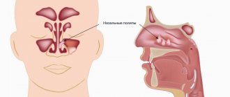
The maxillary (maxillary) sinus is a cavity with bony walls that is located inside the upper jaw. The cavity communicates with the nasal cavity through the anastomosis, which is located on the side wall of the sinus facing the nasal cavity. The lower wall has contact with the upper teeth. Quite often (in about 15% of cases) the apex of the tooth root lies directly under the mucous membrane of the bottom of the maxillary sinus, and there is no bone septum between them.
When an infection enters the maxillary sinus, inflammation of its mucous membrane or sinusitis occurs.
There are two ways of infection:
- rhinogenic - through the nasal cavity. In this case, the infection penetrates from the nasal cavity through natural or artificial (after surgery) communication;
- odontogenic - through the tooth or tissue around it.
Odontogenic sinusitis most often develops slowly against the background of chronic infection in the area of the tooth root. As a result of chronic inflammation, a cyst forms in the root area, which destroys the barrier between the sinus and the tooth. Pathogenic microbes gradually penetrate the sinus mucosa, causing inflammation.
Also, odontogenic sinusitis can occur as a result of the actions of the dentist. Often, after the removal of the upper tooth, the thin barrier between the sinus and the oral cavity may be damaged. As a result, a gateway for dental infection appears. In this case, the anatomical features of the patient with a thin bone septum between the sinus and the tooth root or its complete absence are of key importance.
Infection can occur when cleaning the canals and filling them. In some cases, the filling material gets inside the sinus, causing the formation of fungal sinusitis, and the zinc contained in the filling material promotes the growth of mold fungi (Aspergillus, Mucora). Also, odontogenic sinusitis can develop after the sinus lift procedure and the installation of dental implants in the upper jaw.
What are the maxillary sinuses
The maxillary sinuses (also called the maxillary sinuses) are special cavities on both sides of the nose that are filled with air. Each cavity is connected to the nasal passage by small openings called anastomoses. The cavities are covered with mucous membrane. The function of mucus is to trap bacteria and harmful particles in it, and then remove them from the body through those same anastomoses. When edema occurs, the excretory opening becomes very narrow, as a result of which mucus, along with harmful particles and bacteria, cannot come out and stagnates. At this time, the patient begins to experience bursting pain in the cheek area - this is how inflammation of the maxillary sinus begins. Treatment of the maxillary sinus should not be neglected, since inaction can provoke serious consequences, including sepsis and meningitis.
Classic sinusitis can be bilateral, when both sinuses are affected. In the odontogenic form, the inflammatory process starts in the sinus on which side the diseased tooth is located.
Complications
With odontogenic sinusitis, a chronic inflammatory process occurs. Dental microflora appears in the sinus, not typical for the upper respiratory tract, which can destroy bone tissue. Due to the fact that the paranasal sinuses have contact with the orbit and brain, odontogenic sinusitis can lead to severe complications:
- intraorbital (orbital phlegmon, ophthalmitis, optic nerve neuritis);
- intracranial (meningitis, encephalitis, brain abscess).
Therefore, at the slightest suspicion of this disease, you should consult a doctor.
Solution
After a consultation of dentists (surgeon, orthopedist and therapist), the following treatment plan was adopted:
- removal of the entire prosthetic structure (!), removal of 14, 26, 27 teeth with elimination of the infectious focus.
- in the second stage (approximately a month later) bone grafting in the area of the formed defect
- the third stage (approximately after 3 months) is the implantation of artificial roots in place of teeth 24, 25, 26, 27
- after another 3 months, installation of crowns on these implants.
During this period, the patient will wear a temporary prosthesis to replace missing teeth and undergo therapeutic tooth-preserving treatment of teeth 37, 47, 13.
After presenting this information, the entire team of doctors was present at a very sad scene - an adult respectable man, almost crying, talked about the fact that just 2 years ago, for three months, he was treating his teeth, spent 120 thousand rubles on 15 crowns, and now it turned out that his problem has not only not been solved, but has also worsened and requires new physical and moral efforts and new financial costs. After the first shock passed, he asked us a fair question: why did such complications arise?
Treatment
The treatment of odontogenic sinusitis requires an integrated approach. As a rule, treatment requires the simultaneous participation of an otolaryngologist and a dentist. Isolated antibacterial and conservative therapy lead only to temporary relief of the condition and removal of the severity of the process.
For a complete recovery, it is necessary to eliminate the source of infection - remove or treat the causative tooth while simultaneously sanitizing the inflamed sinus.
In case of foreign inclusions in the sinus (filling material, sinus lifting material, fungal bodies), their complete removal is necessary. For this, endoscopic techniques are used. They allow you to remove these formations through the nasal cavity. If there is a connection between the sinus and the oral cavity (oroantral fistula), it must be closed using special bioinert collagen-based membranes and mucosal flaps.
Sinusitis or tooth – what to treat first?
In this case, many otolaryngologists prescribe tooth extraction.
In our clinic, we collaborate with specialists who treat odontogenic sinusitis or help the patient prepare the sinus mucosa for sinus lifting.
Of course, it is not always possible to prosthetize and treat a tooth with a source of infection in the periodontium in the upper jaw. Therefore, such teeth often still need to be removed.
Tooth extraction surgery is a major source of irritation for the body and the immune system. After it, you need to wait 4-6 months until the porous bone tissue of the alveoli is restored.
In this case, conservative anti-inflammatory therapy, antibiotics, and active copious rinsing of the nasal sinuses are prescribed. Treatment lasts about a month after tooth extraction.
If CT and diagnostics revealed a source of infection (cyst or granuloma) on the tooth, then treatment for odontogenic chronic sinusitis begins immediately after removal. Then the chance to reduce inflammatory changes in the mucosa is quite high.
Otherwise, the patient risks getting large growths, which sometimes occupy more than half the height of the sinus.
This is manifested by nasal congestion, because when we have ARVI, the nasal mucosa swells greatly. And our nasal mucosa is exactly the same as in the sinus: the same columnar epithelium. It swells very much when initially, as a result of constant irritation, the infection has already grown by more than half.
In this case, the half of the nose that is more blocked will be an indicator to check what is wrong with the teeth on the same half of the jaw.
Eternal companion
Every winter I suffer from sinusitis. No matter what I do, no matter how I treat him, he still comes back to me. What is the reason?
Antonina, Vologda
– Alas, in your case, sinusitis has clearly become chronic. For a more accurate diagnosis, an experienced doctor just needs to look at an x-ray of the paranasal sinuses: if the mucous membrane of the maxillary sinus is thickened by more than 6 mm, this is considered a sign of chronicity of the process. Apparently, you have a pathology of the nasal cavity that predisposes you to frequent sinusitis (deviated nasal septum, pathology of anastomosis, etc.). As a rule, acute sinusitis begins with a common runny nose of viral origin. By infecting the cells of the nasal mucosa, the virus causes activation of the microbial flora, causing inflammation and swelling. Various reasons can lead to such a development of events. One of them is a decrease in general and local immunity due to certain stress factors - cold, heat, physical (overload, fatigue), psychological.
Be sure to treat!
What happens if sinusitis is not treated?
Eduard, Rostov
– If acute sinusitis is not treated, it can become chronic, and also cause complications - in the eye socket (as a result of which you can even lose an eye), in the brain (which is fraught with the development of a brain abscess, meningitis). There are (fortunately, rarely) fulminant forms of sinusitis, which develop from several hours to a week and can result in the death of the patient. This happens especially often in people with diabetes, who have high glucose levels in their tissues and are a good breeding ground for microorganisms. So, if you get sinusitis, try to treat it properly.
Patient Reminder
In order to avoid the development of sinusitis, it is important to follow the following rules:
- it is necessary to treat infectious diseases in a timely manner and in no case self-medicate;
- maintain oral hygiene;
- visit the dentist, have your teeth professionally cleaned twice a year;
- avoid hypothermia and strengthen the immune system.
If you still have sinusitis, get advice from a specialized specialist (ENT or dentist) and follow the prescribed treatment tactics. Remember that sinusitis is a temporary contraindication for sinus lifting, as well as for some dental implantation techniques.
Prevention
As for preventive measures regarding perforation of the maxillary sinus, they consist of following the following rules:
- A thorough and comprehensive examination of the patient before undergoing serious dental procedures, including tooth resection or implantation.
- Correct assessment of the anatomical features of the jaw structure of each patient.
- Strict compliance with all technological requirements for complex dental procedures.
Diagnostics
Diagnosis of perforation of the floor of the maxillary sinus during tooth extraction is based on a typical clinical picture. In doubtful cases, as well as when such a complication is suspected during implantation or endodontic manipulations, it is necessary to use instrumental diagnostic methods:
- Probing the socket of an extracted tooth or perforated canal with a thin probe . This allows us to determine that there is no bone bottom in the wound. In this case, the instrument passes freely through soft tissues and does not encounter obstacles along its path.
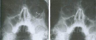
- X-ray of the sinus area . In this case, the pictures can reveal both darkening of the cavity due to the accumulation of blood in it, as well as fragments of dental roots, implants or filling material. Sometimes it is advisable to conduct radiography with contrast, when a contrast agent is introduced into the cavity through a perforation fistula.
- Computed tomography , which allows you to determine perforations and the presence of foreign bodies in the sinus with maximum accuracy.
- If old perforations are suspected, general clinical blood tests , the result of which may indicate the presence of an active source of infection in the body.
Functions of the maxillary sinuses
The maxillary sinuses are large air cavities located on either side of the nose. They begin to develop in utero, but are finally formed only by the age of 14-15. The sinus is bounded above by the orbit, and below by the alveolar process of the upper jaw, in which the teeth are located. On the inside there is an opening that connects the sinus to the nasal cavity. The size of the sinuses and their location vary from person to person and vary from person to person. For example, in some, the roots of the upper teeth may be located a centimeter below the bottom of the sinus, while in others they end in the cavity and lift its mucous membrane. These differences play an important role for surgical work in this area.
The sinuses perform respiratory, protective (warm and humidify the air) and secretory functions, and also take part in the process of speech formation, help with the sense of smell, and regulate intranasal pressure.
Tooth root cyst in the maxillary sinus
Removal of the tooth root is considered the most effective method of treating cysts localized in the apex area. When the patient has not been carefully examined, and the dentist does not have information about the thickness of the bone tissue that separates the cystic wall from the sinus floor, and also in the case when it is necessary to remove a large amount of jaw bone, rupture of the sinus tissue quite often occurs.
Preventing inflammation
In addition to preserving the wound, preventive therapy is carried out aimed at preventing the development of the inflammatory process. Most often, antibacterial drugs and anti-inflammatory drugs are prescribed. In addition, drops are prescribed for instillation into the nasal passages, which have a vasoconstrictor effect. Treatment can be carried out both on an outpatient basis and while staying at home.
If the examination reveals the presence of a foreign body in the maxillary sinus, then treatment is carried out only in a hospital setting. Therapy consists of surgical intervention to open the cavity and remove the tooth root from the maxillary sinus.