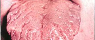
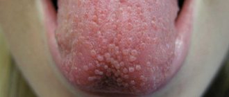
Oral papillomas are benign neoplasms in the oral cavity that grow from epithelial cells. Papillomas are discovered during a dental examination and look like separate growing seals on a small stalk, they are painless and have a white or pale pink color.
This type of neoplasm in the oral cavity is diagnosed most often. About 60% of patients are women aged forty years, about 20% are teenagers of any gender. Often, adults experience the appearance of individual papillomas, while children may experience so-called papillomatosis (multiple papillomas). In half of the cases, papillomas are localized on the mucous membrane of the tongue.
Papilloma in the mouth: causes of appearance
The most common cause of this type of tumor is the human papillomavirus (HPV).
Factors that provoke the appearance of papillomas in the oral cavity are, for example, constant microdamage to the cheeks and tongue. A relatively small damage is enough for viral particles to penetrate inside and trigger the formation of papilloma. In children, the provoking factor is a too short frenulum of the tongue - the lower incisors injure it, creating a gateway for infection.
When analyzing papilloma under a microscope, it can be noted that this neoplasm is a tumor, which consists of many layers of epithelial tissue, which in some places has become significantly keratinized. In some areas, traces of the appearance of a focus of inflammatory infection can be noted.
Symptoms
When the papillae on the tongue become inflamed, tissue sensitivity increases significantly and taste changes. Patients complain of unpleasant discomfort in the mouth. The taste spectrum is distorted and becomes unnatural. In addition, a number of other symptoms appear.
- Noticeable tissue swelling.
- Marked increase in size of the papillae.
- Tingling and itching.
- Burning, mild soreness.
If you do not consult a dentist in time, complications may develop: lingual eruptive papillitis. This disease occurs over a long period of time with an increase in body temperature. Most often the disease affects children. An alarming symptom is the appearance on the surface of the vocal organ of bubbles (pustules) filled with a transparent liquid. When the first signs of illness appear, you should immediately visit the dentist. Timely treatment of the disease minimizes complications and speeds up recovery.
Classification of oral papillomas
Based on the number and concentration of neoplasms, oral papilloma is differentiated from papillomatosis – a massive accumulation of neoplasms in one place.
According to their origin, papillomas are divided into the following types:
- Traumatic (reactive) papilloma. May appear after traumatic effects of a mechanical, chemical or temperature nature. A distinctive and characteristic feature of reactive type oral papilloma is that their growth stops immediately after the irritant that caused them is eliminated.
- True (neoplastic) papilloma. This type of papilloma begins to develop after the mechanism of cell division, growth, and differentiation is disrupted. In most cases, this type of papillomas appears in the distal part of the cheek, in the area located behind the molars and in the area of the pterygomandibular fold.
- Viral papilloma of the oral cavity. May appear after the patient has been infected with the human papillomavirus. This type of infection occurs through direct contact with a carrier of the virus. When the integrity of the oral mucosa is compromised (for example, due to microtrauma), a path for infection appears.
Benign tumors of the tongue
The clinical course of a tongue tumor and the characteristics of its growth are associated mainly with the type of tissue from which it originates. The presence of epithelial, muscle, glandular, and adipose tissue in the tongue structure, as well as the possible entry into the tongue tissue during embryogenesis of the rudiments of other tissue structures (bone, cartilage, thyroid tissue) determine a wide variety of clinical forms of tongue tumors. Most often dentistry encounters vascular tumors of the tongue (angiomas). The second place in prevalence is occupied by papillomas, the third place by fibroids of the tongue.
Papilloma . This tumor of the tongue grows from the stratified squamous epithelium of its mucosa. Most often it occurs on the back and tip of the tongue. Papillomas are multiple or single formations of pale pink color, round or elongated, rarely growing to large sizes. The appearance of keratosis on the surface of the papilloma, as a rule, indicates its malignant degeneration. In some cases, spontaneous involution of papilloma was observed.
Adenoma . Formed from the glands of the tongue mucosa. Cystoadenomas are more often observed on the tip of the tongue. In the area of the root of the tongue, polyps from the heterotopic gastric mucosa may appear.
Botriomyxoma . The tongue tumor is flat or spherical in shape, in rare cases divided into several lobules. Initially it is red in color, but over time it becomes brown. In its growth it can reach the size of a walnut. The surface of botryomyxoma can be smooth or coarse-grained, often covered with crusts. Factors that provoke the formation of this type of tongue tumor include trauma and tongue fissure.
Fibroma . A round tumor of the tongue of elastic consistency, growing from connective tissue cells. Fibroids can grow on a stalk. Its color often does not differ from the color of the mucous membrane, in other cases it has a yellowish or whitish tint.
Retention cyst . Most often located on the lower surface of the tongue in the area of its tip. It has a multiple character. This tumor of the tongue develops from the nunnia glands located in its superficial muscle layer.
Lipoma . A tumor of the tongue developing in the submucosal layer with a lobular structure and a soft-elastic consistency. It is most common on the undersurface at the back of the tongue. Lipoma is characterized by slow growth and painless course.
Myoma . A tumor of the tongue that occurs when the cells of its muscles proliferate. It often has a size of up to 1 cm and a dense consistency, but can grow to a significant size. Covered with mucous membrane. It is usually localized on the upper surface of the tongue. In some cases, small papillary outgrowths are observed on the surface of the fibroid.
Neurofibroma . They develop from the tissues of the nerve branches passing through the tongue, most often in the posterior half of the tongue. This type of tongue tumor occurs in rare cases and is characterized by slow growth. May be accompanied by various pain sensations.
Hemangioma . Tumor of the tongue, originating from the tissues of blood vessels. Associated with a violation of embryogenesis, more often observed in girls. This tongue tumor is usually detected at birth or in early childhood. Capillary hemangioma appears as red spots of various sizes and shapes that do not rise above the surface of the tongue. The spot turns pale when pressed. Cavernous hemangioma is a tumor of the tongue with a bluish-purple color and soft consistency. Often rises above the surrounding mucosa. Characterized by deep germination into the underlying tissues. Pressure on the tumor leads to a decrease in its size, which quickly recovers when the pressure is removed. Vascular tumors of the tongue may be accompanied by bleeding, most often caused by injury.
Lymphangioma
. It grows from the walls of the lymphatic vessels of the tongue and appears in the first years of a child’s life. It can cause diffuse damage to the tongue, leading to its significant enlargement. Local lesions are represented by growths of a warty structure with vesicular elements and are most often located along the upper surface of the root or tip of the tongue. When injured by food or teeth, this swelling of the tongue often becomes inflamed.
Struma of the tongue . A rare tumor of the tongue that arises from cells of thyroid tissue that enter the tongue as a result of impaired embryonic differentiation. It is a node localized at the root of the tongue with a diameter of up to 3 cm.
Treatment of oral papillomas
Diagnosis of this disease includes a collection of the patient’s medical history, as well as a thorough histological examination of removed papillomas.
Treatment of papillomas is only surgical. The neoplasm is excised down to the borders of healthy tissue. Techniques such as electrocoagulation, cryosurgery, sclerotherapy and others are rarely used, since as a result of their implementation it is impossible to conduct a histological analysis of the papilloma removed to the base.
If a large accumulation of papillomatous neoplasms is detected, a combined technique is used: a scalpel is used to dissect the largest number of papillomas accumulated in one place, and single papillomas are removed using electrocoagulation.
If oral papillomas have a viral etiology, antiviral and immunomodulatory therapy is prescribed along with surgical intervention to prevent relapses.
Depending on the etiology of the disease, relapses may occur with greater or lesser probability. So, if there is a human papillomavirus in the body, the risk of papillomas returning after surgery is quite high.
Diagnostics
Basic diagnostic methods in dentistry allow a thorough examination to be made in order to make the correct diagnosis. During the initial examination, the dentist determines the size of the pathological zones, appearance, color, and shape of the organ. The doctor finds out whether there is swelling of the tongue, ascertains plaque and its localization, abrasions, punctures, eczema, ulcers. The health of gum tissue and teeth is important.
In order to determine the type of pathogen and the form of the disease, a number of tests are prescribed:
- Histological smear;
- Sensitivity is determined using a special test;
- A general blood test is informative;
Systemic pathologists are also excluded: HIV, syphilis, hepatitis, AIDS. If necessary, the patient is referred for consultation to a gastroenterologist, dermatologist, endocrinologist, otolaryngologist, or immunologist.
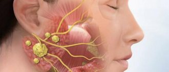
Sialadenitis
Sialadenitis is an inflammation of the salivary gland that affects the sublingual glands. The main reason for the development of the disease is the penetration of infection.
Various microorganisms act as pathogens. Harmful bacteria that cause disease include:
- tuberculosis bacillus;
- virus ;
- causative agent of syphilis;
- various other infectious diseases.
The causes of the disease can be:
- weak immunity;
- infections;
- oncological diseases.
The acute form of sialadenitis is manifested by tissue swelling. After this, compaction occurs, suppuration and, as a final result, necrosis. Sometimes the disease process is inhibited at the initial stage.
In case of illness, symptoms are observed:
- general malaise;
- increased temperature ;
- a characteristic taste in the mouth ;
- pain in the form of attacks in the gland area;
- discomfort and pain while eating;
- swelling of the affected area.
When treating sialadenitis, both traditional medicine and folk methods are allowed. All therapeutic measures are carried out as prescribed and under the supervision of a doctor.
Hemangioma
Hemangioma of the tongue is a benign tumor that is formed as a result of impaired development of blood vessels. This type of tumor is a red or purple bulge that does not grow quickly but is considered unpredictable in its development. Sometimes in a short period of time it can reach its maximum size, which leads to functional disorders of organs.
Why can our articles be trusted?
We make health information clear, accessible and relevant.
- All articles are checked by practicing doctors.
- We take scientific literature and the latest research as a basis.
- We publish detailed articles that answer all questions.
The most common types are: capillary and cavernous hemangioma.
The reasons for its occurrence are various:
- hormonal imbalance ;
- weakened immune system;
- metabolic disorder ;
- injuries in the oral cavity.
The presence of a tumor and its symptoms requires immediate treatment, otherwise the disease can develop many complications, which, in turn, will lead to consequences, including a malignant tumor.
How common are papillomas under the tongue?
Papillomas are a very common disease that most often occurs in children. Seborrheic papillomas predominate in older people (aged 50-60 years). About 50% of adults have papillomas on the arms and legs at least once in their lives. In 7% of cases, papillomas occur under the tongue.
About 25% of papillomas under the tongue disappear spontaneously. It is estimated that 30-80% of all sexually active people will become infected with HPV once in their lifetime. The vast majority of these infections are not detected after a year. Some women develop only temporary immunity. They can get infected again and again. In 10-30% of cases, the infection persists even on an ongoing basis. Women can develop cervical cancer.
Cyst (wound)
A cyst under the tongue appears as a growth in the form of a ball with liquid inside. It is localized on the tip of the tongue, in the sublingual area and on the root. Most often, a cyst forms in the area of the frenulum of the tongue.
The cyst progresses slowly, but as its size increases, it causes a sensation of a foreign body in the oral cavity; there is no pain. A large growth is accompanied by the following symptoms:
- swelling of the lower jaw;
- rupture of the growth shell.
In case of the first manifestations, it is necessary to seek the help of a doctor, since ranula can become infected and cause complications of the following nature:
- displacement ;
- disorder ;
- breathing and swallowing problems
- development of an abscess.
The cyst itself is not dangerous, but additional research is required to establish a diagnosis.
Inflammation of the papillae on a child's tongue
If the child’s body is weakened and he complains of discomfort in the mouth and pain in the muscle organ, parents should immediately show him to the pediatrician and pediatric dentist. The doctor will determine whether his papillae are red and whether there is an inflammatory process in the mouth. Once the diagnosis is made, therapy is prescribed. As a preventive measure, parents should ensure that the baby regularly brushes his teeth and rinses his mouth thoroughly. Children's immunity is not strong enough, so the body cannot cope with the disease on its own. The situation is complicated by the fact that children are curious; they put most objects in their mouths. To avoid serious complications, take your child to the dentist regularly.