Tongue cancer refers to malignant tumors of the oral cavity and oropharynx, which, in turn, are a type of head and neck cancer. This is a rare type of malignant tumor. Oral cancer accounts for about 3% of all cancers. People over 62 years of age are most often affected.
- Causes: what causes tongue cancer?
- Types of tongue cancer
- Symptoms and signs of tongue cancer
- Methods for diagnosing tongue cancer
- Stages of tongue cancer development
- Modern methods of treatment
- How long do people live with tongue cancer? What is the prognosis for recovery?
General information
A malignant neoplasm in the tongue comes from the mucosal epithelium.
Despite the fact that tongue tumors are uncommon, they have an aggressive course, so early detection is important. Distinctive features of this disease are early metastasis (including distant), rapid growth, resistance to treatment, high recurrence rate and mortality. At the time of diagnosis, half of the patients had affected regional lymph nodes. Recurrence is observed in 30-50% of patients. If we consider the localization, then most often a tumor of the tongue occurs on the lateral surface, much less often there is cancer of the root of the tongue, the back and the tip. A neoplasm of the root of the tongue is classified as oropharyngeal cancer. The asymptomatic course of the disease makes early diagnosis difficult. Tumors are either not visible in the early stages or initial changes are regarded as a consequence of trauma to the tongue. Due to the lack of alertness of patients and doctors regarding malignant neoplasms, most cases are diagnosed with a full clinical picture or already in an advanced stage.
The highest incidence is observed after 50 years of age, but this disease also occurs in people under 30 years of age. In men, this pathology is diagnosed 5 times more often. In 95% of cases, tongue tumors are squamous cell carcinoma. Squamous cell carcinoma of the tongue is an aggressive tumor and insensitive to treatment ( chemotherapy ), but radiation treatment .
Stages of the disease
Stages of tongue cancer
We offer you a table of correspondence between tongue tumor stages and clinical TNM classification, which uses the following designations:
- T – indicates the primary tumor: Tx – the primary tumor cannot be assessed;
- T0 – no data on the primary tumor;
- Тis – cancer in situ (pre-invasive stage);
- T1-T4 – the primary tumor is enlarged and/or widespread.
- Nx – regional lymph nodes cannot be assessed;
- M0 – no distant metastases;
Correspondence table for stages of tongue cancer TNM classification
| TNM stage | |
| 0 (carcinoma in situ) | Tis, N0, M0 |
| I | T1, N0, M0 |
| II | T2, N0, M0 |
| III | T3, N0, M0 or T1-T3, N1, M0 |
| IV | 4a: T4a, N0 M0 or T4aN1, M0 or T1-T4a, N2, M0 4b: T4b, any N, M0 or any T, N3, M0 4c: any T, any N, M1 |
Classification
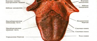
Depending on the location, cancer is distinguished:
- Bodies. These include tumors of the tip, dorsum and lateral surface (most often the middle of the lateral surface). This localization occurs in 70% of cases.
- Root (accounts for 20% of all cases).
- Lower surface (sublingual area).
By growth pattern:
- Exophytic form (this includes papillary and ulcerative forms of tumors growing outward).
- Endophytic (growing inside the tissue of the tongue - infiltrative and ulcerative).
Papillary form a is a dense papillary growth of a mushroom shape. There may also be raised plaque-like growths that have clear boundaries.
Ulcerative (occurs in 50% of cases). Characterized by a superficial ulcer surrounded by a ridge. The ulcer is constantly increasing in size. At first, the ulcer does not bother the patient, but as it grows, pain and bleeding appear. The ulcer can become infected and inflamed, making diagnosis difficult.
Infiltrative form . The tumor grows into the thickness of the tissues, and they become denser. With diffuse growth, the compaction spreads to the entire tongue, which impairs its mobility.
The infiltrative-ulcerative form is characterized by thickening of the tongue with the presence of deep ulcers.
According to histological composition:
- Adenocarcinoma.
- Squamous cell carcinoma.
Classification according to the TNM system.
- Tis "cancer in place."
- T1 Tumor no more than 1 cm. Not accompanied by complaints, discovered by chance during examination.
- T2 ≤2 cm. There are ulcers and areas of compaction.
- T3 >4 cm. The tumor is equal in size to half the tongue. Metastases to the occipital lymph nodes, postauricular.
- T4 Locally advanced cancer that occupies the entire tongue and invades the tissues of the mouth and face. Metastases to internal organs (brain, liver, heart) and bones.
Symbol N - the presence of metastases in the lymph nodes:
- N0 - No lymph node involvement.
- N1 One node on the side of the tumor is affected.
- N2 Metastasis: one metastasis no more than 6 cm, several up to 6 cm on one side or both sides up to 6 cm.
- N3 Metastases larger than 6 cm.
With T1 tumors, lymph nodes are affected in 40% of cases, with T4 stage in 85%. A reliable factor for metastasis is the depth of invasion - 4 mm is considered a critical value. Most often, metastases are found in the submental, submandibular and cervical nodes (upper third).
Histopathological differentiation:
- G1 High degree.
- G2 Medium.
- G3 Low.
Kinds
In 70% of cases, cancer of the body of the tongue is detected, in 20% - damage to the root, and in 10% - to the lower surface of the organ. If we divide diseases according to cell characteristics, we can distinguish the following forms:
- Papillary. It looks like a dense growth with papillary outgrowths and plaque-like formations.
- Ulcerative. It is observed in approximately 50% of cases. Ulcers develop over time, can bleed, and often become infected, thereby masking the root cause of the disease.
- Infiltrative. Cancer grows inside the tongue and hardens its tissues. The form can be diffuse or spread throughout the entire organ.
If we talk about microscopic analysis, then in 95% of cases we are talking about a squamous cell form of tongue cancer, other options are much less common.
Causes
- Bad habits. Among which, smoking is of particular importance. The influence of carcinogenic substances in tobacco in the development of cancer has long been proven. The risk increases with duration and intensity of smoking. The risk increases further with alcohol consumption.
- Occupational hazards (contact with radiation, work in the oil industry).
- Constant traumatization of the mucous membrane (poor-quality dentures and damaged teeth, poor treatment of fillings, and constant biting of the tongue are important).
- Chewing mixtures (betel nut, non-smoking tobacco), consuming spices that have a carcinogenic effect.
- Oral infection.
- Pre-tumor conditions ( leukoplakia , Bowen's disease , ulcerative-erosive form of lupus erythematosus , papillomatosis , erythroplakia , leukokeratosis ).
- Action of oncogenic viruses. The connection of this pathology with chronic human papillomavirus infection and HIV . The oncogenic effect of viruses is associated with blocking or the influence of tumor suppressor genes.
- Long-term use of immunosuppressive drugs.
Forms of cancer of the oral mucosa
Oncological diseases of the oral cavity are conventionally divided into three types, differing in etiology and external signs:
| Form of cancer of the oral mucosa | |
| Name | Description |
| Knotty | Seals with clear edges are observed on the tissues. The mucous membrane either has whitish spots or remains unchanged. Neoplasms in the nodular form of cancer quickly increase in size. |
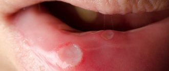
| Ulcerative | The neoplasms look like ulcers; they do not heal for a long time, which causes severe discomfort to the patient. Pathology in the ulcerative form is rapidly progressive. Compared to other species, it affects the mucous membrane much more often. |
| Papillary | The neoplasm has a dense structure. It is impossible not to notice, since the tumor literally sags into the oral cavity. The color and structure of the mucosa remain almost unchanged. |
Papillary
Knotty
Ulcerative
Symptoms of tongue cancer
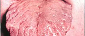
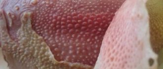
During the course of the disease, the initial stage, developed and advanced, is distinguished. It is important to know and identify the symptoms of the initial stage. Tongue cancer initially has an asymptomatic course. What does the neoplasm look like?
It may manifest itself:
- whitish spots similar to plaque;
- compaction and slight redness;
- painless nodules;
- superficial ulcers or cracks.
Most often, such changes are located on the lateral surface and are mistaken for glossitis (inflammation of the tongue). It is important that conservative treatment (treatment with antiseptics, sea buckthorn oil) does not help. It is rare, but it still happens that already at an early stage discomfort (burning or tingling) appears when eating and pain.
Photo - initial stage of cancer of the lateral surface of the tongue
Signs of tongue cancer at an advanced stage are characterized by pain, which is observed in 100% of cases. It has different intensities, can be local and diffuse, and also radiate to the temple, various areas of the mouth and ear. Patients are bothered by a feeling of numbness. When an infection occurs, bad breath appears. If the tumor begins to disintegrate, necrosis products irritate the mucous membrane and cause salivation. Bleeding of the tongue may also occur.
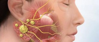
The advanced stage is characterized by an aggressive course and rapid invasive growth. Signs in an advanced stage are massive destruction of the tumor and surrounding tissues, including bone structures, and metastasis to local lymph nodes (occipital, submandibular, cervical and mental). With this disease, distant metastases occur in the lungs, bones, brain and liver. As the tumor disintegrates, the smell from the mouth increases, difficulty swallowing, difficulty speaking, numbness of the tongue and bleeding are observed. Common symptoms include weakness, weight loss and exhaustion.
Symptoms of tongue root cancer
This localization, which is observed in 20% of cases, is characterized by a rapid and aggressive course. There is a sore throat and discomfort when swallowing as the tumor reaches a large size. It often spreads to the auditory nerve. Patients have impaired tongue mobility.
Modern methods of treatment
Approaches to the treatment of malignant tumors of the body and root of the tongue are somewhat different.
Treatment of tongue body cancer
If the tumor is less than 4 cm in size, it is removed surgically. In some cases, removal of regional lymph nodes is also indicated. Usually no other treatment is required.
Typical tactics for advanced cancer of the body of the tongue (the tumor is more than 4 cm or has managed to grow strongly into the surrounding tissues) involves a combination of three treatment methods:
- Surgical removal of the primary tumor, as well as regional lymph nodes.
- Radiation therapy. Before carrying out it, a consultation with a dentist and sanitation of foci of infection in the oral cavity are required.
- Chemotherapy.
In case of relapse, as well as in the last stage of tongue cancer with distant metastases, chemotherapy becomes the main treatment method.
Treatment of cancer of the base (root) of the tongue
If the tumor diameter is less than 4 cm, it is removed, and regional cervical lymph nodes are also excised. If the risk of relapse is high, radiation therapy to the neck area or chemoradiotherapy (radiation combined with chemotherapy) is prescribed after surgery.
If the tumor is larger than 4 cm or has grown into surrounding tissues, different treatment options are possible:
- chemoradiotherapy;
- removal of part of the tongue and surrounding tissues, regional lymph nodes, followed by a course of chemotherapy or radiation therapy;
- only radiation therapy.
The type of surgery for cancer of the body and root of the tongue depends on the location of the tumor, its size and the degree of invasion of surrounding tissue. Chemotherapy or radiation therapy may complement surgery. When used as a stand-alone treatment, they are usually prescribed for palliative purposes. They will not completely destroy cancer, but will help slow its progression, improve the patient’s condition, and prolong life.
In some cases, the targeted drug cetuximab (Erbitux) and the immunodrug nivolumab are prescribed.
Tests and diagnostics
To clarify the diagnosis, patients are prescribed:
- Biopsy of a tumor formation (or scraping and impression smears from the surface of erosions).
- CT and MRI of the affected area with intravenous contrast. This study makes it possible to assess the extent of the tumor and the depth of invasion into the tongue tissue.
- CT scan of the facial bones with contrast if there is a suspicion of tumor spread to the bones of the jaw and skull.
- Ultrasound of the lymph nodes of the neck.
- Puncture of the affected cervical nodes with cytological examination.
- Ultrasound of the abdominal cavity to exclude distant metastatic process.
- Chest X-ray to exclude metastases.
- Osteoscintigraphy for metastatic lesions of skeletal bones.
How is the disease diagnosed?
Along with common diagnostic methods, leading Israeli clinics are increasingly using molecular diagnostics - determining the presence of mutations in malignant cells that determine their sensitivity to a variety of treatment methods. With this innovative type of diagnostic, called Caris Target Now, it is possible to establish the resistance of malignant cells to a particular treatment method, avoiding unnecessary waste of time.
A comprehensive diagnostic examination takes no more than three days in Israeli clinics.
- The first day
- Second day
- Day three
At the initial consultation, the treating otolaryngologist oncologist gets acquainted with the patient’s medical history, establishes the probable causes of the pathology, conducts a physical examination and prescribes the necessary examinations.
Completing prescribed examinations, including:
- general and biochemical blood tests;
- biopsy of the tumor followed by cytohistological analysis;
- puncture of the cervical lymph nodes with cytological analysis of the obtained material;
- ultrasonography;
- endoscopic examination of the tongue;
- visual research methods (radiography, CT, MRI).
A medical council, which includes the treating oncologist and a number of highly specialized specialists, reviews the results of the examinations, makes a final diagnosis and develops a treatment program.
Diet
Diet for tongue cancer
- Efficacy: therapeutic effect for a month
- Timing: constantly
- Cost of products: 1300-1500 rubles per week
Difficulties with swallowing in the patient before and after surgery force the installation of a nasoesophageal tube. Through it, broth with meat and eggs beaten in a blender, sour cream, cream and other high-calorie liquid products are introduced. A nutritional mixture must be prescribed in addition to the main diet . This is a liquid high-calorie and high-protein mixture enriched with micronutrients. This can be Nutrizon Advance , Nutrizon Protein Advance , Nutrizon Energy , Nutrizon Protein Intense , Supportan Mixture for enteral nutrition, Fresubin original , Fresubin Energy , Nutricomp . With the transition to natural nutrition, a diet table with liquid dishes is organized - puree soup, boiled fish and meat (veal), poultry (turkey, chicken), whipped in a blender, meat and fish soufflé, liquid omelet, yoghurt, milk, cauliflower, broccoli in the form of mashed potatoes, mashed potatoes.
Prevention
- Quitting bad habits (smoking, alcohol).
- Elimination of tongue injury (high-quality treatment of fillings after their installation, correct selection and correct installation of dentures, timely treatment of dental chips).
- Regular oral hygiene, including professional.
- Timely detection of precancerous diseases of the tongue ( leukoplakia , dyskeratosis , papillomatosis ).
- Diet rich in vitamins . It has been proven that vitamin A and β-carotene are important in the prevention of neoplasms. With a deficiency of carotenoids, there is a risk of squamous cell carcinoma .
Forecast
With early diagnosis and radical treatment, the prognosis for recovery from tongue cancer is more favorable. It is believed that complete recovery from cancer is impossible and the goal of treatment is to achieve long-term remission of 5-8 years. Thus, with this pathology, the five-year survival rate ranges from 65 to 85%. Complete remission within 5 years after surgery and radiation therapy with T1 is achieved in 80% of patients, with T2 in 60% of patients, and with T3-4 does not exceed 35%. Metastases in the lymph nodes are an important prognostic factor and, if present, survival rate is halved.
How long do people live with stage 4 tongue cancer or with relapse after chemotherapy? Such patients have a poor prognosis: they live no more than 3.5 months while on maintenance treatment. The addition of cetuximab to chemotherapy increases survival by 2 times.
List of sources
- Alieva. S.B., Alymov Yu.V., Kropotov M.A., Mudunov A.M., Podvyaznikov S.O. Cancer of the oral mucosa. Oncology. Clinical recommendations / Ed. M. I. Davydova. – M.: Publishing group RONC, 2015, pp. 27-37.
- Romanov, I.S. Features of regional metastasis of squamous cell carcinoma of the oral cavity, detected during preventive lymph node dissections. / Romanov, I.S., Yakovleva L.P., Udintsov D.B., Dzhumaev M.G., Tsiklauri V.T. // Dentistry. – 2012. – T.91, No. 4. — P. 28-31.
- Romanov I. S., Yakovleva L. P. Issues in the treatment of oral cancer. Farmateka 2013; (8):59–63.
- Paches A.I. Tumors of the head and neck. 5th ed., supplemented and revised M.: Practical Medicine, 2013. P. 119‒146.
- Romanov I. S. Prospects for the use of cetuximab in the treatment of squamous cell carcinoma of the head and neck / Oncology. Hematology. Chemotherapy. - 2015. - No. 17/1.