Causes Danger of fistula Diagnostics Treatment Prevention
A fistula on the gum is a pathological condition, the result of purulent inflammation near the root of the tooth with perforation of the gum tissue. This is a channel in the thickness of the gums through which pus comes out from the source of inflammation.
The disease is diagnosed and treated by dentists: a therapist, a surgeon, and sometimes a maxillofacial surgeon.
What is a fistula, what does it look like, symptoms
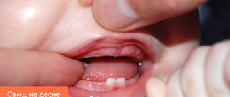
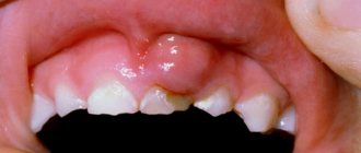
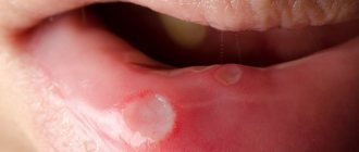
Externally, the fistula is a convex formation with a noticeable white spot on it. This means that the pathological process is accompanied by the formation of pus in large quantities.
There are two types of fistula:
- External, when the gum fistula comes to the surface;
- Internal, when the process remains hidden inside the gum, due to the fact that the fistula channel does not reach its surface. The inflammatory process can only be detected by x-ray examination.
Symptoms of a dental fistula are detected primarily by visual examination. There is a noticeable bulge on the gum, from where pus comes to the surface. There are other signs:
- The pain is cutting, stabbing, spasmodic. Becomes stronger with impact (for example, when chewing), weakens slightly after some of the purulent contents come to the surface;
- Putrid odor;
- Foreign tastes in the mouth;
- Tooth mobility with pathology;
- Swelling of the gums in the area of inflammation;
- Discharge of mucus with pus;
- Increased temperature, symptoms of intoxication of the body;
- Inflammation of the lymph nodes.
Fistula (Fistula)
A common sign of the disease is the presence of a pathological communication between two or more cavities or a body cavity and the environment. With external fistulas, a hole (course) is revealed during the examination. The edges of the hole are inflamed and macerated due to skin irritation. The amount and nature of discharge vary depending on the type of fistula and the characteristics of the pathological process.
Internal fistulas are manifested by the presence of atypical discharge from any natural opening. Significant losses of fluid and nutrients in some types of fistulas cause exhaustion and gross disturbances of all types of metabolism, which can lead to the death of the patient.
Digestive system fistulas
They can form throughout the gastrointestinal tract, from the esophagus to the rectum, and can be external or internal. Gastric fistulas are usually of artificial origin (gastrostomy), esophageal fistulas are provoked by a pathological process, intestinal fistulas are artificial (colostomy, ileostomy, cecostoma) and pathological. The first place among the causes of the formation of esophageal fistulas is occupied by tumors, intestinal - anastomotic failure.
The formation of external fistulas is accompanied by the appearance of infiltration. With internal fistulas, the clinical picture resembles a perforation of a hollow organ. With tracheoesophageal anastomosis, pieces of food are coughed up from the upper respiratory tract. Internal gastrointestinal fistula is manifested by fecal vomiting and fecal odor from the mouth. With an externally formed fistula, a passage with esophageal, gastric or intestinal discharge is found on the skin; with an unformed fistula, a purulent wound is found, at the bottom of which a loop of intestine and the wall of the stomach are visible.
Diarrhea, vomiting, and autointoxication are typical. The rate of exhaustion and development of metabolic disorders depends on the diameter of the fistula - the wider it is, the faster the symptoms worsen. Possible dehydration, exhaustion, protein-free edema, anemia. Intoxication and metabolic disorders lead to changes in the liver and kidneys. In severe cases, kidney and liver failure develop.
Gallbladder fistulas
Biliary fistulas usually become a complication of cholelithiasis, can be external or internal, and can connect the bile ducts with the stomach, intestines, pleural cavity, and bronchus. External fistulas are accompanied by the formation of a hole with bile or purulent discharge, internal fistulas are accompanied by symptoms of cholangitis, diarrhea, weight loss, and intoxication.
Rectal fistulas
Includes various forms of paraproctitis and rectovaginal fistulas. Paraproctitis occurs with purulent inflammation of the intestinal wall and pararectal tissue, manifested by pain, foreign body sensation, and general symptoms of intoxication. Pus is released from the anus or a hole with purulent discharge is found on the skin of the perianal area. With rectovaginal fistulas, gases and feces are released from the vagina. Pain in the perineum, inflammatory diseases of the genital organs and urinary tract are common.
Urogenital fistulas
Formed between the genitals and the urethra, ureters or bladder. They develop after difficult and complicated childbirth, operations, and radiation therapy. The most common are urethro-vaginal and vesicovaginal fistulas. Less common are vesicouterine, ureterovaginal and other variants.
Accompanied by leakage of urine from the genitals, the appearance of blood in the urine during menstruation. Usually painless. They do not pose a threat to life, but significantly reduce its quality and cause social maladjustment. They are of great clinical importance due to their high prevalence (0.6-2%).
Bronchial fistulas
More often they occur after operations on the lungs, and are characterized by the appearance of communication with the skin, pleural cavity or hollow organs - the esophagus, stomach, intestines, gall bladder. Manifested by shortness of breath, cough, discharge of pus or atypical contents (food, bile) from the respiratory tract, weakness, sweating, increased body temperature.
Ligature fistulas
Ligature fistula is a common complication (up to 5%) of surgical interventions, especially during operations on the abdominal and pelvic organs. It develops due to the rejection of non-absorbable threads that are located deep in the tissue. During the formation period, pain and intoxication are noted; after the formation of a fistula tract, the condition returns to normal. Fistulas are prone to a chronic relapsing course until the thread is removed or comes out on its own.
Causes of a fistula on the gums in an adult
A fistula is formed due to an infectious purulent process in the dental canal. Pathology can occur for the following reasons:
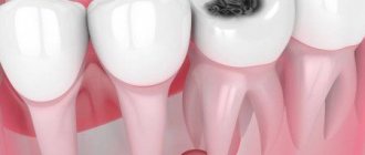
- Deep caries and untimely treatment;
- Newly emerging periodontitis and periostitis with the formation of pus;
- Chronic periodontitis, damage to the walls of the tooth root;
- Cystic formations;
- Oncology;
- Pulpitis, damage to the tooth root;
- Granuloma.
Fistula also occurs after tooth extraction. This is a common pathology that occurs as a result of infection in the wound.
A fistula after dental treatment can form, for example, due to infection as a result of errors in the treatment of caries. Improper filling of a tooth can also lead to the development of pathology. In this case, a tooth root fistula is formed when pus, having passed through the dental canal, breaks into the surrounding tissue. The most common fistula of the upper and front teeth occurs.
Alcohol, smoking, stress, exacerbations of chronic diseases, abuse of carbohydrates, lack of nutrition, chronic diseases - all these factors increase the root causes of the disease.
Pressure ulcers in elderly patients: utmost attention is required
Bedsores in older people can appear even if the person uses a stroller and moves around in it. Bedsores are also considered to be abrasions on the inner mucous membrane of the mouth, which can appear from dentures. That is why constant attention to patients and monitoring of the slightest changes in their condition is so important. Such care is possible only with a permanent professional nurse who will be with the patient constantly.
Treatment of pressure ulcers in older people is more difficult than in young and mature patients. In old people, all metabolic and regeneration processes proceed much more slowly. To this we must add age-related diseases - diabetes, hypertension and many others, which complicate the treatment of the skin and general condition. Inattention to problems can ruin the patient, and hiring an experienced foster nurse is the direct duty of relatives.
A special bandage has been developed in the USA, which is put on the patient and sends signals from electrodes on the body. Healthy tissues do not conduct current, but patients give electrical impulses and warn that bedsores are beginning to form in a person. There are no such innovations in Russia yet, and the only thing that really helps is the professional care of the nurse, her constant communication with the doctor and the strict implementation of his recommendations.
All these services can be provided to its clients by the social support patronage service. Our nurses with experience and medical education provide effective treatment of bedsores of any location and stage of development.
How dangerous is a fistula? Consequences
The fistula itself does not pose a danger, since through it the pus is removed from the source of infection, and accordingly, the degree of tissue damage is reduced. But the presence of a fistula indicates that the process is serious, and various types of complications may arise, such as:
- Loss of teeth in the affected area;
- Attachment of a secondary infection - inflammation of the lymph nodes, ears;
- Sinusitis;
- Sinusitis;
- Damage to bone tissue;
- Sepsis as a result of infection entering the general circulatory system;
- The appearance of cystic formations at the roots of the tooth;
- Heart complications, bacterial endocarditis.
Sometimes it happens that the pus drains almost completely, the fistula heals, and the patient thinks that everything is gone. But in fact, the source of inflammation has not been eliminated, so relapse of the disease is inevitable. Repeated abscesses and fistulas are more difficult to treat.
Discussion
Treatment for fistulas of the stumps of the main bronchi has two main objectives: sanitation of pleural empyema, which, as a rule, develops with BS, and elimination of the fistula. If the first problem is solved technically quite simply (drainage, thoracoscopic sanitation, thoracostoma), then the problem of eliminating the fistula is still very far from being resolved. In this direction, three fundamental approaches can be distinguished: conservative management, surgical techniques and endoscopic treatment. Conservative treatment does not directly affect the fistula, but is aimed at sanitizing the pleural empyema and correcting the patient’s homeostasis. If you successfully solve these problems, you can count on a favorable result. Thus, according to our data, healing of the ulcer occurred with conservative management in 111 (45.1%) of 246 patients who underwent pneumonectomy for lung cancer in the period from 1964 to 2013 [1]. We emphasize that the vast majority of these were patients with point (less than 3 mm) fistulas.
When choosing a method of radical surgery for BS, the size of the fistula, the length of the bronchial stump, the side of the lesion, and the general condition of the patient are taken into account [2, 3, 7—9, 14]. The last condition is sometimes decisive in the question of whether the operation is possible at all. So, according to O.O. Yasnogorodsky [15], radical surgery for BS was possible only in 33.7% of the observed patients with BS, according to P. Hollaus et al. [14] - in 68.7%. In the remaining patients, this was prevented by the uncorrectable severity of the general condition and the activity of the purulent process. Regarding the choice of surgical access to the stumps of the main bronchi, recent publications indicate that three options are still used: homolateral [16], transsternal [7-9] and contralateral, used for left-sided fistulas [3, 9]. The effectiveness of these operations, according to the authors, is 81.8% [14], 82.3% [7], 89.8% [8], 86.7% [2]. Postoperative mortality, according to the same authors, is 3.8% [2], 8.2% [8], 11.7% [7], 18.2% [14]. Thus, along with fairly high efficiency, reconstructive operations for fistulas of the stumps of the main bronchi represent a serious surgical injury due to the difficulty of surgical access to the bronchial stump, whether access through the post-pneumonectomy cavity, or transsternally-transpericardially, or through the opposite pleural cavity. It is emphasized that such interventions are accompanied by a certain surgical risk and, therefore, as we saw above, cannot be performed in every patient with BS [3, 17].
The introduction of endoscopic technologies for the treatment of patients with BS is absolutely justified and logical. In this regard, attempts to occlude the BS using a mediastinoscope deserve mention. The first publication dates back to 1996 [17]. O.O. demonstrates the greatest experience. Yasnogorodsky [15]: cervical-mediastinal mediastinoscopic occlusion of the main bronchus stump was performed in 27 patients with an efficiency of 88.9%. S. Groth et al. [19] describe a combined mediastinoscopic cervical approach with the formation of three incisions in the neck into which a mediastinoscope, telescope and stapler were inserted. The method was successfully used in one patient. One should pay tribute to the surgical skill of the above authors, but one should share the opinion of P. Moreno et al. [3], indicating the technical complexity of mediastinoscopic occlusions of the stumps of the main bronchi. The technique is applicable almost only to the treatment of right-sided fistulas. With a short stump, it is also difficult to count on success here. For this reason, the reproducibility of the method is difficult, and we did not find further publications on this topic in the literature available to us.
Bronchological techniques using silver nitrate, collagen and other adhesive compositions are attractive due to their low morbidity and virtually no contraindications, but healing was achieved only for small fistulas (up to 3 mm) [2, 11, 13, 14, 20, 21]. To occlude the fistula opening, spongycalfbone was used in combination with adhesive compositions [14].
The advent of vascular occluders (plugs) undoubtedly opened a new chapter in the endoscopic treatment of B.S. The very design of the occluder, which has a “waist,” ensured strong fixation of the occluder in the fistula opening, and the fouling of the occluder “umbrellas” with granulation tissue and scars ensured reliable tightness. The greatest experience is presented in the article by O. Fruchter et al. (2011) [10] - 10 cases of successful use of the Amplatzer Vascular Plug occluder in BS after pneumonectomy for lung cancer. The remaining publications include isolated observations. Some surgeons combine fistula occlusion with a plug with adhesive compositions [11, 17, 22, 23], believing that this contributes to more reliable fixation of the plug. We do not have such experience, but we can agree that strengthening the plug with glue is rational if the diameters of the fistula and the plug do not fully match. However, even the persistence of slight air leakage around the occluder during straining and coughing, which can last up to several months, gradually regresses as the occluder epithelializes and becomes overgrown with connective tissue. In two of our patients, where the pleural cavity was drained, washing the cavity was accompanied by the injection of an antiseptic into the opposite bronchus through the fistula. After installing the plug, the casting stopped, which made it possible to more intensively wash the cavity and fully sanitize it. Thus, the possibility of installing a plug in conditions of empyema significantly increases the value of this technique. One condition is indispensable - the presence of a formed fistula with rigid edges, which protects against tearing and further divergence of the stump during installation of the plug. The only failure in our series was the loss of the plug 3 months after occlusion in a patient in whom, against the background of treatment interruption, the tuberculosis process progressed, accompanied by complete failure of the stump of the main bronchus and the patient’s subsequent death.
Diagnostics
First of all, this is a visual inspection. An external fistula is visible to the naked eye. If the fistula in the jaw is hidden, an x-ray is taken. With the help of X-ray examination, the focus of inflammation and the depth of the pathological process of damage to the periosteum are accurately identified. It is important to distinguish a fistula from gum tissue, purulent tissue inflammation and other pathologies.
Self-medication of such a disease at home and self-diagnosis are unacceptable. Treatment is carried out under the supervision of a doctor, which he prescribes based on the diagnostic results.
What can you do
Of course, treatment of such a deviation requires mandatory consultation with an experienced doctor. Indeed, if treated untimely and incorrectly, a fistula can lead to serious complications that can endanger a person’s life.
But, despite this likelihood, adherents of alternative medicine still use numerous folk methods to close the resulting fistulas. Let's look at some of them in more detail.
Treatment of ligature fistula with aloe
To prepare the medicine, you need to take 10-12 fleshy arrows from the presented plant, and then wash them in warm boiled water. Next, the aloe needs to be finely chopped and placed in a liter jar. Pour 300 g of any honey into the container, cover loosely and place in a dark place for 7-10 days. In this case, after 4-5 days it is advisable to mix the mass well. Finally, the tincture must be strained several times and taken a dessert spoon three times a day.
Ointment for external fistulas
This ointment is good for healing and treating fistulas of the vagina and rectum. For this we need water pepper grass, oak bark, lard and flax flowers. All imposed plants must be chopped, and then placed in some container and immediately poured with melted lard. In this case, the ratio of fat and herbs should be one to two.
After these steps, you need to place the filled dish in the oven and turn on low heat. It is advisable to heat the ointment for at least 7-11 hours. Finally, the medicine must be cooled at room temperature. The method of treatment with this ointment is quite simple. To do this, you need to make a cotton swab, generously lubricate it with the product, and then apply it to the fistula opening. It is advisable to change dressings every five hours.
Decoctions for external fistulas
Infusions and decoctions of medicinal chamomile are used only externally - for washing holes. To prepare them, you need to take 1 large spoon of dried flowers, brew them with one glass of boiling water, and then leave for 60 minutes and filter.
Also, decoctions made from calendula are used to wash fistulas and create compresses.
Treatment of fistula on the gum
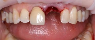
There are several ways to treat a dental fistula: surgery (the most common) and methods for treating a dental fistula without tooth extraction. When surgically removing a fistula on the gum, the inflammatory cavity is opened, cleared of pus, and then drainage is installed to ensure the outflow of purulent contents. Surgical treatment is clearly indicated for a large area of damage, when a fistula occurs over a tooth covered with a crown, or when there is damage to the periosteum.
In the medical treatment of gum fistula, conservative methods are used.
It could be:
- Endodontic therapy (teeth preparation; cleaning of canals, elimination of inflammation, filling);
- Drug therapy (used at all stages of work to eliminate pathology) involves taking antibiotics, NSAIDs, antihistamines;
- Physiotherapy. It is never prescribed separately, only in combination. Increases the effectiveness of any treatment methods.
Fistula due to root perforation
Working with canals requires the dentist to be careful, attentive, and precise, controlled movements. The slightest carelessness can lead to the formation of a non-physiological hole (perforation), which will subsequently cause inflammation and, as a consequence, the appearance of a fistula.
A common mistake that leads to perforation and inflammation is the fixation of the pin when it is installed not along the canal, but its removal into the bone tissue.
With perforation, treatment of the fistula is the most problematic of all cases of its formation. Moreover, the main difficulty is not in medical procedures, but in the behavior of dentists who do not inform patients about the complication that has arisen, so as not to question the professionalism of themselves and their colleagues. As a result, a person who goes to the hospital for dental treatment returns again due to pain and inflammation: often the process becomes so advanced that it is no longer possible to do anything other than remove the tooth.
But still, perforation is not a death sentence, and the tooth can be saved. The sooner the patient applies, the greater the chance that the treatment will be completed successfully. True, this requires expensive materials (Pro Root MTA is often used), with the help of which the perforation area is sealed. Sometimes filling is performed directly through the canals, sometimes a surgical incision is required to gain visual access to the damaged area.