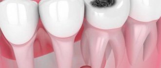
Every day we subject our teeth to serious tests: physical activity when eating tough and hard foods, eating at different temperatures, excess sweets, as well as tea, coffee and other coloring drinks. No wonder that dental caries considered the most popular dental disease. But in such conditions, not only the teeth suffer, but also the mucous membrane.
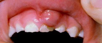
Not all adults are familiar with such a phenomenon as an ulcer on the gum , because it occurs most often in children. However, no one is immune from the disease, so everyone should take care of oral hygiene and visit the dentist in a timely manner!
Causes of development of ulcers on the gums
- Poor oral hygiene
- Genetic predisposition
- Lack of useful minerals and vitamins in the body
- Weakened immunity
- Injuries to the mucosa (for example, due to improperly installed prosthesis or braces)
- Caries and dental diseases
- Allergy
- Breathing through the mouth (the mucous membrane dries out, and as a result, wounds appear on the gums)
- Poor diet (too much sweet and sour foods)
- Diseases of the thyroid gland and liver
- Hormonal imbalance
- Dehydration
- Bad habits (cigarettes, alcohol)
- Diseases of the gastrointestinal tract
- Diseases: tuberculosis, cancer, syphilis, HIV
- Eating dirty foods (vegetables, fruits, berries)
Gingivitis and periodontitis
Cause
Gingivitis is an inflammation of the gums, and periodontitis is a more severe form of the disease when not only the gums, but also the jaw bone are involved in the inflammatory process. Both diseases are often chronic and do not cause the patient much discomfort, but during periods of exacerbation more severe symptoms appear and the patient consults a doctor.
There are many causes of gum inflammation:
- hard and soft dental deposits;
- sharp edges of crowns and fillings, which constantly injure the mucous membrane;
- improperly restored contact points between teeth (due to which food accumulates in the gap, puts pressure on the gums and is an excellent place for microorganisms to settle);
- malocclusion (crowding of teeth, incorrect position in the arch);
- general diseases (diabetes, vascular problems, infectious diseases and others) can cause gum inflammation;
- changes in hormonal levels (during pregnancy, for example).
These are not all possible causes of gum disease, but most patient visits have one (or several) of these factors predisposing to inflammation in the gums.
Constant trauma (from food, the edge of a crown, tartar), microbial toxins, and impaired blood flow in the gums lead to the development of an inflammatory process. Usually, pus as such is not visible, but during periods of exacerbation, noticeable suppuration often occurs from periodontal pockets or its accumulation in the thickness of the mucosa (this formation is called a periodontal abscess).
Symptoms of gingivitis (periodontitis) in the acute stage
Patients with this diagnosis complain of:
- pain in the gums near a group of teeth;
- redness of the gums, swelling, pain when touched;
- it hurts to brush your teeth and eat;
- pus (sometimes with bloody veins) is released from the periodontal pocket or from the fistula tract (a fistula can form if the pockets are very deep);
- with gingivitis, the teeth usually do not suffer, but with periodontitis, tooth mobility, exposure of roots, pain from cold and sweet foods may be observed (since the root is more sensitive to irritants than the crown part of the tooth);
- Sometimes there is a deterioration in general health: fever, weakness, headache.
Exacerbations most often occur against the background of a general decrease in immunity (with acute respiratory infections or other diseases). In addition, periodontitis is characterized by seasonality (in spring and autumn, exacerbations occur more often).
Diagnostics
How to distinguish periodontitis from periodontitis? Outwardly, both cases may look the same: swollen gums, redness, a fistulous tract from which pus is released. In both cases, it hurts to bite on the teeth. In addition, one disease does not exclude another (there are cases when a tooth is surrounded by inflamed bone on all sides and it is almost impossible to say with certainty what is primary: periodontitis or periodontitis).
An x-ray is required to make a correct diagnosis and choose treatment tactics. In the image with gingivitis, we will not see any changes at all, but with periodontitis, areas where there is no bone tissue will be visible, while the teeth will be either healthy or well treated.
Treatment
It must be said right away that gingivitis is a reversible process, and periodontitis cannot be completely cured in most cases (it can be stopped, put into remission, but growing back the height of the bone is very problematic, and in many cases it is completely impossible).
If in the case of periodontitis we were dealing with microbes inside the root canal, then here we also have a microbial infection, only it is localized in the gums and bone. Treatment should be aimed at combating the cause, so the following measures can be taken:
- mandatory professional hygiene – removal of soft and hard dental plaque, polishing the surface of teeth, removal of granulations from periodontal pockets;
- correction of fillings and orthopedic structures, creation of normal contact points between teeth;
- if there is mobility of the teeth, they need to be splinted (limit movement);
- active anti-inflammatory therapy (rinses, dressings, ointments with antimicrobial action, in severe cases - antibiotics);
- general therapy (at the doctor’s discretion and depending on the severity of the process, antihistamines, painkillers, immunostimulants, vitamins, etc. can be added).
Usually this is enough to stop suppuration and relieve the acute process. To prevent exacerbations in the future, it is imperative to maintain hygiene (remove tartar at least once every six months), restore the dentition (if some teeth are missing), correct the bite (if there are problems with this) and maintain general health (if the cause of periodontitis is some common diseases).
First aid
An ulcer on the gum is not the most pleasant disease that requires medical attention. But, there are situations when it is impossible to get an appointment with a dentist (for example, you are on a hike with friends, and the nearest clinic is tens of kilometers away, you went to the country, and the next train to the city is only tomorrow evening). How to relieve discomfort and pain now? In such situations, you should rinse your mouth with a soda solution or tincture of oak bark, chamomile, or calendula. Some people advise applying grated raw potato gruel to the wound. But remember, you shouldn’t get carried away with self-medication: traditional medicine only helps to temporarily relieve symptoms; only a professional doctor can cure the disease!
Possible diseases
If you find an unknown formation in the oral cavity, you should immediately contact a specialist. The formation of an ulcer on the gum may indicate that the patient has:
- stomatitis of various forms (candidal, aphthous or herpetic);
- periodontal diseases;
- infectious diseases;
- avitaminosis;
- systematic damage to a certain area of the soft tissues of the oral cavity.
Depending on the cause of the ulcer, the optimal treatment for the disease is selected.
Treatment
To get rid of an ulcer on the mucous membrane, it is necessary to find out the cause of its appearance. If it’s all about diseases of the body, you will need the help of more than one specialist. If the cause is diseases of the oral cavity, the dentist will treat the teeth under anesthesia , remove plaque and stones, and in order for the wound to heal faster, prescribe special antiseptic solutions for mouth rinsing, gels or ointments with a healing effect. If the pain is severe, your doctor may prescribe a pain reliever.
During the treatment period and after it, the toothbrush must be replaced! Diet will also play an important role: try not to eat acidic foods and be sure to wash food before eating. Pay special attention to fresh vegetables, herbs and fruits. And to strengthen your immune system, take a complex of vitamins and minerals. A healthy body means a healthy and beautiful smile!
How can a fistula on the gum be cured?
Of course, we treat it, we deal with it, we work for it. In order to cure a fistula on the gum, first of all, we should eliminate the reason due to which the fistula occurred. We should not
fight the pimple itself, smear it with various ointments and lotions, but must eliminate the cause of its occurrence.
We must
eliminate the chronic inflammation itself that led to this fistula.
The first treatment option for a fistula is endodontic
For such treatment, we provide the patient with high-quality endodontic treatment under a microscope when:
- the tooth canal itself is visible,
- its branches are visible
- and you can see if there are cracks in the tooth canal.
Our doctors perform minimally invasive treatment procedures under a microscope at a high level. Treatment of processes that led to a fistula, to a fistulous tract on the gum, to complications of various periodontitis. In the video - endodontist of the Scientific Research Center, winner of many international competitions in endodontics and dental restoration Melikov Azer:
In the clinics of the German Implantology Center, special materials are used that seal the apical part very well and for a long period, which are biocompatible with the tissues surrounding the tooth and which give a very good result.
That is, we have a therapeutic method of treatment - this time
. When highly qualified endodontists, using microscopes, special equipment and special materials, carry out canal therapy and eliminate the cause of the fistula.
The second treatment option for fistula is mixed
Second method
- This is a mixed treatment, when treatment is carried out jointly by a therapist and a surgeon. If the lesion is already quite strong, lysis of a certain part of the root apex has occurred, when the process has already been going on for quite a long time, then the therapist cleans the canals:
seals the apical part of the root with a special material, and the surgeon opens, makes surgical access and polishes the apical part of the tooth root.
We do not perform dental resections when the tooth is cut down in half. You often think, I often have patients come to me who have had a resection and you think the person has had ⅔ of the root cut off and this tooth is no longer tenable, it can barely hold on, and why did it have to be done? We do it minimally invasively. We polish the apical part of the root with special ultrasonic tips after the therapist has carried out the treatment, and we get very good long-term results.
The third treatment option is surgery.
There is also a surgical method - this is the third method
treatment when the inflammation is already chronic according to the principle “No tooth - no caries, no periodontitis, no nothing.”
That is, the tooth is removed
, the source of inflammation is removed:
and an implant is placed
:
Subsequent rehabilitation of the patient takes place on the implant.
The fourth treatment option is autotransplantation.
Autotransplantation is a type of third, surgical treatment option. But the patient’s own donor tooth acts as an implant. As a rule, wisdom teeth, which are not involved in the process of chewing food and are actually the body’s reserve for such cases, are excellent for this role. Wisdom teeth transplantation is suitable for posterior teeth and premolars (fourth, fifth, sixth and seventh teeth).
Advantage
This method of treatment is that the patient’s own tooth is transplanted, the patient’s own tissue, which is not foreign to the body, even in conditions of the inflammatory process with a fistula, significantly reduces the risk of tooth-implant rejection.
The specialists of the German Implantology Center have accumulated many years of clinical experience in such operations. You can see how the autotransplantation operation takes place in the following video. There, at the end of the film, the patient shares his impressions of the dental transplant (this is a review 1 year after
the transplant operation):
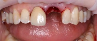
When white plaque is a harbinger of disease
It also happens that a whitish coating indicates the development of alveolitis after tooth extraction. This means that the tooth socket has become infected. In this condition, the patient is concerned about:
- severe pain in the area of the removed unit;
- increased body temperature;
- weakness, decreased performance;
- enlargement of the submandibular lymph nodes;
- headache;
- unpleasant putrid odor from the mouth and the same taste in the mouth.
If you experience these symptoms, you should make an appointment at the dental clinic as soon as possible.
Recommendations after tooth extraction
There are recommendations that, if followed, significantly speed up the gum healing process. Not only the speed of wound healing depends on them, but also the absence of possible complications. The doctor’s main recommendations in the postoperative period may be the following:
- you need to avoid foods that are too hot, spicy, or irritating to gum tissue;
- in the first days you need to carefully avoid damaging the clot; It is necessary to be extremely careful in maintaining oral hygiene;
- about 3 hours after the tooth extraction procedure you need to refrain from eating;
- in the next three days you need to eat only soft food, without consuming sweets, alcohol, or hot drinks;
- on the first day after the procedure, it is recommended to sleep on a high pillow;
- During the week, it is recommended to avoid visiting the sauna, solarium, sunbathing on the beach, and reduce physical activity;
- on the first day, it is forbidden to brush your teeth to avoid damaging the blood clot;
- Do not try to pick the clot with your finger, toothpick or tongue;
- a cold compress should be applied to the surgical area for 20 minutes, every 2 hours;
- It is recommended to use oral baths and then rinse with antiseptic agents.
The postoperative period requires careful attention to the patient's health status. A complication resulting from neglect of the rules will require much more time, money and effort to heal the wound.
Content:
- When white plaque is the norm
- When white plaque is a harbinger of disease
- Why do complications occur after tooth extraction?
- How to recognize the problem
- Diagnostic measures
- Treatment of alveolitis
After tooth extraction, it is important to monitor the condition of the mucous membrane.
Very often a white coating appears in the extraction area. It frightens patients because it seems something strange to them, and to some it resembles an accumulation of purulent masses. It should be noted that white plaque in the socket after tooth extraction can be either normal or a complication. In the first case we are talking about natural regeneration, in the second - about alveolitis.
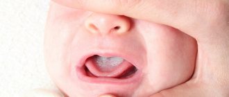
Problems in childhood
Children can also develop ulcers in the mouth. The reasons for their appearance may be:
- oral injury;
- infectious diseases;
- burns of the mucous membrane.
A baby can damage the mucous membrane, for example, by putting a hard toy in his mouth. A fall or blow can also cause damage to the mucous membrane, which can result in stomatitis. Also, the development of stomatitis can be triggered by a viral infection or taking any antibacterial drugs (candidal stomatitis appears).
Gum inflammation: patient mistakes
Working as a periodontist, I observed thousands of patients with gum inflammation, and in most cases, patients came with severe forms of the disease and complications that could have been avoided (if treated in a timely manner). Mostly patients make the following mistakes:
1) When periodontitis or gingivitis develops, only antiseptic rinses and applications of gels to the gums are used, but they ignore the need to remove dental plaque using ultrasound at a dentist’s appointment. This situation leads to chronic inflammation, and if the patient only had gingivitis, then thanks to self-medication it will turn into periodontitis. If the patient already has periodontitis, this only threatens to worsen its shape, cause tooth mobility and pose a threat of the need to remove them.
2) Patients often think that antiseptics and antibiotics are enough to control gum inflammation, and therefore use them in regular courses (ignoring the need to remove the cause of inflammation). Remember that even long-term use of antiseptics and antibiotics does not allow complete “disinfection” of dental plaque, periodontal pockets or root canals in the tooth. All this acts only superficially and has a temporary effect.
3) With slight inflammation of the gums in the area of 1-2 teeth, patients often do not go to the dentist at all, but constantly rinse their mouths with antiseptics and smear various gels on the gums. Please note that if inflammation of the gum is caused by the presence of “supercontacts” or injury to it by the overhanging edges of fillings/crowns (we are talking about a localized form of periodontitis), then anti-inflammatory therapy will not stop the process of bone destruction around the tooth at all - until the factor traumatic to the gums is eliminated. Those. You still have to go to the dentist.
4) If you experience periodic swelling of the gums in the area of 1-2 teeth due to periodontitis, then anti-inflammatory drug therapy (including antibiotic therapy) is completely useless. In this case, inflammation is caused by an infection in the root canals, and until the root canals of this tooth are properly filled, the inflammation will not go away.
Important: therefore, it is very important to understand that treatment at home can be effective - only in terms of conducting a course of anti-inflammatory therapy for gingivitis and periodontitis (after removing plaque from the dentist). Home treatment can also be used to strengthen the gums - during periods between the main courses of anti-inflammatory therapy, but in this case, products that do not contain antiseptics and antibiotics should be used.
How to distinguish stomatitis from other diseases
Since stomatitis can appear for a variety of reasons, and the forms of its manifestation can be very different, it is highly not recommended to self-medicate, since in this case you can only aggravate the course of the disease and cause the appearance of new symptoms. Due to such diversity, methods of treatment and prevention of this disease can also be very different.
Manifestations of stomatitis may be similar to a number of diseases, from each of which a doctor, when examining a patient, will definitely be able to distinguish this ailment:
- Sore throat or stomatitis: you can determine a sore throat, which may be similar to stomatitis, by damage not only to the mucous membranes of the mouth, but also to the pharynx. It is very painful for the patient to swallow.
- Herpes or stomatitis: In the case of herpes, the rashes first look like blisters and appear later.
- Cancer or stomatitis: the first disease is distinguished by the fact that ulcers appear, which grow together over time. As a result, one ulcer forms and begins to grow.
- Thrush or stomatitis: when examining the mouth, if there is a disease such as candidiasis, a yellow or white coating is detected, redness, the wounds are round in shape, and the ulcers are small.
- Syphilis or stomatitis: the first disease is distinguished by the nature of the ulcers. They appear in the form of eosis and do not hurt. Sometimes there is a depression in the center of the wound.
The doctor selects the treatment for stomatitis based on the cause of the disease, the complexity of its course, the form of manifestation, the individual characteristics of the body, and so on - in general, you will have to take into account a lot of things in order to choose the correct and most effective treatment. A specialist usually easily determines that it is stomatitis and not another disease.
Clinical trials
According to the results of clinical trials of the ASEPTA rinse after 3 weeks of use of the rinse, gum bleeding is reduced by 28.3%, inflammation is reduced by 32.3% and the hygienic condition of the oral cavity is improved by 33.5%.
Sources:
- Clinical experience in using the Asepta series of products Elena Ivanovna Fuchs, assistant at the department of therapeutic and pediatric dentistry. State budgetary educational institution of higher professional education Ryazan State Medical University named after Academician I.P. Pavlova of the Ministry of Health and Social Development of the Russian Federation (GBOU VPO RyazSMU Ministry of Health and Social Development of Russia)
- The use of adhesive balm "Asepta®" in the treatment of inflammatory periodontal diseases L.Yu. OREKHOVA, Dr. med. Sciences, Professor, Head of Department V.V. Thermal Power Plant, Dr. med. Sciences, Professor, Head of Department S.B. ULITOVSKY, Dr. med. Sciences, Professor A.A. LEONTIEV, dentist A.A. DOMORAD, O.M. YAKOVLEV SPbSMU named after. acad. I.P. Pavlova, St. Petersburg - *Department of Therapeutic Dentistry, Department of Microbiology
- The use of new anti-inflammatory drugs in the complex of therapeutic and preventive measures for periodontal diseases (E.D. Kuchumova, A.A. Leontyev, O.V. Kalinina, L.Yu. Orekhova, S.B. Ulitovsky) E.D. Kuchumova, Ph.D., Associate Professor, A.A. Leontyev, dentist, O.V. Kalinina, dentist, L.Yu. Orekhova, Doctor of Medical Sciences, Professor, Head of Department, S.B. Ulitovsky, Doctor of Medical Sciences, Prof. Department of Therapeutic Dentistry of St. Petersburg State Medical University named after. acad. I.P. Pavlova
- Evaluation of the effectiveness of treatment of chronic generalized periodontitis of mild and moderate severity using Asepta antibacterial agents (S.I. Gazhva, A.I. Voronina) S.I. Gazhva, Doctor of Medical Sciences, Prof., Head of Department A.I. Voronina, PhD, dentist. Department of Dentistry FKPV GOU VPO "Nizhny Novgorod State Medical Academy"
Gum condition
Pain in the gums may occur after the anesthetic drug wears off, that is, after 3-4 hours. After the procedure, the patient is often worried about the return of painful sensations and the release of ichor (for 4–6 hours. After the operation, the wound looks quite scary, especially if a wisdom tooth was removed.
In the absence of pathology, the healing process occurs in several stages.
How to recognize the problem
How to understand that white plaque signals a problem?
If the gums heal well, then within 2-3 days after surgery the pain becomes very weak and barely noticeable. The wound closes with a dense clot and practically does not bother the person. With the development of alveolitis on days 3-5, the pain, on the contrary, intensifies. It's pulsating. It can become unbearable. Sometimes it radiates to the entire half of the face. Body temperature rises, lymph nodes enlarge, appetite decreases, salivation becomes stronger. The face may also become swollen.
In such a situation, staying at home is dangerous - inflammation can spread to the deep tissues of the jaw. You need to immediately go to the dental clinic.
Possible reasons
- Mycoses. Most often, red dots in the mouth are symptoms of oral candidiasis. Fungi of the genus Candida are considered opportunistic. They live in the intestines and do not cause harm to the body. However, when they enter the oral cavity, they quickly multiply and provoke inflammation of the mucous membrane. Spots of different shades of red can form on the gums, palate, cheeks, tongue, and tonsils.
- Bacterial infections. One of the main pathogens is streptococci. They change the structure of the mucous membrane and lead to inflammation of the soft tissues. They provoke the development of pharyngitis and tonsillitis.
- Viral diseases. Acute respiratory viral infections can also be accompanied by the formation of red spots on the oral mucosa. Pathogens enter the oral cavity through the nose. The gums and palate become inflamed, local hyperemia appears. If the spots become covered with a white coating, we can talk about a fungal or bacterial infection.
- Neoplasms. Suspicious spots on the gums may turn out to be benign tumors. The most common include papillomas and granulomas. Also, the risk of malignant neoplasms cannot be excluded.
Gum healing process
The speed of gum healing depends on several factors:
- individual characteristics of the patient’s immunity;
- the success of the operation;
- location of the extracted tooth.
If the tooth is in a hard-to-reach place, has crooked roots, or its crown has been significantly damaged, then the removal procedure becomes more complicated. During manipulation, the tooth may begin to crumble, leaving fragments in the gum. In this case, it is necessary to cut the gum tissue, detach it from the bone, remove the tooth in parts, and use a drill. These traumatic procedures prolong the period of gum healing after surgery.
Slight swelling of the gums is considered normal. The temperature may rise slightly (due to an immune reaction). The swelling usually subsides within three days.
Noticeable swelling of the gum is also observed after cutting it. This swelling goes away in about one week.