Perforation of the maxillary sinus during the treatment of upper teeth is damage to its shell when trying to treat root canals without prior examination. The penetration of infection provokes inflammation with symptoms of sinusitis, which cannot be treated with medication.
For more than 20 years, Doctor Levin has specialized in eliminating complications after unsuccessful dental manipulations in the area of the maxillary sinuses. Surgical treatment is performed by maxillofacial surgeons with ENT training.
Causes of sinus perforation during dental treatment
The maxillary sinuses or maxillary sinuses are located inside the upper jaw. They are separated from the oral cavity by a septum and the alveolar process, which contains the roots of the teeth. Treatment of the canals of such teeth is complicated by the risk of sinus perforation for a number of reasons:
- Anatomically thin septum Sometimes the height is only 1 mm. It happens that the teeth penetrate the cavity with their roots and only the sinus mucosa separates.
- Treatment of teeth with inflammation on the roots If there is inflammation on the roots of the teeth (periodontitis, cyst), the surrounding bone melts and becomes thinner.
- Excessive efforts by the doctor If the dentist does not calculate the efforts when passing the canals, the canal may rupture, damage the root, or perforate the bottom of the sinus.
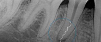
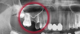
Without a preliminary diagnosis, the endodontist, having no idea about the size of the sinus septum and the location of the roots, may not calculate the effort and damage the tooth and the thin bone layer that lies between the maxillary sinus and the root. As a result, infection penetrates into the sinus cavity, and fragments of instruments and filling material may fail. In our practice, there are cases when a foreign body in the sinus attracts secondary infections with the development of neoplasms - fungal colonies, cysts, polyps.
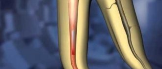
A fragment of an endodontic instrument in a tooth canal
Anatomy and purpose of the maxillary sinuses
The maxillary sinus is a cavity in the upper jaw that is filled with air. The sinus has bony walls that are in contact with the orbit, oral and nasal cavities. The nasal sinuses regulate intracranial pressure, adapting it to atmospheric pressure, clear small particles and warm the inhaled air, and create an individual sound for the voice.
The sinuses communicate with the nasal cavity through small openings. If the holes are closed for some reason, the sinuses are deprived of ventilation and the possibility of self-cleaning. Then microbes accumulate inside the nose and provoke inflammation.
How to prevent perforation
If patients with anatomical features of the location of the roots in the sinus are treated according to the standard protocol, the threat of rupture of the membrane is very high
The doctor must have the necessary information about the anatomical and topographical features of the patient and be prepared for an emergency situation.
- To prevent perforation of the sinus membrane, computed tomography is required. After studying the CT image, the doctor selects the optimal treatment tactics. But not every equipment allows you to identify all the nuances. In our Center, diagnostics are carried out using a modern 3D tomograph Sirona Galileos with ENT mode settings. Provides high quality images, allows you to evaluate the structural features of the upper jaw and sinus, calculate the height of the septum, find out the location of the roots, their anatomy.
- When working with the canals of teeth located in the sinus, all manipulations must be extremely careful. The dental canals are very thin, sometimes tortuous, the doctor must control every action. Endodontic dental treatment in our Center is carried out using a Seiler dental microscope. Multiple magnification expands the view, allows you to examine the entire length of the dental canal in great detail, and prevent the filling material from leaving the root.
- The experience of a specialist is of great importance so that he can give an adequate assessment from a CT image, choose tactics, and calculate efforts. Our Center employs highly qualified endodontists, with excellent clinical thinking and honed manual skills. Qualifications and skills allow us to prevent complications and act promptly in the event of an emergency situation.
We have minimized the likelihood of complications after dental treatment
. Advanced CT diagnostics in ENT mode and work using a dental microscope allow us to select competent tactics to prevent perforation of the bottom of the maxillary sinus.
Levin Dmitry Valerievich Chief physician and founder of the Doctor Levin center
After endodontic treatment, control CT diagnostics is mandatory! This preventive measure allows you to timely identify perforation (even if the doctor did not notice it during treatment) and take measures to eliminate the consequences.
If a piece of instrument or filling material gets into the sinus, removal of the foreign body must be prompt and urgent ! Otherwise, there is a high risk of infection, development of inflammatory processes and neoplasms.
Removal of sinus cyst without surgery
If a sinus cyst is detected, treatment without surgery is carried out using decongestant sprays, antibiotics, painkillers, mucolytics, steroids and antihistamines. They treat concomitant diseases - allergies, sinusitis, inflammatory processes of the gums, teeth, nasal mucosa. In combination with these drugs, various means for rinsing the nasal cavity, regenerating and restorative sprays are used. Treatment is prescribed by the doctor based on the results of the patient’s examination.
Signs if perforation was left without the attention of a doctor
Symptoms of inflammation after unsuccessful treatment occur in different ways - after a few days, weeks or even years. Depends on the sterility of the foreign body in the sinus, its ability to cause inflammatory processes, and the characteristics of the patient’s immune system. The following manifestations are possible:
- feeling of heaviness;
- pain when chewing;
- purulent and serous discharge from one nostril;
- elevated temperature;
- impaired sense of smell;
- pain when lightly tapped under the eye and in the nose area.
These are typical signs of odontogenic sinusitis caused by poor quality dental treatment. The key difference from rhinogenic sinusitis (when sinus infection occurs from the side of the nasal passages as a result of ARVI or influenza) is that signs of the inflammatory process are detected only on one side, where treatment was carried out.
Reasons for the development of sinusitis
Since the pathology is inflammatory in nature, its main causative agents are microorganisms and viruses that seek fertile ground among patients with weak immunity. The main causative agents of the disease include streptococci, staphylococci, chlamydia, fungi, viruses, mycoplasma, and hemophilus influenzae.
Often sinusitis develops as a secondary pathology after:
- acute and chronic forms of rhinitis;
- acute respiratory viral infection;
- chronic tonsillitis and pharyngitis;
- against the background of caries of the upper teeth;
- dental surgery;
- curvature of the nasal septum;
- with congenital narrow nasal passages.
Like colds, sinusitis most often appears in the autumn-winter season, when a natural decrease in immunity occurs.
What are the dangers of sinus injury?
Since the complication does not always manifest itself immediately, symptoms of sinusitis that arise after a few years force the patient to consult a regular ENT doctor. Treatment, as a rule, does not bring results - in most cases the cause is not found. You start going from one doctor to another, and precious time is lost. The longer a foreign body is in the sinus, the higher the risk of tumor formation. If sinus perforation is left unattended, it can lead to:
- chronic sinusitis and sinusitis
- inflammation of the roots of adjacent teeth
- osteomyelitis of the jaw
- encephalitis and meningitis
Treatment: additional and mandatory
It is mandatory to use vasoconstrictor drugs in the treatment of sinusitis, not only in drops, but also in gels, aerosols or tablets. Oxymetazoline or xylometazoline preparations quickly eliminate swelling and restore the patency of the anastomosis. It is almost impossible to dose nasal drops correctly, and aerosol sprays have the advantage of being able to administer a limited single dose.
It is recommended to use vasoconstrictor drugs for the nose for no more than a week due to the possibility of developing a “ricochet”, when instead of reducing nasal swelling they lead to its increase. The tablets do not initiate a “ricochet”, but have a general effect on the body in the form of increased blood pressure and heart rate. It can be argued that sinusitis will not be cured in a week, therefore, it will not be possible to do without vasoconstrictor drops. True, but a consultation with an ENT doctor will help solve this problem.
Why you should entrust your treatment to the ENT Department of Dentistry
ENT dentistry is a comprehensive approach to the treatment of complications in the maxillary sinuses after dental treatment
The symbiosis of two areas - dentistry and otolaryngology - makes it possible to identify the cause, assess the situation, and select competent treatment tactics.
The ENT department of the Doctor Levin Center has been providing assistance for many years when problems arise after treatment and removal of teeth located on the border with the maxillary sinus. Surgical treatment is carried out by candidates of medical sciences, maxillofacial surgeons with otolaryngological training .
Patients come to the Center after a painful search for a solution to the problem. Repeated treatment by an ENT doctor in the hospital does not bring results. But a thinking patient should understand that if there is concern on the side of the sinuses where endodontic treatment once took place, you need to contact an oral and maxillofacial surgeon with ENT training. Only in this case can a comprehensive assessment of the situation be made and an adequate treatment plan drawn up.
Organization of treatment
In Germany, the latest technologies are used, the best doctors in the world work, and the most modern operations are performed, including fenestration of the paranasal sinuses. Many patients prefer to go to this country for treatment, as this allows them to achieve better results, reduce the risk of relapse of chronic sinusitis to a minimum, and also minimize the likelihood of complications.
If you want to go to Germany for treatment, ]Booking Health[/anchor] will help you with this. We are leaders in the field of medical tourism. When you contact us, you get many advantages:
- We will choose for you exactly the clinic that specializes in this type of surgery and has the best statistics on relapse-free recovery
- We will help you establish communication with the clinic administration and your attending physician
- The cost of treatment is reduced by 50%, because we will agree on a favorable price for the operation, all treatment and rehabilitation measures, without surcharges and coefficients for foreign patients
- When the disease is diagnosed, you will not need to re-take tests and undergo instrumental examinations that were carried out earlier
- You will be able to undergo treatment in Germany in the near future - we will reduce the waiting time for medical care
- We will help you purchase and send medications to your country
- The treatment program is under our complete control
- If necessary, we will organize additional diagnostic and treatment procedures
- We will ensure communication with the clinic doctors after completion of the therapeutic program to monitor the results of treatment
- We organize high-level service: transfer from the airport to the clinic, booking airline tickets and hotels
All financial relationships are transparent. You will be able to find out at any time what procedures your funds are being spent on. Anything not spent will be returned to your bank account.
Diagnosis of perforation
Having an accurate understanding of the situation in the sinus, the maxillofacial surgeon chooses the correct tactics and method of eliminating the problem
As part of diagnostic activities, our Center carries out:
- X-ray allows you to assess the condition of the causative tooth and the quality of canal treatment
- Computed tomography in ENT mode allows you to get a complete picture of the condition of the maxillary sinuses
The modern Sirona Gallileos CT scanner guarantees high quality images , in which you can determine the location and size of the perforation, identify the presence and localization of foreign bodies, cysts and polyps (if they form), and assess the size of the inflammatory process.
CT and x-ray of the maxillary sinuses
X-ray examination is an effective method for diagnosing the human body, including the condition of the ENT organs. If a person has a stuffy nose, there is discharge from it, or the timbre of his voice has changed, then this is an indication for an examination. X-rays are considered one of the most effective ways to detect sinusitis. With its help, it is possible to diagnose various pathological changes even at an early stage of their development, which is extremely important when prescribing appropriate treatment.
An examination is prescribed if the following symptoms are present:
- Nasal discharge (sometimes with pus).
- Problems with nasal breathing. As a rule, only one nostril is blocked.
- Frequent headaches.
- General fatigue, trouble sleeping and loss of previous activity.
- Nosebleeds.
- Soreness and swelling of the forehead.
If these symptoms are present, the doctor may suspect the presence of an inflammatory process, but it is necessary to determine the exact cause of this inflammatory process, because similar signs exist with polyps and a deviated septum. Therefore, an x-ray of the maxillary sinuses is a mandatory procedure, which allows us to determine the degree of development of the pathology, the presence of neoplasms and other aspects.
Content
- Why do you need an x-ray of the nose for sinusitis?
- How can you see sinusitis on an x-ray?
- How is an x-ray of the maxillary sinuses performed?
- X-ray in childhood
- How to determine sinusitis in an adult patient from an image?
- Computed tomography for sinusitis
- Is it possible to determine sinusitis without a picture?
Why do you need an x-ray of the nose for sinusitis?
Vivid symptoms will help the doctor make an appropriate diagnosis, but without an accurate diagnosis, further treatment will be impossible. It is important to first examine the condition of the sinuses, and then determine the medications and physiological procedures that can get rid of the disease.
When diagnosing, it is extremely important to know what sinusitis looks like on an x-ray. With this examination method, soft tissues are not visible, but bone structures are clearly visualized. If in the picture the shade of the paranasal sinuses and eye sockets is identical, then there is no inflammation. If there is purulent content, then visually it looks like large darkened areas.
An x-ray of the sinuses with sinusitis allows you to determine a number of pathological changes:
- Localization of lesions. They appear as dark spots.
- Presence of cystic formations. They have fairly clearly defined boundaries.
- The inflammatory process and the degree of its development. In the picture they are visible as white spots. Accordingly, the brighter they are, the stronger the inflammatory process.
- Various changes. You can see the level of filling of the nasal sinuses with pus, thickening of the mucous membrane and other pathological changes.
How can you see sinusitis on an x-ray?
Below you see a picture of sinusitis before and after treatment. The radiograph is interpreted by the attending physician or radiologist.
In the absence of pathological changes and abnormalities, the image will show the following information:
- The nose is in the form of a triangle of a light shade, in the middle of which there is a septum.
- On the side of the nasal cavity, the maxillary sinuses are located in the form of triangular clearings with not blurred boundaries.
- Symmetrical arrangement of the nasal passages on both sides of the nasal cavity.
- The frontal sinuses are located above the eye sockets. In the picture they look like clearings of various sizes.
The pathology develops from inflammation of the lateral sinuses. Further, it is localized in the region of the frontal lobes. This will be clearly visible in the darkened areas above the eye sockets and nose. Simultaneous inflammatory processes in the frontal and lateral sinuses indicate not only the presence of sinusitis, but also frontal sinusitis.
A specialist who takes pictures of the nose for sinusitis always determines the condition of the ethmoid bone. Infiltrative fluid often accumulates in the maxillary sinuses:
- mucous;
- catarrhal;
- purulent.
It is quite easy to notice - the liquid appears as a clearly formed white area. This is a clear sign of pathology, which is one of the grounds for making a final diagnosis.
How is an x-ray of the maxillary sinuses performed?
X-ray of the nasal sinuses for sinusitis is a completely standard diagnostic procedure. Before the examination, you must remove all metal objects and removable dentures. If implants, crowns or pins are installed, the radiologist should be notified about this.
To obtain an accurate picture of the disease, a photograph of the nasal sinuses with sinusitis is taken in several projections:
- Chin. Based on this image, the specialist analyzes the actual condition of the maxillary sinuses.
- Nasomental. This projection clearly shows the presence of an inflammatory process in the mucosa.
- Nasofrontal. In this picture you can see the state of the lattice labyrinth.
The examination takes 10-20 minutes, after which the doctor can clearly see the presence or absence of pathology.
X-ray in childhood
Experts agree that X-ray examinations are advisable from the age of 14. Here we are not talking about potential harm to the child’s body, because a single dose of radiation during an examination is absolutely safe for the child. The fact is that at an early age the paranasal sinuses are not yet fully formed, so the examination result cannot be 100% accurate.
In addition, x-rays require maintaining complete stillness throughout the entire examination, which, for obvious reasons, is very difficult to achieve in childhood.
How to determine sinusitis in an adult patient from an image?
Pictures of sinusitis in adult patients are quite characteristic, especially when compared with radiographs of people who do not have this pathology. X-ray will show the following changes:
- The eye sockets and sinuses will be of different shades.
- The presence of infiltrative fluid (a clear white spot against a darkened background).
- Thickened walls and blurred boundaries of the sinuses.
- Cystic formations of varying sizes with clearly defined boundaries.
The only thing that cannot be seen in a photo of the nose with sinusitis is the type of fluid that has accumulated in the sinuses. The study only shows its presence in the form of a white spot, but cannot show what kind of infiltrate (purulent, mucous or catarrhal fluid).
Computed tomography for sinusitis
In some cases, in addition to X-rays, a CT scan of the maxillary sinuses is used. This is a more informative diagnostic method, which has its advantages:
- This is the most reliable way to examine bone structures.
- The result of the study is a three-dimensional image that allows you to view the examined area from different angles and in all planes.
- The procedure takes the least time (when compared with X-rays and MRIs).
- The picture turns out to be as detailed as possible.
- The technique allows you to accurately determine the location and amount of liquid.
When answering the question of what is better to do for sinusitis - CT or MRI, you need to definitely make a choice in favor of computed tomography. This examination can be carried out even in the presence of crowns and implants, which is not possible with magnetic resonance imaging. In addition, MRI has the greatest diagnostic value when examining soft rather than bone tissue.
Is it possible to determine sinusitis without a picture?
If there are significant symptoms, the doctor collects an oral history and conducts an examination. The forehead area and around the nose are palpated for pain. The nasal passages are also examined, the purpose of which is to assess the condition of the mucous membrane.
Based on the manipulations performed, an experienced doctor can diagnose sinusitis without x-rays. But a visual examination does not give a clear picture of the disease, so in order to draw up a competent treatment regimen, it is necessary to take a picture.
X-ray - have been conducting X-ray examinations since 2015. Modern equipment allows you to obtain images of high accuracy and detail. We clearly understand how important diagnosis is for further therapy, therefore we are strict in fulfilling our professional duties.
You can make an appointment with us using the contact phone number listed on the website. Contact us for more detailed advice.
Sign up for a study by phone
+7 (812) 332-52-54
How is the treatment carried out?
We respect the patient’s personal time and strive to carry out all activities comprehensively, in one day :
- Professional hygiene Preparation of the oral cavity to ensure sterility during surgery to avoid secondary sinus infection
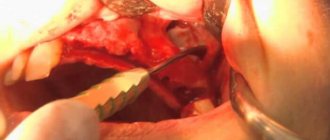
- Surgery to eliminate inflammation Performed while you sleep; the method of access to the sinus is selected depending on the location of the foreign body and the presence of tumors
- Temporary prosthetics If the causative tooth had to be removed, a temporary crown or immediate prosthesis is installed to mask the defect
Surgical operations in our Center are performed under sedation. The patient does not feel anything, fears and worries are excluded. Sedatives put you into a controlled drug-induced sleep without falling into unconsciousness - this is not general anesthesia! They do not contain toxic components, act gently, preserving reflexes. The artificial lung ventilation device is not connected, hospitalization is not required.
The surgical intervention is completed by a mandatory CT examination , which is necessary to assess the quality of the work performed.
After 10-14 days, the patient is invited to the clinic for suture removal and a control CT image. A schedule of professional inspections is drawn up.
Antibacterial and anti-inflammatory drugs in the treatment of sinusitis
For severe and moderate sinusitis, today it is recommended to carry out antibacterial therapy for up to 12–14 days, preferably by injection, and even better to perform a culture of nasal discharge for sensitivity to antibiotics. But this is not a quick process, so it is more often necessary in case of a chronic process and relapses of sinusitis. For mild cases, antibiotics are also used, but only the doctor will indicate the duration and regimen, as well as the specific drug.
Mostly antibiotics are prescribed empirically, that is, according to the suspected type of bacterial pathogen. Standard schemes for the first and subsequent lines of application have been developed. Co-trimoxazole (Bactrim, Biseptol), lincomycin and gentamicin are definitely not recommended for the treatment of Russian sinusitis - the domestic bacterial flora that leads to sinusitis is not sensitive to them.
A special local glucocorticosteroid (fenspiride) is used as an anti-inflammatory, because conventional non-steroidal anti-inflammatory drugs for the treatment of sinusitis are rather weak. The glucocorticosteroid reduces swelling, corrects immune defects and prevents bacterial colonies from growing further.
To reduce the viscosity of the secretion, mucolytics with acetylcysteine (ACC) are used, which thins the viscous gel and facilitates easier flow, clearing the cavity of both pus and bacteria.
We will call you back, leave your phone number
Message sent!
expect a call, we will contact you shortly
To remove the causative agent, it is recommended to rinse the nose with saline solutions or sea water; an ENT doctor will teach you how to do this correctly.
The first symptom of sinusitis is swelling of the nasal mucosa, which significantly complicates breathing. In case of an acute viral infection, it is difficult to combat swelling; vasoconstrictor drugs instilled into the nose, on the one hand, relieve swelling, but irritation of the mucous membrane causes obsessive sneezing, followed by a second wave of swelling. The effect of the drops is very short-lived and in the acute period is low, but without them there is almost no nasal breathing.
After 3-5 days of such suffering, the feeling of nasal congestion decreases somewhat, but there is a feeling of an increase in the area of congestion and the secretion released from the nose changes, it thickens and its color can change from transparent yellowish to cloudy greenish.
The following symptom appears - pain under the eyes inside the cheekbones, if the maxillary sinus is involved in the swelling, and in the bridge of the nose, if the frontal sinus is inflamed. The pain may radiate to the teeth and intensify when blowing your nose.
The density of the secretion increases, it is difficult to blow out, but is extremely abundant. At night, secretions flow down the back wall of the throat; in the morning, a cough with greenish sputum may appear, which is actually just “snot” that has run dry at night.
Chronic hypoxia - starvation of the brain due to constant nasal congestion leads to headaches and a feeling of “brain swelling.” The temperature may be normal or low-grade.
The sense of smell and even the taste of food may be impaired, and the ears may become blocked. The process is most often bilateral, but not symmetrical in flow. When vasoconstrictor drugs are instilled into the nose, the swelling of the nasal mucosa decreases, which facilitates the evacuation of secretions from the diseased sinus.
Chronic sinusitis also begins with ARVI, but its course is not so acute. Due to the constant presence of bacterial flora, the discharge changes earlier from mucous to purulent. Only chronic patients with sinusitis can independently determine which sinus is affected; in an acute process, clinical manifestations in different sinuses give the same type of symptoms.
All troubles in the sinuses begin with inflammation of the upper respiratory tract, usually together with an acute respiratory viral infection (ARVI), often rhinovirus. Therefore, people suffer from sinusitis during the same period as ARVI, at least in winter, early spring, early and late autumn. The mucous membrane affected by the virus swells, the secretion of the glands increases, and the outflow of the secretion produced is impossible due to swelling of the sinus-nasal junction.
In 95 out of a hundred people with ARVI, CT or MRI will show inflammatory swelling of the sinuses, but this does not mean that all of them are at risk of bacterial inflammation of the sinuses, and this will cause sinusitis. Only one or two will be unlucky; the rest will resolve well - with a full recovery. Only every year adults suffer from 3 to 6 acute respiratory viral infections, so the likelihood of getting sinusitis appears repeatedly.
No hospitalization required
Operations in our Center are performed by operating teams of experienced maxillofacial surgeons with ENT training in a sterile operating room. The treatment is as gentle and minimally traumatic as possible; a 24-hour hospital stay is not required; you will go home the same day .
The intervention lasts about an hour, under sedation. After completion, the patient quickly returns to clear consciousness without unpleasant consequences or risk of complications. After 30-40 minutes you can safely go home. For patients with concomitant cardiovascular diseases, a day hospital is provided . You can lie down for the time necessary for recovery under the supervision of our anesthesiologist-resuscitator.
Accelerated rehabilitation
After the operation, you will receive free medications and instructions with rules of behavior during the rehabilitation period. We have collected the necessary set of drugs to improve your well-being at home. Please take them as prescribed by your doctor and follow the recommendations to avoid unpleasant consequences.
For quick recovery after surgery, our Center offers its own method of rehabilitation in 1-2 days. The procedures allow us to minimize the formation of possible hematomas, swelling, and eliminate pain. The program uses:
Microcurrent therapy
A weak pulse current normalizes metabolic processes, activates ATP production, and improves tissue nutrition. Regeneration starts, healing accelerates, swelling decreases. Muscle spasm goes away.
PRP plasma therapy
Injections of purified and platelet-enriched plasma from one’s own blood trigger cellular regeneration processes using the body’s internal reserves. The manifestation of edema and hematomas is reduced.
Biostimulation of the face
Lymphatic drainage drugs D-NUCLEO and MesoSculpt C71 help reduce swelling. Active anti-inflammatory components improve blood flow, increase tissue nutrition, and improve healing.
Main sinus cyst: symptoms, treatment
A cyst of the main (sphenoid) sinus develops more often in young people and rarely in older people. The cavity of the main sinus of the nose is covered with mucous membrane. The sheath glands secrete mucus; disruptions in functioning lead to blockage of the gland ducts and the formation of a cyst of the sphenoid sinus. Inflammatory processes, injuries, and allergic reactions have a negative effect on the mucous membrane. A growing cyst of the sphenoid sinus is accompanied by a feeling of fullness in the nose, nausea and dizziness may occur, a headache that radiates to the back of the head, and visual disturbances rarely develop.
If a sinus cyst is detected, treatment is carried out after a complete examination. The doctor prescribes diagnostic tests for the patient using:
- X-rays to determine the location of the cyst, changes in the condition of the facial bones and nasal septum.
- Magnetic resonance imaging, which determines the condition of the skull bones, tissues, and blood vessels. A more informative study than radiography. Computed tomography has the same capabilities. Research is carried out as prescribed by a doctor.
- Research using an endoscope allows you to examine the nasal cavity and perform a biopsy of nasal tissue.
After the studies, conservative or surgical treatment may be prescribed. Conservative treatment is aimed at pain relief, relieving an allergic reaction, and treating the inflammatory process. If conservative treatment is ineffective or the cyst is large, the patient is referred for removal of the formation.