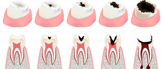
- Symptoms of sinus integrity violation
- Treatment Options
- Consequences and complications
Tooth extraction is considered by dentists to be a simple operation, but sometimes, due to the anatomical features of the patient’s jaws, complications occur that require further treatment. Such complications include perforation of the maxillary sinus (perforation). Such a nuisance can occur when removing upper molars and premolars, especially if the treatment is complicated by periodontitis, in which bone tissue is resorbed.
The upper teeth are separated from the maxillary sinus by a thin plate, approximately 0.1-0.2 mm. It happens that the roots of the upper teeth protrude into this plate and when removed, the mucous covering the root ruptures, creating a hole in the plate that separates the maxillary sinus from the bone tissue. There are two options here:
- The tooth is removed intact.
- A piece of root remains in the sinus.
Treatment is suggested depending on which option befalls the patient.
Features of the maxillary sinus
The maxillary, or maxillary, sinus is a cavity inside the skull, bounded on all sides by bone tissue. In adults it is quite voluminous, sometimes reaching 10 cm3. Its shape and location in the bones determine the unique pronunciation inherent in humans. The cavity, resonating, forms the timbre of sound.
The paranasal sinuses are lined from the inside with a thin mucous membrane and are connected to the nasal cavity by thin tubules for drainage of mucus and ventilation. If the holes become clogged, inflammation develops - sinusitis.
There are a number of known features that explain the appearance of a hole in the bottom of the maxillary sinuses:
- The size and thickness of the walls separating the maxillary sinuses from the oral cavity are individual for each person. With thinned bone (up to 1 mm thick), the sinus is easily perforated when the dentist works. This is also facilitated by the proximity of the roots of the front teeth.
- It happens that the roots of distant teeth penetrate into the sinus area and are separated from it only by a thin film of mucous tissue. As long as the tooth is healthy, everything is fine. If the doctor begins to manipulate the canals of such teeth, the mucous membrane breaks through and penetration occurs.
- Even healthy bone can atrophy and become thin and weak with persistent periodontal and periodontal diseases. Or the trabeculae of the upper jaw - the plates that form its skeleton - are insignificant from birth. It is easy to perforate such an obstacle with sharp instruments.
Therefore, it cannot be unequivocally stated that the doctor is always to blame for the development of complications.
When does perforation of the maxillary sinus floor occur?
In ordinary life, perforation is impossible; it always becomes a complication of any dental manipulations with the upper teeth. Perforations occur during root removal, during installation of implants, and during the treatment of pulpitis. At the same time, a gross violation of treatment tactics by the doctor, as well as the special structure of the skull and the anatomy of the teeth can become a source of trouble for a person.
For example, the risk for the patient increases many times if preliminary radiography reveals too small a distance between the apex of the tooth root and the bottom of the maxillary sinus.
There is also danger if the doctor tries too hard to expand the root canals or fills them with excessive compaction. The filling material, transforming, is able to go beyond the apex of the tubule; subsequently, the sinus is perforated in almost one hundred percent of registered cases.
A breakthrough can also occur when installing a pin or inserting an implant. In the latter cases, it is always the dentist's mistake. Such an accident significantly complicates further procedures when performing prosthetics. The jaw bones of patients who have lost their teeth long ago and applied late for implantation are extremely vulnerable. If a tooth is removed, the processes of tissue degeneration accelerate. The doctor must take this feature into account when determining the size of the pin and performing pre-implantation preparation.
Perforation also occurs during root resection if the dentist did not bother to carefully study the patient’s examination data in advance and does not know the size of the bone plate that separates the maxillary sinus and the inflamed cyst on the tooth. Inaccurate movement and perforation occurs. It can also be caused by surgery to remove a significant amount of jaw bone.
Trigeminal neuritis
Today, traumatic neuritis of the trigeminal nerve is considered one of the most common causes of pain syndromes in the maxillofacial area.
As a complication, it occurs during the removal of permanent molars of the lower jaw, due to damage to the lower alveolar nerve in the mandibular canal. The apices of the roots of the lower molars are in close proximity to the mandibular canal and in some cases may be located in the canal itself. Sometimes, due to chronic apical periodontitis, the bone between the root apex and the wall of the mandibular canal is resorbed. When a tooth is loosened by an elevator, the lower alveolar nerve can be injured, which will lead to partial or complete disruption of the functions of the third branch of the trigeminal nerve. The result is pain in the jaw, numbness of the lower lip and chin, decreased or absent sensitivity of the gums, decreased electrical excitability of the dental pulp on the affected side. Usually all these phenomena gradually disappear after a few weeks.
Electroodontometry
Electroodontometry is the most effective method for assessing the functional state of the trigeminal nerve when it is damaged. EDI is based on a study of the reaction of the teeth of the lower jaw to electrical stimulation. The method is performed on all teeth of the lower jaw, with preserved pulp both in the affected area and on the opposite healthy side.
S.N. scale Fedotova (1997) to assess the severity of damage to the inferior alveolar nerve based on electrical odontometry data:
- mild degree - reaction of teeth with preserved pulp on the side of nerve damage within 20-40 μA;
- moderate severity – reaction of teeth to currents from 40 to 100 µA;
- severe degree - complete loss of pain sensitivity, reaction of teeth to currents above 100 μA
The use of EDI to diagnose traumatic injuries of the inferior alveolar nerve is impossible if:
- lower jaw teeth endodontically treated
- lower jaw teeth are covered with orthopedic structures
- metal elements of splinting structures are fixed on the teeth
- missing teeth
Locations for measuring electrical excitability of facial skin
Assessment of the area of paresthesia in traumatic neuritis of the inferior alveolar nerve
The zone of paresthesia is identified - impaired sensitivity of the skin based on a tactile test, photographed, followed by an assessment of the area of the paresthesia zone: points are drawn on the border of areas of normal sensitivity of the skin, the red border of the lips and the paresthesia zone, which are then connected by a continuous line. Zones of hyper-, hypo- and anesthesia were marked with different colors.
I - vertical lines:
- midline,
- a line passing through the outer edge of the philtrum,
- a line passing through the outer edge of the wing of the nose,
- pupil line;
II - horizontal lines:
- lip line,
- a line running along the lower edge of the red border of the lips,
- the border line between the chin and lower lip,
- a line drawn along the most protruding part of the chin,
- line of the border of the chin and submental areas.
Schematic representation of the areas for measuring facial skin paresthesia.
Each of the 12 resulting squares was assigned a score depending on the nature of the sensitivity disorder:
0-sensitivity is not impaired;
1-skin hyperesthesia
2-hypoesthesia of the skin;
3-anesthesia of the skin.
Next, the sum of points was calculated and divided by 12 (quadrants).
With the results:
3.0-2.1 – severe sensitivity impairment was diagnosed;
2.0–1.1 – moderate severity;
less than 1.0 – mild severity of the pathology being studied
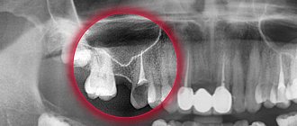
Symptoms of perforation
When the integrity of the maxillary sinus is violated, clear symptoms develop:
- Air bubbles are visible in the blood coming from the bottom of the tooth socket. The doctor asks the patient to exhale sharply through the nose. If the intensity of the bubbling increases, the diagnosis is obvious;
- There is bleeding from the nostril on the side where the perforation occurred;
- The timbre of the voice acquires a characteristic nasal quality;
- Periodically, there is a feeling as if air is passing through the cavity of the tooth, “bursting” in the sinus near the nose, pressing from the inside.
Due to the anesthesia performed, the patient is not able to feel the pain that occurs at the moment of tissue breakthrough. But the doctor is able to suspect the phenomenon based on characteristic signs:
- The moment of sinking of the endodontic instrument is felt after overcoming a tangible obstacle;
- The doctor's delicate instruments have changed their position from their recent position;
- The tooth socket is bleeding, air bubbles are visible in the liquid.
Unnoticed perforation inevitably causes the development of severe complications within the closed space of the maxillary sinus. An infection enters the cavity, causing sinusitis. A person begins to have nasal discharge with pus, breathing is impaired due to the development of swelling of the mucous membrane, and pain of a high degree of intensity is felt, which increases with pressure on the area where the nasolabial folds begin. Sometimes the temperature rises, the patient experiences weakness, chills
Diagnostics
If the therapist diagnosed sinusitis, and the person had his teeth treated shortly before, the cause-and-effect relationship is quite clear. Instrumental research will help establish an accurate picture and locate the source.
The endodontist probes the hole left after tooth extraction, determining the presence or absence of a bone bottom in it. The procedure is performed extremely carefully.
X-rays of the nasal sinuses will provide significant assistance. If darkening in the cavity is visible on the film, it means that there is an accumulation of blood inside. Also in the picture it is possible to see fragments of the tooth roots, the location of the pins, and the protruding material of the intracanal filling. X-rays using contrast will help dispel doubts.
The disadvantage of X-rays is that they are two-dimensional. Stereo images are easy to obtain when undergoing computed tomography. The doctor will review the scan result using a special program, examine the tooth from all sides and choose a treatment plan.
If a long-standing perforation is suspected, a general blood test will show the presence of a number of pathological indicators in the results, indicating the presence of a focus of infection in the patient’s body. It is considered exclusively in conjunction with other studies.
Preparation
Before prescribing a maxillary sinusotomy, the patient is advised to undergo a thorough examination. It includes:
- examinations by a therapist, otolaryngologist, dentist;
- general blood and urine tests;
- coagulogram;
- bacteriological culture of maxillary sinus discharge;
- fluorography;
- ECG;
- radiography of the sinuses;
- CT or MRI of the skull.
Such surgical interventions are usually carried out as planned and are scheduled for the morning. The patient should not eat for 8-12 hours before surgery.
Treatment of perforation of the maxillary sinus
Non-surgical intervention is possible only in the case of instantly detected perforation, when it is diagnosed during the procedure of tooth extraction or treatment of pulpitis. The doctor immediately sends the patient for an x-ray and, based on the results, determines whether an infection of the maxillary sinus has developed.
For example, a cavity wall was perforated during tooth extraction. The dentist noticed the defect immediately. In this case, the doctor makes efforts to preserve the blood clot in the hole where the tooth was previously located. The therapy involves the application of tampons with iodine, and the tampon is tightly fixed in the formed depression and is not removed from there during treatment - so as not to disturb the clot that seals the wound. It usually takes up to a week for the breakout to granulate and disappear.
The option of fixing a plastic barrier separating the axillary cavity from the oral cavity is allowed. This plate is held on adjacent teeth with special clasps. Meanwhile, the perforation heals on its own.
If it was not possible to avoid the penetration of foreign bodies into the maxillary sinus or the infection began to spread, the sinus will need to be opened and cleaned. The resulting wound is closed plastically.
How we treat
Treatment is carried out only comprehensively and strives to be carried out in one visit , simultaneously:
- Hygienic cleaning and dental treatment We prepare the oral cavity for sterile surgical work. We treat compromised roots. Operations are performed only in a sanitized oral cavity to avoid re-infection.
- The operation is carried out in the surgical department, subject to all conditions of sterile operating rooms. Ultrasound removes inflammatory processes, removes roots that provoked or supported inflammation, eliminates all inflammatory elements, removes cysts, polyps, mucoceles, and foreign bodies.
- Orthopedics Temporary crowns or any other orthopedic elements are fixed to mask the work performed. We do everything to avoid sending the patient home without teeth!
We do not welcome radical and punitive surgery!
Rest assured that after surgery we will never keep you in the hospital unless absolutely necessary.
Levin Dmitry Valerievich Chief physician and founder of the Doctor Levin center
Old perforations
It happens that the patient does not go to the clinic after treatment due to accidental perforation. At first the pain is sharp and pronounced, but gradually its character changes and the sensations smooth out. A fistulous tract is formed between the affected sinus and the outer part of the gum. Everything that happens is accompanied by the development of symptoms of typical sinusitis: pain from the perforated intramaxillary cavity, constant nasal congestion, outflow of pus from the nasal passages (and sometimes the fistula on the gum begins to fester). Some patients complain of swelling of the cheek on the side where the sinus is damaged.
If after treatment a person develops strange sensations, as if he feels the movement of air through the gums when sneezing, coughing, and drunk liquid enters the nose, there is a direct indication of an old perforation of the maxillary sinus.
Unfortunately, non-invasive techniques will not help here. An operation is required to remove foreign matter and foci of infection from the sinus. The fistula is cut out, and the opened defect is repaired. After the procedure, a course of antibiotics is prescribed with accompanying physiotherapeutic treatment and the use of drugs that reduce the risk of allergic reactions.
Branded rehabilitation
For those who want to recover faster, we offer an accelerated rehabilitation program - quickly relieves swelling and eliminates bruises
Injection procedures are performed immediately after surgery while the patient is still under sedation. Physiotherapy procedures are usually prescribed on the third day, when postoperative discomfort worsens and the patient understands what difficulties he has.
Microcurrent therapy
Pulsed low-frequency currents restore metabolism at the cellular level. Relieves swelling, muscle spasms, reduces hematomas.
PRP plasma therapy
Injections of enriched blood plasma increase the ability of cells to regenerate, lymph and blood flow. Minimize swelling and cosmetic defects.
Biostimulation of the face
Biomodulators D-NUCLEO and MesoSculpt C71 accelerate recovery, have a lymphatic drainage effect, an anti-inflammatory effect, and reduce swelling.
Consequences of perforation
The patient should not expect that the perforation will heal on its own; such a wound should not be perceived superficially. Attempts at self-treatment with folk remedies or opening a fistula at home are prohibited. The consequences of careless intervention look ominous:
- Loss of healthy teeth in close proximity to the perforation site;
- Penetration of infection into other intracranial cavities;
- Development of abscesses and phlegmons with extremely serious health consequences;
- The risk of developing meningitis or meningoencephalitis due to the proximity of the brain to the inflamed lesions inside the maxillary sinus. With each act of sneezing, being under great pressure, the pathological flora scatters inside the cavities and is able to penetrate into areas from where it is easy to affect the meninges. All this can lead to death.
Therapy of old perforations overgrown with fistula tracts is usually complex and time-consuming. Relapses with new breakthroughs from the sinus to the gum surface are common. Fistulas are not at all harmless formations; they are not easy to cure.
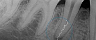
What is an apexectomy
Stages of tooth root resection
This is a surgical procedure that allows you to get rid of the source of infection located inside the gums without removing the tooth unit. Otherwise called resection of the apex of the tooth root. Often, infectious foci form in the area of the tooth root, and apexectomy allows you to cut out and remove the affected part of the root without affecting healthy tissue. This treatment method is considered the best for granuloma, periodontitis and similar serious pathologies, in which the affected teeth were removed in the past.
Preventive actions
The patient is unable to prevent a perforation of the maxillary sinus, and if this happens, he will not be able to overcome what happened without the help of a doctor. But an experienced dentist has the right to take measures to prevent an undesirable scenario. To do this, you will need to conduct a high-quality preliminary examination of the anatomical features of the person applying to the clinic and strictly follow the method of medical manipulation.
Having caused perforation, the doctor is obliged to draw up a treatment plan and bring the patient to a favorable outcome. And a person who experiences disturbing sensations after tooth extraction, treatment of pulpitis, or in other cases, should not endure and remain silent. A timely visit to the clinic will prevent the development of serious complications.
Recovery period
Home medication is prescribed - a course of antibiotics and anti-inflammatory therapy. To prevent the patient from running to pharmacies in a postoperative state and to avoid purchasing uncertified products, we provide the entire package of necessary medications free of charge .
The medicine package contains a detailed list of recommendations and a card with a 24-hour support number.
The service operates 24/7 , if you have any problems, call the number provided.
After 10-14 days, the patient is invited to have the stitches removed. A follow-up CT scan is performed and an examination date is set to monitor the situation.