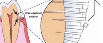
In a healthy oral cavity, only the crown part of the tooth is visible. However, for a number of different reasons (age-related changes, periodontal diseases, etc.), over time, gum recession may occur and develop - a decrease in the volume of its tissue, leading to exposure of the tooth root - not only the dental crown, but also the tooth itself becomes its visible part. root. At the symptomatic level, the problem may manifest itself in the following signs and symptoms:
- visual lengthening of teeth due to the appearance of a bare neck and root;
- growing spaces between teeth due to gum recession;
- change in the color of the tooth, since its cement part becomes visually open;
- the appearance of caries below the gum level;
- increased sensitivity of teeth;
- bleeding gums when brushing teeth.
Why is the tooth root exposed?
The reasons for the development of gum recession can be different, and in some cases there is a combination of several factors at the same time.
- aggressive brushing with a toothbrush - there is such a thing as brush gum recession. They occur when there is excessive pressure on the toothbrush when brushing, or when using a brush of unsuitable stiffness (hard). If you have increased tooth sensitivity when brushing, if your toothbrush becomes deformed over time (the villi move in different directions), you should consult a dentist about proper self-hygiene.
An example of brush gum recession on the upper jaw.
- incorrect position of the tooth in the dentition. Sometimes even a small bite problem can lead to gum pathology. This occurs because the tooth bears excess load when chewing or when teeth are closed, which over time leads to atrophy of the tissues of the supporting apparatus of the tooth, including the gums. In addition, excessive protrusion of the tooth root from the dentition also leads to gum atrophy.
Multiple gum recessions caused by malocclusion.
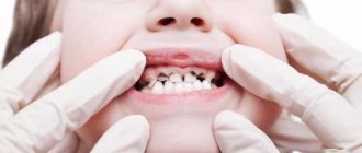
- caries - in cases of gingival caries, gum loss often occurs due to the presence of an infectious focus in the immediate vicinity of the gingival margin.
Gum recession caused by tooth decay in the immediate vicinity of the gum margin.
- Tartar can also cause root exposure.
- anatomical features - for example, a thin biotype of gingival tissue in combination with other factors can lead to recession. Shallow vestibule of the oral cavity (in which the muscles of the lips attach very close to the gingival margin and gradually “pull back” it, exposing the roots of the teeth). In addition, improper attachment of the lip and tongue frenulum can contribute to gum recession.
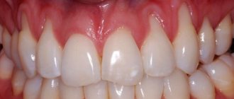
The frenulum of the lower lip, a thin tissue biotype, caused the exposure of the lower incisor root
- bad or professional habits - for example, chewing a pen/pencil, biting threads (for a seamstress), pinching metal objects with teeth, etc.
- lip or tongue piercing - constantly touching the gums and regularly injuring it, metal jewelry can lead to exposure of the roots of the teeth.
- periodontitis - with inflammatory diseases of the gums, the roots are often exposed due to the resorption of the tissues surrounding the tooth, including gum tissue. Unlike “normal” gum recession, periodontitis is treated differently and the prognosis for such recessions is somewhat worse (as a rule, it is impossible to restore the volume of lost gum by completely covering the exposed root).
Why are my gums swollen and sore?
In this article, we will help you figure out why your gums around the tooth are swollen, and how exactly it will be necessary to treat this tooth. Below we will list the symptoms that will allow you to make the correct diagnosis, but first take a close look at the teeth adjacent to the swelling in the mirror. In most cases, the gums swell precisely in the projection of the causative tooth. You need to define the following −
- whether toothache in one of the teeth preceded the appearance of gum swelling,
- does pain appear in one of the teeth when biting on it,
- whether the tooth has decay, a filling or a crown,
- Is this tooth movable?
- does pus discharge from under the gums - when you lightly press on the swelling.
Next, by comparing your symptoms with the symptoms characteristic of periodontitis or periodontitis (which we described below) - you can understand the cause of the inflammation and what treatment you will need in this case.
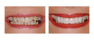
How to treat gum recession?
To cover the exposed root, a small operation is performed in the gum area.
There are many methods of gum plastic surgery, but they are fundamentally divided into 2 types:
- gum plastic surgery with local tissues (when the existing gum is “stretched” or moved to the root in a certain way so that the exposed area is closed, and fixed with small sutures, which are removed after healing).
- gum plastic surgery using a “patch” (the patch can be a special collagen-based material or the patient’s own tissue from the hard palate or the area of the far upper tooth - gum transplantation).
The choice of method is at the discretion of the doctor, according to each specific situation.
Symptoms and consequences
Changing the level of the gingival margin
and visual lengthening of the teeth is the most obvious symptom, which becomes more noticeable as the problem progresses. Usually the disease begins with one or a few units and, if left unattended, can cover the entire or almost the entire series.
In addition, increased sensitivity
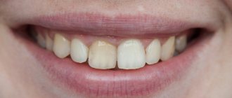
for hot, cold, sour or sweet. Sometimes gaps appear between the teeth in the root zone. They also cause discomfort as food gets stuck in them. In some cases, it is noticeable that the tooth at the gum edge seems to be of a different color - this becomes visible to the part not protected by enamel.
The consequences of significant loss include not only high tooth sensitivity, but also the risk of caries formation
in the root area. In advanced cases, bleeding, inflammation, tooth mobility, and ultimately even tooth loss are possible. Therefore, at the first signs of a recession, you should consult a doctor.
Is gum grafting painful?
No . Operations to eliminate gum recession are performed under local anesthesia; the surgical intervention is 100% painless. In the postoperative period, especially with gum plastic surgery using local tissues, patients often do not experience pain at all. If pain occurs, it is usually only on the day of surgery, a couple of hours after the intervention, when the effect of the local anesthetic wears off. During this period, the pain is perfectly eliminated with painkillers prescribed by the doctor. During the entire rehabilitation period, on average, 1-2 painkiller tablets are required.
In cases of gum plastic surgery using a “patch,” unpleasant sensations may be added due to the presence of a donor zone, which, according to patients, resembles the feeling “as if you grabbed hot tea,” but this does not always occur. The presence/absence of pain is determined by the area from which the “patch” is taken. If anatomical conditions allow, doctors can remove the graft from the donor area absolutely painlessly.
How to fix the problem?
To achieve 100% gum regeneration, you need to consult a doctor as soon as you notice a shift in the gingival margin. The type of treatment for gum recession depends on the stage of the disease.
If the drooping is insignificant, does not exceed 2-3 millimeters, the roots are not exposed, then they speak of an early stage. In this case, drug treatment of gums is indicated - protein preparations based on amelogenins are prescribed, which help restore damaged and form new healthy dental structures. Thus, the natural forces of the body are used. The advantage of drug treatment for gum recession is its safety and good effectiveness, but in severe forms it is useless.
Another method of treating gum recession without surgery is to inject collagen into the affected tissue. Collagen is a natural substance for our body and does not cause allergies or rejection. It stops inflammation and promotes tissue restoration at the initial stage. Collagen treatment is most effective for gum recession caused by severe inflammation. If the cause of prolapse is injury, then collagen will not help here.
In the moderate and severe stages - the tissues drop to 6 mm - treatment is carried out only surgically. There are 3 ways:
- Transplantation of a flap of your own healthy tissue onto the affected area. It is used in most cases and is highly effective. The disadvantages include discomfort in the area where the flap was taken and the risk of rejection.
- The placement of fibrin membranes formed from the patient’s own blood allows for complete tissue restoration, significantly reducing the risk of relapse, reducing postoperative swelling and shortening rehabilitation time.
- Implantation of separation membranes between gingival and dental tissues. A rigid membrane promotes better bone tissue restoration and eliminates prolapse by more than 70%. The disadvantage of this method is that later you will have to undergo a second operation to remove the membrane.
How and how to treat gum recession is decided only by the treating dentist. The cost of treating gum recession depends on the severity of the disease and the amount of work performed.
What are the features and complications after gum recession surgery?
With a mild course of the rehabilitation period, patients do not note any peculiarities . But sometimes it is possible:
- the appearance of white plaque on the gums (this is normal, one of the healing options after gum surgery).
- swelling of the soft tissues of the face (from minor swelling to significant) depending on the extent of the surgical intervention (number of teeth involved), the size of the recession, the method of its closure, anatomical features and the patient’s body.
- hematomas on the facial skin (rarely encountered with gum surgery).
- increase in body temperature.
- pain (relieved by painkillers).
Swelling of the gums due to periodontitis -
In case of poor-quality root canal treatment, due to tooth trauma, or in the absence of timely treatment of caries and pulpitis, inflammation occurs at the apex of the tooth root, which dentists call “periodontitis.” Sometimes the terms “granuloma” or “cyst” are used to refer to periodontitis, which you may well have heard. Such names are due to the fact that the focus of inflammation at the apex of the tooth root in these cases has the appearance of a purulent sac.
Symptoms - usually periodontitis has a chronic asymptomatic course, or there is only slight pain when biting on this tooth. But sometimes periods of exacerbation of inflammation occur, and in this case, acute pain may first occur in the causative tooth (especially when biting on it), and a little later, swelling of the gums usually appears in the projection of the causative tooth. But sometimes pain may be completely absent, and patients complain solely that the gums near the tooth are swollen (Fig. 1-3, 7-9).
Please note that with periodontitis, the source of inflammation is located at the apex of the tooth root, i.e. quite deep in the bone tissue. Therefore, swelling of the gums during periodontitis usually develops not just in the projection of the causative tooth, but most often in the projection of the apex of the root of the causative tooth. And in multi-rooted chewing teeth - no less often in the projection of the bone interradicular septum. But periodontitis is not characterized by swelling of the interdental papilla or the gingival margin adjacent to the neck of the tooth.
In general, if you have swelling on the gum in one of your teeth, pain occurs when you bite on it, and there is a crown, filling or carious destruction on the tooth, and also if, in addition to swelling on your gums, you also have swelling of soft tissues face (again in the projection of the causative tooth) - you can definitely make a diagnosis of “Exacerbation of chronic periodontitis.” An X-ray of this tooth will allow you to accurately determine the cause of periodontitis and the required amount of treatment.
What treatment may be required -
As we said above, if the gums are swollen and painful, then in most cases the reasons are poor-quality filling of the canals, or the lack of timely treatment of dental caries and pulpitis. Only according to official statistics, root canal fillings are performed poorly by dentists in at least 60-70% of cases. The main errors during treatment are underfilling of root canals, poor obturation of root canals with filling substances, breakage of instruments, perforation of the tooth root...
As a result of this treatment, a focus of purulent inflammation appears at the apex of the tooth root. Moreover, in the absence of timely treatment of the tooth for caries and pulpitis, exactly the same focus of inflammation will appear at the apex of the tooth root, but only against the background of unsealed root canals. In Fig. 10-12 you can see how the inflammation at the apex of the tooth root looks like during periodontitis (on the diagram, x-ray and on the root of the extracted tooth) -
Below we describe several treatment options that may be possible for patients with gum swelling due to dental periodontitis. In any case, the doctor will first be forced to take an x-ray. The image will allow us to determine whether this tooth can be saved at all, the size of the inflammatory focus at the apex of the root, and whether root canals have been filled in this tooth previously. The treatment tactics will depend on the latter, and below we will tell you how to cure a tooth and remove swelling from the gums so that it does not appear again.
1) If the channels are not sealed -
If root canal treatment has not previously been carried out on this tooth, then standard treatment of periodontitis is carried out with mechanical treatment of the root canals and treatment of the source of inflammation behind the root apex. On your first visit, they will open your tooth, expand the root canals to allow pus to drain out through them, and leave the tooth open for several days, prescribing antibiotics and anti-inflammatory therapy.
If necessary, your dentist may then refer you to an oral surgeon to make a small incision in the gum to allow additional drainage of pus. After about 3-5 days, the doctor makes an appointment for a second appointment and, if the inflammation subsides, completes the mechanical treatment of the root canals and seals them - either first with a temporary medicinal paste, or immediately with gutta-percha (the latter depends on the size of the source of inflammation at the apex of the tooth root). You can read more about the treatment of periodontitis at the link above.
How the gum incision is made - significant swelling of the gums, or if your gums and cheek are swollen at the same time - indicates the formation of a large purulent abscess, which will require not only opening the root canals of the causative tooth, but also making an incision in the gums to release the pus. The incision is made under local anesthesia, the wound is then washed with antiseptics and a drain is inserted into it (see video below).
2) If the channels are sealed poorly -
If an x-ray shows that canal treatment has already been carried out previously and the cause of inflammation was incomplete filling of the root canals to the apex of the tooth root, then there are 2 treatment options: either standard conservative therapeutic treatment, or an option associated with resection of the root apex.
- Conservative therapeutic treatment - on the first visit, the filling/artificial crown is removed from the tooth, poorly filled root canals are unsealed, washed with antiseptics, and antibiotics are prescribed.
If necessary, the patient is referred to a dental surgeon to make an incision along the gum. Thus, to the treatment we described in the previous section, only 1 point was added here (unsealing the root canals). Then everything is the same - after the inflammation subsides, temporary or permanent filling of the root canals is carried out. If the source of inflammation is small, the canals are usually filled immediately and permanently with gutta-percha, and a permanent filling is placed at the next visit. If the inflammation is large, the canals are sealed with temporary medicinal paste for 1-3 months, and a temporary filling is placed. And only after this time the canals are filled with gutta-percha + a permanent filling or crown is placed. - Resection of the root apex (Fig. 13) –
This surgical method is much simpler and much cheaper than conventional therapeutic treatment, and it allows you to avoid removing the crown from the causative tooth in order to unfill and reseal the root canals.
However, this method is only suitable for those teeth in which the root canal was poorly filled only at the very apex of the root (and throughout the rest of the length the canal should be filled normally). This simple surgical operation is carried out within 25-35 minutes and consists of cutting off the apex of the root with the unfilled part of the root canal using a drill. To do this, an incision is made along the gum and a flap of the mucous membrane is moved back to give access to the bone tissue in the projection of the apex of the tooth root. Next, a small hole is made in the bone with a drill, through which the apex of the root is cut off and removed from the wound along with the granuloma/cyst at the apex. The wound is sutured and antibiotics are prescribed. Pros: cheap, simple, no need to remove the crown and re-treat the tooth.→ How is root resection surgery performed?
What are the recommendations and restrictions after gum recession surgery?
- Do not disturb the wound surface (with tongue, food, foreign objects, etc.) this is the main condition for good healing.
- Do not bite (food eaten should be soft or finely chopped).
- Do not brush your teeth in the surgical area with a toothbrush (hygiene in this area should be maintained only as permitted by the attending physician).
- Do not eat or drink hot foods.
- Avoid physical activity.
- Follow the recommendations of your doctor and take prescribed medications.
What happens if gum recession is not treated?
- root caries (the root, unlike the crown of the tooth, is not covered with enamel - a dense “pearl” shell, and therefore is more vulnerable). Normally, the root is covered by the gum and is not exposed to the aggressive environment of the oral cavity (acids, humidity, microorganisms, enzymes). With gum recession, the exposed root is susceptible to destruction.
- abrasion of root tissues (again, due to the lack of enamel, tooth root tissues are not resistant to abrasion).
- increased sensitivity of teeth.
- the transition of a recession to a class that is not subject to complete closure.
- more plaque (the surface of the root is rougher than the surface of the tooth crown, which means that plaque is retained more when the roots are exposed).
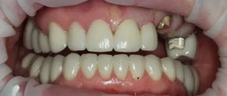
Large gum recession, caries of the exposed tooth root, an abundance of soft plaque on the root surface.
Types of gum exposure
Why does gum recession occur? There are three ways of development of pathogenesis:
- genetic theory;
- exogenous theory;
- endogenous theory;
Genetic says that recession begins as soon as teeth erupt.
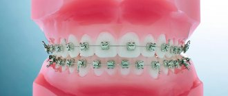
Exogenous begins with the influence of external circumstances, plates, braces. The development of caries is provoked by insufficient oral hygiene.
The endogenous theory begins its development with internal causes, occlusion, and the overall density of teeth.
Stages of development
X-rays can accurately determine the class of recession. There are 4 stages
.
First
When the gum recession is insignificant, the neck of the tooth is only slightly exposed.
Second
, the process of decline occurs 2-4 mm towards the root, while the tissue between the teeth becomes pale and white.
Third
The stage is accompanied by complete exposure of the neck of the tooth. This is noticeable and brings discomfort to the patient in terms of aesthetics. Painful sensations may occur.
Fourth
leads to complete receding of the gums, and there is a loss of mucous and bone tissue. The neck is exposed along with the root. The space between the teeth becomes wide.