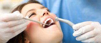
5936
One of the serious complications encountered during prosthetics with crowns is the separation of the edge of the artificial tooth from the gum tissue.
The process indicates developing inflammation and requires immediate treatment to avoid crown loss and the development of other oral pathologies.
Causes
When fixing the crown, a small indentation is left between its base and the gum tissue. This is done to ensure ease of use and quality of hygiene of artificial teeth.
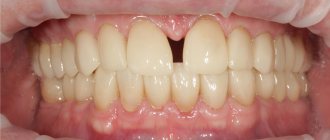
When the prosthesis fits correctly, the size of the gap is no more than 1 mm, however, there are situations when the rinsing space expands, causing discomfort and pain.
Factors that directly affect the human oral cavity, so-called local causes, can provoke an increase in the gap between the crown and the gum.
The main ones are:
- an error by the orthopedist when fixing the crown - the specialist could not have fastened the artificial tooth tightly enough to the base or, conversely, pressed it too hard;
- improper manufacturing of the orthopedic structure - discrepancy in size or shape;
- poor oral hygiene , resulting in the accumulation of bacterial plaque, leading to the development of inflammatory processes in the gum tissue;
- the occurrence of an allergic reaction to the materials from which the crown is made;
- poor-quality measures to sanitize the elements on which the prosthesis is fixed. This can lead to the destruction of stump tissue and the development of an inflammatory process in the periodontium;
- the occurrence of galvanic syndrome due to intolerance to prostheses made of metals;
- regular mechanical trauma to the gums as a result of performing hygienic procedures with a brush with hard bristles or installing an excessively high filling;
- change in the position of teeth adjacent to the prosthesis as a result of the development of periodontal disease or periodontitis.
There are also a number of common reasons that can cause an increase in flush space:
- diseases of the endocrine system, in particular diabetes;
- hormonal changes in the body caused by pregnancy, menopause;
- development of immunodeficiency states.
What does the smell from under the crown indicate, and how to get rid of it.
Visit here to learn about the indications for using half-crowns.
At this address https://www.vash-dentist.ru/protezirovanie/nesemnyie-p/koronki-np/pochemu-na-implante-shataetsya.html we will explain why the crown on the implant is loose.
Crown contour and soft tissue aesthetics - relationship between parameters
The production of aesthetic crowns requires mandatory consideration of the biology of the surrounding soft tissues in order to achieve the most effective restoration results. This task, however, is not quite as simple as it might seem at first glance, and requires close cooperation between the dentist and the dental technician.
The free gingival margin is a section of soft tissue that has a knife-like parabolic shape and covers the terminal region of the alveolar ridge from the vestibular and lingual side, reaching up to the enamel-cementum border of the tooth. The most apical border of the marginal gingival margin, called the zenith, is important in restoring the harmony of white and pink aesthetics when performing various types of restorations.
Normally, the zenith point is 1 mm distal to the middle of the tooth, which is most clearly visualized in the area of the central incisors of the upper jaw, while this ratio decreases slightly from the area of the lateral incisors towards the molars. The proximal surfaces of the cemento-enamel interface of the teeth are parabolic in shape, and their contour is slightly concave. The interdental papilla, occupying the proximal area apical to the point of interdental contact, in turn, is a very sensitive structure, the correction of the parameters of which depends on the volume of the interdental space, the position of the interalveolar bone septum, the condition of the periodontal or peri-implant tissues around the tooth or the installed dental implant.
In the clinical cases illustrated below, an algorithm for managing the tissues of the dental papilla is described, thanks to which the doctor can correct its vestibular and proximal contours by correcting the profile of the aesthetic restoration. Understanding the biology of soft gum tissue in itself can significantly help in achieving the most effective result of complex rehabilitation of patients using various orthopedic structures, including those supported by titanium implants.
Possibilities of coronal migration of the free gingival margin
Gingival recession, similar to that illustrated in the first clinical case, can be provoked by the result of iatrogenic intervention with a violation of the biological width. An example of damage to the area of connective tissue attachment by the edge of a fixed crown is shown in Photo 1.
Photo 1. The edge of the crown extends into the area of connective tissue attachment. The probing depth did not exceed 1 mm and, due to the intact parameters of the periodontium, it is possible to ensure coronal migration of the gums through a “gradually creeping attachment.”
The clinical condition of the underlying bone tissue was checked during probing, and given its intactness, it could be assumed that the vestibular gums were still capable of migrating coronally during crown correction. To make provisional crowns, plaster models were obtained. After removing the old metal crown, it turned out that the edge of the crown was too apical, and the preparation area was unreasonably expanded in the axial direction (photo 2).
Photo 2. After crown removal, excessive reduction of hard tissues in the axial direction was detected.
Treatment began with the removal of old cement from the core surface using a 16-sided carbide bur, followed by correction of the preparation area to create a gingival margin coronal to the cemento-enamel junction of the adjacent central incisor (Figure 3).
Photo 3. Correction of the previous core design: the gingival margin of the preparation has been moved coronal to the cemento-enamel junction of the adjacent central incisor.
The provisional crown was deliberately made to be insufficiently contoured at the gingival margin to allow for the “gradually creeping attachment” phenomenon that allows for coronal gingival migration. This approach has already proven its stable long-term results, and the attachment formed in this way is resistant to bacterial contamination.
After 6 weeks, the optimal level of the vestibular gingival profile was formed, suitable for fixation of the final restoration (photo 4). After impressions were taken and a series of laboratory steps were completed, the final metal-ceramic crown was fabricated and cemented. During the control clinical examination 1 year after the primary intervention, a stable healthy condition of the soft tissues and an optimal aesthetic contour of the gingival papilla from the vestibular and proximal sides were recorded (photo 5).
Photo 4. View with provisional crowns.
Photo 5. View with permanent crown one year after treatment: complete stability and symmetry of the soft tissues in the area of the central incisors.
Crown contour and soft gingival tissue profile
The free gingival margin can be moved more apically only by crown convexity without any additional surgical manipulation, provided that the distance from the edge of the underlying bony ridge to the unattached gingiva is greater than 2 mm.
This parameter allows you to adjust the position of the marginal region of the gums without disturbing the space of biological width. An important criterion for achieving effective results in soft tissue management is the condition and position of the bone tissue relative to the overlying gingival contour.
The patient who sought dental care had an asymmetry in the position of the gums in the area of the front teeth (photo 6) as a result of impaired passive eruption of the right incisor. To correct aesthetic parameters, the manufacture of a ceramic crown was indicated, which would help form an adequate aesthetic profile of the smile. When probing the periodontal sulcus, its parameters in the area of the left incisor corresponded to 1 mm, and in the area of the right – 2.5 mm (as a result of impaired passive eruption). Therefore, the marginal gingival margin can be moved 1.5 mm more apically without disturbing the epithelial attachment of the ligamentous apparatus of the tooth. Before correcting the gum level, an unimpregnated retraction thread was placed in the area of the periodontal sulcus (photo 7), with the help of which it was possible to simulate the position of the gums in the area of the problem tooth, similar to that of the adjacent incisor.
Photo 6. Inadequate restoration in the area of the right incisor and the presence of caries on the palatal surface argue for the need for a full crown for aesthetic rehabilitation.
Photo 7. Installation of an unimpregnated retraction cord in the periodontal sulcus.
On the model, a similar procedure was carried out by removing a certain level of plaster until an identical position of the gums was achieved in the area of both frontal incisors (photo 8). The convex contour of the simulated cemento-enamel interface formed on the ceramic crown helped to correct the position of the gums to the most optimal aesthetically acceptable level. A control clinical examination of this patient one year later confirmed the stability and effectiveness of the initial treatment results (photo 9).
Photo 8. Imitation of the gingival margin on a plaster model: the shape of the convexity of the facial edge of the crown determines the contour of the soft tissues of the gums.
Photo 9. View 1 year after fixation of the final crown: stability of the gingival contour and absence of signs of inflammation.
Restoration of the interdental papilla
Although clinicians report the excellent effectiveness of most interdental papilla repair techniques, a systematic review of the literature by Blatz and Hurzeler suggests that most proposed clinical approaches are unpredictable. But in truth, the potential for coronal migration of the interdental papilla is quite enormous with adequate underlying bone support.
One recent case presentation regarding the problem of interproximal soft tissue restoration reported that increasing papillary height through soft tissue augmentation is virtually impossible without additional damage to the adjacent gingiva around the surgical site.
Photo 10 shows a clinical case in which the patient had a deficiency in the height of the interdental papilla in the area of the frontal incisors, which compromised the overall aesthetic appearance of the smile. Replacing the defective right incisor with a metal-ceramic crown made it possible to achieve optimal parameters for the height of the interdental papilla, thanks to the appropriate contours of the crown. When analyzing the diagnostic wax-up, it was determined that the difference in mesio-distal tooth dimensions could be caused by the presence of a feldspathic restoration on the mesial side of the left incisor. The vital state of the underlying bone provided conditions for adequate migration of soft tissues.
Photo 10. Deficiency of papilla tissue between the central incisors and discrepancy between the mesio-distal dimensions of the teeth when smiling.
The dental technician fabricated a ceramic crown for the right incisor and an adequate restoration for the left anterior tooth. The contour of the preparation imitated the natural irregularities of the incisal crown to camouflage the area of the labial margin (Figure 11), and the proximal contour of the restoration, in turn, determined the shape of the interdental papilla (Figure 12). 1 year after the intervention, the patient had an optimal aesthetic smile profile with a stable corrected position of the interdental papilla (Figure 13).
Photo 11. Restoration of a defective crown and ceramic restoration with new structures with restoration of adequate mesio-distal dimensions.
Photo 12. Contour of final restorations: filling the proximal area with soft tissue of the papilla.
Photo 13. View of the smile 1 year after treatment.
Contour of crowns supported on dental implants
Although implant-supported crowns can very successfully imitate natural tooth tissue, if the parameters of the “pink” component are inadequate, even such designs are not sufficiently aesthetic (photo 14). Due to the fact that the implant platform is completely flat and not parabolic, like the contour of the enamel-cement junction, it is clinically quite difficult to achieve optimal soft and hard tissue parameters in the periplant area. When installing an implant adjacent to a natural tooth, the height parameters of the interdental papilla depend on the vertical position of the periodontal attachment in the area of the natural tooth (photo 15). If the height of the soft tissues on the proximal side of the tooth is still not enough, then 5 mm of soft tissues relative to the base of the bone tissue of the alveolar ridge will be just enough to predict and correct the position of the interdental papilla. To achieve successful treatment results, it is important that the intervention is carried out in an intact state of the crestal ridge, dentogingival and dentoperiosteal fibers of adjacent natural teeth.
Photo 14. Deficient papillary height in the peri-implant area before crown fixation.
Photo 15. X-ray of the implantation area: intact bone tissue provides conditions for restoration of the soft tissue of the papilla.
In this clinical case, the position of the cemento-enamel border of the right central incisor of the maxilla was duplicated on a plaster model. The contour of the cement-enamel junction of the ceramic crown determines the position of the vestibular and proximal profile of the gingival papilla, which in some cases requires several months to restore its position. 1 year after installation of the zirconium abutment and ceramic crown (Figure 16), the optimal position of the gingival papilla was achieved, which remained stable over a long period of time.
Photo 16. Soft tissue profile 1 year after treatment.
Another clinical case demonstrates the problem of asymmetry of the free gingival margin relative to an implant-supported crown replacing the right central incisor and an all-ceramic crown in the region of the left central incisor (Figure 17). The linear inclination of the crown supported on the titanium infrastructure (Figure 18, left) provoked apical migration of the free gingival contour. The contour of the final superstructure with the corresponding root concavity and crown convexity determines the formation of the optimal soft tissue profile (photo 18, right). The final restorations were not only able to closely mimic the contour, color, shade and overall appearance of natural teeth, but also provided adequate positioning of the free gingival margin and surrounding tissue as documented by clinical photography (Figure 19) and radiographic findings (Figure 20).
Photo 17. View of the soft tissue profile in the area of the crown on the implant (right) and the natural tooth.
Figure 18. The facial contour of the crown determines the position of the soft tissues: the linear transition from the implant platform to the crown provides coronal gingival migration, and the more vertical contour and convexity in the area of the enamel-cementum interface provides the gingival profile on the labial and proximal sides.
Photo 19. View of the final restorations with optimal gingival profile.
Photo 20. X-ray during the period of fixation of the final crown.
Discussion and conclusions
Performing adequate aesthetic restorations in the anterior area is a very difficult clinical task, the implementation of which requires close cooperation between the dentist and dental technician. Thanks to the teamwork of specialists, optimal soft tissue parameters can be achieved even without any surgical interventions.
However, a thorough understanding of the behavior of soft and hard tissues adjacent to the area of the prosthetic structure is mandatory both to avoid possible inflammation as a reaction to iatrogenic interventions and to prevent excessive apical migration of the free gingival margin.
Assessment of the condition of bone tissue, as well as the vitality of periodontal structures, is mandatory for an adequate prognosis of the outcome of therapeutic intervention. Changes in the vestibular gums in the area of the anterior teeth of the upper jaw can be very variable: the parameters of the papillae can vary from 2 to 5 mm. The convexity of the tooth neck also plays an important role in the formation of the optimal gingival profile. Therefore, imitation of the convexity of the enamel-cement border with the volume of ceramics of the orthopedic superstructure is also mandatory to achieve successful results of aesthetic rehabilitation and the most acceptable relationships between the elements of pink and white aesthetics.
Authors: Richard P. Kinsel, DDS; Bryan I. Pope, DMD, MSD; Daniele Capoferri
First signs
An increase in the gap between the artificial tooth and the gum tissue may be accompanied by the following sensations in a person:
- itching in the area of the pathological process;
- pressure of an artificial tooth on the gum;
- insufficiently tight fit of the prosthesis, its protrusion from the general row;
- increased rate of tartar formation.
The pathological process is often accompanied by a change in the color of the soft tissue of the oral cavity - acquiring a reddish or bluish tint. In some cases, the gums may swell, bleed, and fester.
If the widening of the gap is associated with improper fixation of the crown, the patient may experience the following complaints:
- difficulties in pronouncing certain sounds that are not related to the functioning of the language;
- the formation of whistling sounds during a conversation due to the penetration of air into the cracks formed due to incorrect installation of an artificial tooth.
How the gum bed for an implant crown is formed
During implantation there is a need to form soft tissues. For this, a shaper is used. The design is necessary to create the correct gingival contour, on which the final aesthetic result will depend.
The forming element has the form of a threaded screw equipped with a cylindrical head. Made from hypoallergenic metal. Installed after complete engraftment of the titanium rod. Designed to create a recess around the future crown. This will allow the artificial tooth to fit tightly .
Possible complications
Increasing the flushing space can lead to a number of negative consequences. First of all, this is the emergence of difficulties in the process of eating, associated with the penetration of food particles and bacterial plaque into the resulting gap.
The situation over time leads to the development of serious dental diseases, such as:
- Gingivitis is an inflammation of the gum tissue that does not affect the teeth and periodontium.
The disease is characterized by swelling, bleeding of the gums, and changes in their color. In the absence of timely treatment, gingivitis is fraught with ulceration of the gum tissue, the formation of abscesses on it, and the penetration of infection into the periodontium. - Stomatitis is an inflammatory process that occurs on the oral mucosa, accompanied by pain, swelling of soft tissues, and the formation of ulcers.
As a rule, eliminating the pathology does not cause difficulties, however, there are cases of secondary infection. - Periodontitis is an inflammatory disease that affects the tissues surrounding the tooth.
As a result of inflammation of the ligamentous apparatus, there is a risk of developing pathological mobility of the elements of the row, which, if left untreated, can lead to their loss.
Elimination tactics
When a patient complains of an increase in the gap between the artificial tooth and the gum, the dentist conducts a thorough examination of the prosthetic area. X-rays may be required to clarify the diagnosis.
The treatment plan depends on the degree of complexity of the pathology. If there is slight deviation of the gums from the crown, therapy consists of performing the following procedures:
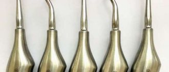
- Removal of mineralized plaque. Using ultrasound or a special device, the specialist cleans the tooth surface from bacterial plaque, after which he performs closed curettage of the periodontal canals.
The procedure allows you to remove subgingival stone using dental instruments without resorting to cutting the gums. - Carrying out anti-inflammatory and antibacterial therapy. The treated areas of soft tissue are treated with special preparations, such as Chlorhexidine, Miramistin.
In the case of an acute inflammatory process, broad-spectrum antibiotics are prescribed.
In advanced cases of expansion of the lavage space, therapeutic methods of treatment are ineffective. To eliminate the pathology, surgical intervention is required.
There are two options for performing the operation:
- Open curettage. The procedure is performed by opening the periodontal pocket to a depth of 4-5 mm.
A specialist removes a flap of gum in the affected area, followed by deep tissue cleaning using special devices. After treatment with an antiseptic drug, the flap is fixed in its original place using suture material. - Flap surgery. The operation involves separating a flap of gum using several incisions, cleaning open areas of pathological material and treating with antiseptic agents.
After this, the previously detached gum flap is applied and fixed using surgical sutures.
The postoperative period requires the patient to be careful when caring for the elements of the oral cavity. To avoid opening of periodontal canals, you should refrain from brushing your teeth and using mouthwashes for 10 hours after surgery.
In the future, it is advisable to use a brush with soft bristles and avoid contact with the operated area.
Causes of allergies to dental crowns and how to get rid of them.
In this publication we will look at the features of Shofu metal ceramics.
Here https://www.vash-dentist.ru/protezirovanie/nesemnyie-p/cad-cam-sistemyi-v-stomatologii.html we’ll talk about the advantages of using Cad Cam technology in dentistry.
How to eliminate the gap between the gum and the prosthesis?
Prosthetics when installing crowns are non-removable. Therefore, it is not enough to simply remove the denture, wash the clogged area and replace it properly. If the gap increases, you should definitely consult a doctor.
The only way to eliminate the gap if it increases mechanically is to reinstall the system. The prosthesis is removed and either completely replaced, or installed again, but now in compliance with the installation rules.
For minor inflammatory processes that occur after dental intervention, the use of Metrogyl Denta ointment is prescribed for 7-14 days until the tissue is completely restored and the gap heals. In case of serious inflammation, the prosthesis is removed and the primary cause of the gap is eliminated.
Prevention measures
In order to prevent pathological expansion of the washing space, it is necessary to carefully follow the specialist’s recommendations regarding the care of the oral cavity after fixing the prosthetic structure.
They are based on compliance with the following principles:
- It is advisable to brush your teeth at least twice a day, and preferably after each meal. Particular attention should be paid to the area where the gum tissue meets the crown.
- To effectively remove plaque particles from the subgingival space, it is necessary to use not only a brush, but also additional devices - a brush, irrigator, dental floss.
- To avoid mechanical expansion of the gap, it is important to avoid foods that are prone to crumbling, since small food particles can penetrate under the gum. Products that you should avoid eating include cookies, chips and crackers, and products with poppy seeds.
- If gum tissue is prone to developing inflammation, you should regularly treat the oral cavity with antiseptic drugs recommended by your doctor, and use rinses.
In addition, you need to pay attention to specialized toothpastes designed for sensitive gums.
In addition, dentists note that it is extremely important to regularly sanitize the oral cavity, carry out professional teeth cleaning and promptly eliminate developing diseases without self-medicating.
In the video, a specialist will answer the question: should the gums fit tightly to the crown.
Reviews
The presence of a barely noticeable gap between the crown and the gum is a natural situation. It is important that the patient is not bothered by pain, swelling, discoloration of the mucous membrane and other unpleasant sensations.
If you suspect an increase in the flushing space, you must immediately seek specialized help, since it is impossible to eliminate the problem yourself.
If, after fixing the crown, your gums were moving away from the base of the prosthesis, share with readers the reasons for this phenomenon and how to eliminate it in the comments section.
If you find an error, please select a piece of text and press Ctrl+Enter.
Tags crowns prosthetics
Did you like the article? stay tuned
Previous article
The whole truth about restoring the functionality of the dentition using composite inlays
Next article
Algorithm for selecting abutment teeth for fixing dentures
Symptoms
The normal size of the flushing gap is no more than one millimeter. When the prosthesis is installed correctly, it helps to increase the degree of its fixation, improve oral hygiene, protect against many pathological processes and extend the service life of the structure.
Causes for concern may be the appearance of significant discomfort and pain, a strong change in the location of the crown, increased sensitivity to various irritating factors, for example, cold or hot food. Also, if the situation worsens, a person may complain of itching and a burning sensation, impaired diction, whistling when speaking, etc.
All of these symptoms can be localized both at the site where the crown is installed and far beyond it. It is very important to immediately seek advice from your doctor when you notice the first signs of a deviation from the norm. If there is a suspicion that the problem arose due to the lack of qualifications of the specialist, it makes sense to visit another clinic.