Tooth root removal – one of the most labor-intensive and complex procedures in modern dental practice. The indication for its implementation is the preservation of the root part of the tooth in the gum, subject to complete destruction of the crown (for example, in severe forms of caries, as a result of injury or illiterate dental treatment). Untimely operations to extract such units is a factor contributing to the development of a whole range of diseases (periodontitis, periostitis, etc.). That is why “problem” roots must be removed as soon as possible.
Procedure for removing tooth roots
The tooth root extraction operation is performed under local anesthesia. Before starting the procedure, the surgeon carefully studies the results of the X-ray examination. The information obtained from analyzing the image helps the doctor plan the course of the operation and select the necessary tools.
During surgery the following can be used:
- forceps;
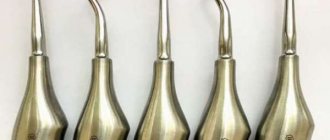
- elevator;
- dental bur.
Extracting a tooth root using forceps
In the first case, the operation begins with careful separation of the gums and other tightly adjacent tissues from the root remaining in the jaw tissue. Then the doctor places forceps on the root part of the tooth and pushes their cheeks under the gum tissue, trying to grasp the unit to be extracted as tightly as possible. A well-fixed root is dislocated using rocking or rotating movements and carefully removed from the alveolar socket.
Carrying out operations using elevators
Elevators are used in cases where there are no conditions for removing it with forceps (for example, when it is located deep in the alveolar socket). The doctor carefully peels away the tissue adjacent to the remaining tooth, inserts an elevator into the space between the alveolar wall and the unit to be removed and, using the tool as a lever, gently dislocates the root. When the root part becomes mobile, the surgeon removes it from the socket using forceps.
Cutting out the root with a bur
If there are no conditions for root removal using standard instruments, alveolotomy is performed - a surgical intervention that involves destruction of the outer wall of the alveolar socket.
This operation is carried out in several stages and includes:
- incision of the gums and subsequent delamination of the mucoperiosteal tissues in the area of surgical intervention;
- cutting the outer wall of the alveolar socket with a bur and exposing the root;
- insertion into the resulting passage of the elevator and careful dislocation of the remaining tooth;
- root extraction from the alveoli;
- grinding the pointed edges of the alveolar socket;
- careful suturing of the wound.
The duration of the tooth root removal operation can vary from several minutes to 2-3 hours.
Sick teeth: save or remove
First of all, I want to say that every doctor should understand that preserving the tooth itself is, for many reasons, better than removing it. Implantation is not a panacea. You can always get an implant – in any case and at any age, when there is no other option. But trying to save your tooth is an art!
Today, many doctors say that conservative treatment of large cysts is impossible. They are removed and a resection of the root tips is performed - that is, the cyst is removed along with part of the root. Then artificial bone material is placed there. To do this, you need to cut the gum. That is, the treatment of such pathologies in the “standard order” is surgical. The life expectancy of such a tooth is 5 years. After this, a relapse very often occurs, that is, the inflammatory process begins again.
I am a supporter of endodontics, in other words, I try to treat teeth where a cyst or granuloma has formed conservatively through a canal, without cutting the gum. And I, of course, have my own technique in these cases: in fact, it is enough to rinse the canal well and remove the infection with therapeutic drugs. In this case, of course, it is necessary to know anatomy, chemistry, and also microbiology - that is, to understand what microflora and what to kill with, because the success of treatment depends on this. For the doctor, in theory, it is no secret that in the case of cysts and granulomas, the causative agent of infection is gram-positive and gram-negative cocci. And believe me, if the doctor understands these things, then there will be nothing magical in endodontic treatment. True, such treatment takes from 1 month to six months, depending on the size of the cyst. Today there is an opinion that such teeth can be treated in one visit, without prior calcium treatment. Based on my own experience, I believe that it is difficult to achieve positive dynamics in this case, we’ve been through it - we know! After all, it also happens: the tooth seems to be improving, and then suddenly there is a sharp relapse. Therefore, once a month the patient visits the clinic, I rinse the tooth, apply medicine, perform an X-ray control and... put a temporary filling. And so on until a positive result is confirmed by the results of a computed tomography scan. After six months, a crown can be placed on the tooth. In difficult cases, we install a temporary crown for six months, after which we perform another computed tomography scan and after that we cover the tooth with a permanent crown. After this treatment, I have no relapses in 100% of cases.
Or here’s another example: a fracture of the crown of a tooth below the level of the gum (gingival attachment). You can, of course, remove the tooth, as is usually the case. But I prefer to clinically lengthen the neck of the tooth due to additional tissue located in this area. How? I'll try to explain. The gum has a movable layer that covers the neck of the tooth, and there is also a layer partially attached to the bone tissue. Using a laser (that is, surgically), I remove the gum around the tooth, thus lengthening its neck and providing space for the installation of an orthopedic structure designed to protect the tooth. I know that for many this sounds like something unreal. Fortunately, I can afford to save teeth with a crown fracture even below the gum attachment, but only if this fracture does not reach the level of the interradicular septum. If the interroot septum is affected, then infection will constantly get there. As a result, the tooth will still have to be removed.
Yes, such treatment is not for one day. After all, it takes 2-3 weeks just to form a new gum. In general, the entire process can take from 1 to 2 months - in complex cases (more often when the painters break, that is, large molars in the area of the palatal or lingual wall), in simple cases - 2-3 weeks. But during all this time the patient will need to come to me 2 times. First time I lengthen my neck. The second is when I restore the wall of the tooth, so that the gums grow correctly: this will provide enough space where you can then put a stump inlay, and put a crown on top. But this is the work of an orthopedist...
With root fractures, a lot depends on how badly the root is damaged. Root fractures in single-rooted teeth are not dangerous, especially if the root is broken, for example, 4 millimeters vertically. In these cases, we combine surgical and orthodontic treatment approaches. And then we restore the tooth using orthopedics. But if the crack is deep, then there is no point in saving such a tooth, because complications may arise later.
Perforations of teeth can be closed in any part of the roots, provided that the tooth is sterile (not periodontitis): with a competent approach, the prognosis of such a tooth will be 100% successful. But if the perforation is located in the area of the interradicular septum, a large cyst has formed there, then this perforation can, of course, be closed, but the success of treating such a tooth will be 50 to 50. It is important to seal the periodontium hermetically so that infection does not get there. I close the perforations with a special cement that was created for this purpose and is also used to close wide apical foramina. It seals the perforations hermetically, hardens quickly, and behaves well in a humid environment. However, I would like to make a reservation right away: working with perforations is only possible under a microscope - after all, I need to see how I close the hole! This is the job of an endodontist. Perforations are treated in one visit, meaning you don’t have to wait several weeks or even months for results. But if the bottom of the cavity is severely broken by a bur, when the tooth is not only perforated, but also the area of the interradicular septum is open and infection constantly gets there, then closing such perforations may be impractical.
When removing an instrument from a tooth root, it is also necessary to use a microscope. Although often, for example, when an instrument is broken in a bend in a canal, even a microscope is powerless. What can I say here? We use... intuition and think through every step well. Sometimes it is necessary to use instruments that were not originally designed for work in the canals, or even several instruments, creating “in-house” designs that ultimately allow the removal of the recalcitrant fragment. In general, everything “that comes to hand” is used - in the good sense of this expression. Why not if it can help? When doing this work, it is very important not to overexpand the channel. In principle, I work without expanding the tooth canal - I work only with the tool! The channel remains intact on all sides. There is also the so-called by-pass technique - its essence is that we “bypass” the instrument broken in the canal, leaving it in the tooth canal system. If there is full access to the canal and the instrument can be bypassed by properly rinsing the canal with aggressive solutions, cleaning it and passing it with “cement,” then the foreign body no longer causes any harm to the tissues, since it ceases to be a source of additional infection. And this is also considered a positive result of treatment! To be honest, many doctors look and do not understand how I manage to remove instruments from the apical foramina - for them it is a mystery, shrouded in darkness =).
Complications after surgery
The most common complications encountered by patients who have undergone tooth root removal surgery are severe bleeding and alveolitis. Factors that can trigger bleeding include high blood pressure or decreased blood clotting. In turn, the main cause of alveolitis is non-compliance with the postoperative regimen, leading to the washing out of the protective blood clot from the socket. The development of the disease is accompanied by the occurrence of an inflammatory process, suppuration of the wound formed during the removal of the tooth root, the appearance of pain and bad breath.
What diseases does it treat?
Among the main diseases treated by a dental surgeon:
- periodontitis;
- diseases of the TMJ (temporomandibular joint);
- periostitis;
- osteomyelitis;
- tumors and cysts of the oral cavity;
- diseases of the salivary glands;
- sinusitis;
- abscesses;
- trigeminal nerve diseases;
- phlegmon and others.
A doctor of this profile also diagnoses some specific systemic diseases that manifest themselves in the oral cavity. Among them: syphilis, actinomycosis, scurvy, tuberculosis, etc. For such diseases, treatment is prescribed to the patient by another doctor (infectious disease specialist, virologist, pulmonologist).
Features of postoperative care
There are a number of recommendations that can significantly speed up the healing process of a wound formed during surgery and avoid the development of complications. In particular, dentists advise:
- during the first 24 hours, avoid too warm baths, do not go to the baths, refrain from any, even minor, physical activity, apply ice to the cheek on the side of the removed root every half hour;
- give up alcohol and smoking, too hot and spicy food for 2 days;
- take analgesics if pain occurs;
- for several days, try not to chew food on the operated side, give up the habit of biting your lips or sucking in your cheeks (this creates a vacuum in the mouth that can displace a blood clot formed on the wound);
- undergo a full course of treatment with antibacterial agents if prescribed by a dentist.
If signs indicating the development of complications appear, it is necessary to abandon attempts at self-medication and seek help from a doctor at the 24-hour A.Dent dentistry as soon as possible.
Dentist recommendations after tooth extraction
Upon completion of the extraction, the dentist places a cotton swab containing medicinal contents into the socket (the cavity remaining after tooth extraction). The cotton swab must be kept in the mouth for 20 minutes. Then you should make sure that the wound is not bleeding and remove the tampon. Sometimes, the dentist advises not to remove the cotton wool for 30 minutes to avoid continued bleeding. You remove the cotton swab yourself. It is important to keep your hands clean. After removing the cotton wool, a small blood clot forms in the hole, which further prevents microbes from entering the healing wound.
It is necessary to refrain from eating and drinking liquids and smoking for two hours. Until the wound is completely healed, stick to a diet that is dominated by liquid and puree foods that are not capable of injuring the wound. It is better to chew carefully so that food does not get into the wound, and carefully ensure that no pieces of food remain there. To avoid pain, take fluids and foods at medium temperature. Cold and hot foods can injure exposed gums. The next day, you are allowed to brush your teeth only on the side that has not undergone extraction. Complete healing of the gums occurs after four days. After this time, you need to make a second visit to the dentist to monitor further treatment. This is also necessary for timely detection or prevention of inflammation.
Removal methods
There are two main types of extirpation - a simple removal method and a complex method.
Complex removal method
It is carried out in cases where there is no access to the tooth, application of forceps is impossible and additional access must be made through an incision in the gums. During a complex operation, the tooth is not removed immediately, but in parts.
For this, a whole set of special tools is used:
- laser,
- saw,
- scalpel,
- burs.
Complex removal may be indicated in the case of a cystic formation under the causative tooth, fistula, dystopic wisdom tooth, or retention. Contraindications to the procedure are pregnancy, allergies to anesthetic drugs, mental illness, problems with the endocrine system. But if certain conditions are created, extirpation can be carried out even in the presence of these contraindications.
Simple operation
This method does not require additional preparation; it is removed by applying standard forceps, after which the bleeding stops and further measures are taken to restore the dentition.
Preparing for your appointment
Before going to the dentist, you need to have a hearty snack - after surgical procedures, you should not eat food for several hours. On the eve of the visit, drinking alcohol is contraindicated - it can negatively affect the effect of anesthetics (painkillers will not work). Immediately before visiting a doctor, you need to perform thorough oral hygiene.
Patients who have encountered acute infectious diseases of the ENT organs, rashes in the oral cavity, or herpes on the lips should avoid visiting a specialist. Women are not recommended to undergo surgical procedures on menstruation days, since at this time the body is in a weakened state.
You must bring your medical history, reports from previous examinations, and all x-rays (if available) with you to your appointment.
Diagnostic methods
In surgical dentistry, instrumental and laboratory diagnostic methods are used. These include:
- taking anamnesis;
- visual and manual inspection;
- temperature diagnostics;
- X-ray examination (x-ray and orthopantomogram);
- electroodontic diagnostics;
- general blood test, etc.
If oncological processes are suspected, the patient is prescribed a biopsy and a series of cytological studies. With their help, you can determine the type of neoplasm, as well as confirm or refute its benignity.
If there are concomitant diseases that are not within the competence of the dental surgeon, the patient may need the help of other specialists. In particular:
- orthodontist;
- orthopedist;
- gastroenterologist;
- oncologist;
- infectious disease specialist;
- allergist, etc.
Wisdom teeth during orthodontic treatment
Removal of wisdom teeth before orthodontic treatment
During the period of orthodontic treatment, when the bite is being corrected, the figure eights are also most often removed. Why?
Before orthodontic treatment, we need to remove teeth that are preventing all the remaining teeth - 28 teeth - from moving into place. As a rule, wisdom teeth do not provide freedom for such activities. We remove them and then perform orthodontic treatment.
In what cases are wisdom teeth not removed during orthodontic treatment?
In the case of large jaws, when the wisdom teeth are in an absolute row and they close correctly, in the correct occlusion, we do not need to remove them. We can leave them, we can also treat them, take care of them, like other teeth.