Tooth canal treatment: when is it necessary and how is it carried out?
Tooth canal treatment is a procedure that is carried out for certain indications and allows you to stop active inflammation in the internal tissues of the tooth and avoid the removal of a dental unit. Treatment of tooth canals has its own specifics and is complex; only an experienced and competent specialist can carry out the procedure efficiently. In the article we will look in detail at the process of root canal treatment: we will find out under what circumstances the procedure is carried out, how it goes, what methods are used for treating tooth canals.
Preparation for the procedure
Before trephination, a diagnosis is carried out, including an assessment of the visible part of the tooth and root canals. For this purpose, the following is carried out:
- examination and history taking;
- electroodontometry (pulp condition);
- x-ray or computed tomography.
Important! Often, before starting treatment, sanitation of the oral cavity is required. This procedure should not be neglected.
Where is the tooth canal located, how is it structured
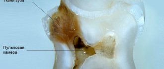
Human teeth have almost the same anatomical structure, including three main parts: crown, neck and root. Inside the tooth there is a pulp chamber, from which canals originate and extend all the way to the root. The pulp chamber contains the pulp or nerve of the tooth, which is a bundle of nerve fibers and blood vessels. Nerve fibers and vessels also pass through the entire internal space of the dental canal. The shape of the tooth canals can be either straight or curved, and their number varies depending on the type of tooth. Thus, the canines and incisors of the upper jaw have one canal, the canines and incisors of the lower jaw have two. There may be two or three canals in molars, and the maximum number of canals is observed in wisdom teeth - from three to five. The exact number of dental canals is determined using radiographic examination.
As mentioned above, the shape of the canals can have bends and turns, which complicates the process of their high-quality cleaning and treatment. Meanwhile, when treating canals, it is important to thoroughly clean their internal space - otherwise it will not be possible to stop the inflammatory process and save the diseased tooth.
How is removal carried out?
Before the operation, an x-ray is prescribed and a diagnosis is made based on laboratory tests. From the image, the doctor determines a certain type of growth, an abnormal location of the wisdom tooth. It also examines infectious and inflammatory foci in this area and prescribes preliminary treatment. The dental surgeon performs the operation under local anesthesia, which begins to take effect within 10 to 15 minutes. After exposure to anesthesia, he begins to excise the excess mucous membrane hanging over the tooth with a scalpel. Removing a wisdom tooth hood involves several main steps:
- Disinfection of gums and segments of the oral cavity;
- Excision of the “hood” with a scalpel;
- Rinsing the cavity with an antiseptic;
- Stopping bleeding and disinfection with iodoform turunda;
- The use of anti-inflammatory drugs, wound healing ointment.
Indications for dental canal treatment
Tooth canal treatment is a specific dental procedure that is advisable to perform in the following clinical cases:
When an active inflammatory process is detected inside the root of a dental unit. The inflammatory process in this part of the tooth can lead to gradual tissue necrosis. If tissue necrosis begins, the diseased tooth will have to be removed.
The condition is diagnosed by radiography;
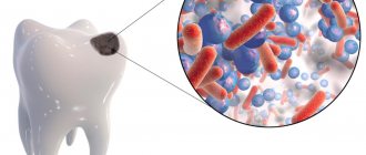
Treatment of tooth canals is required for pulpitis - inflammation affecting the pulp bundle;
Endodontic treatment is carried out for periodontitis, a disease affecting the apical part of the tooth root;
In some cases, cleaning and treatment of dental canals is required for advanced forms of caries.
Dental canal treatment is also indicated for abscesses, the onset of an inflammatory process under an old filling, or a fractured tooth root. The need for measures to treat tooth canals may be indicated by such symptoms as: severe, excruciating pain in the tooth, occurring mainly at night, swelling of soft tissues, discoloration of the gums and tooth enamel.
In some cases, tooth canal treatment is carried out before prosthetics. The nerve is removed from the tooth, and the canal cavities are cleaned, treated with antiseptic drugs and filled.
Indications
Manipulation is prescribed in cases where it is necessary to gain access to the inside of the tooth for:
- vitalization or removal of the nerve;
- canal obturation;
- pin installation, etc.
As a rule, trepanation is performed for deep carious cavities in the coronal part with a thinned septum between it and the pulp chamber. Pathogenic microorganisms can penetrate through the thinned wall, which leads to infection of the dental nerve. Trepanation is also indicated for pulpitis in chronic or acute form, or any form of periodontitis.
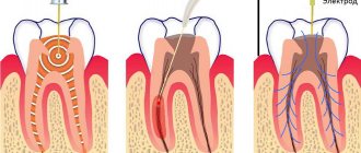
Main stages of root canal treatment
Treatment of tooth canals is a complex, specific process that includes several successive stages. Below we will consider all stages of the treatment process in detail.
Diagnostics
During the initial visit to the dentist, the doctor will conduct a thorough examination of the patient’s oral cavity and will also prescribe x-rays. An image of the tooth will allow you to assess the condition of the dental canals, see their number, determine the stage of development of the inflammatory process, and also select adequate treatment for a specific clinical case.
Anesthesia
To gain access to the canals of a tooth, the dentist will drill out the coronal part of the tooth to reach the pulp chamber, behind which the canals are located. Both the pulp and the canals are penetrated by nerve fibers and therefore the specialist’s actions can cause some discomfort to the patient. To relieve a person of discomfort, local anesthesia is performed before starting root canal treatment. The drug is selected individually for each patient.
Moisture insulation of the area of the diseased tooth
To prevent pathogenic microorganisms that may be contained in saliva from entering the internal space of the tooth, the area of the diseased tooth is first protected from moisture by covering with a special latex bandage - a rubber dam.
Reaming a tooth
To gain access to the dental canals, the doctor will drill the tooth, removing all tissue affected by caries. The access hole is made either on the chewing surface of the tooth (when treating molars) or on the inside (when treating incisors and canines). After drilling, the doctor will remove either the entire pulp bundle or only part of the tissue affected by the inflammatory process.
Treatment of dental canal cavities
For high-quality treatment of dental canals, a specialized tool is used - files. Before the cleaning process, it is important to obtain detailed information on the structure of the dental canals, as well as their length. To do this, a photograph of the diseased tooth is taken, and a special type of device is used - an apex locator. The dentist will use the files to clean the canal cavities, and then rinse them with an antiseptic solution and treat them with antibacterial agents. During cleaning and antiseptic treatment, the dentist will expand the cavity of the canals, clean and smooth their internal surfaces. Then the cavities of the dental canals are thoroughly dried with paper points.
Next, the doctor puts medicine into the canal cavity, places a temporary filling on the tooth and sends the patient home. After a few days, you should come back to the dentist to evaluate the results of the treatment. If the image shows that the inflammation has been stopped, the canals are filled.
Sealing
Gutta-percha pins are used to fill dental canals. It is important that the dental canal is sealed correctly and efficiently, otherwise a recurrence of inflammation is possible and the tooth cannot be saved. After filling the canals, the crown part of the tooth is restored with a photopolymer filling or orthopedic structures. It will be useful to know that mild pain in the tooth within 14 days after the procedure is considered normal by dentists. Taking painkillers prescribed by your doctor will help eliminate discomfort. However, if the pain does not go away, its character is sharp, and does not subside even after taking anesthetics, you should immediately contact the dentist!
Dental caries:
To exclude pulpitis, a thorough curettage of the periodontal pocket is carried out and hygienic measures in the oral cavity are recommended. The cessation of pain after curettage, which most often happens in the presence of a slit-like gap, confirms the diagnosis of inflammation of the gingival papilla - papillitis. If pulpitis is suspected, the reaction of the pulp to cold and hot is determined by EDI. —Treatment is reduced to removing the previously applied filling and filling using a contour matrix, which ensures the creation of a contact point at the equator level. In some cases, it becomes necessary to replace seals on two contacting surfaces. A prerequisite for successful treatment is the correct fixation of the matrix using a wedge, which should be inserted between the teeth with force, which ensures displacement of the tooth by the thickness of the matrix.
In the case of tooth displacement and an increase in the interdental space, it is not possible to create full contact. In such cases, the anatomical shape of the tooth is restored, leaving an interdental gap of significant size, which eliminates the possibility of food retention.
10. Overhanging edge of the filling.
This is a common mistake when filling, which occurs when the matrix is applied incorrectly, if the wedge does not press the matrix tightly to the tooth surface or is not used at all to fix the matrix. However, most often an overhanging edge of the filling occurs, sometimes occupying the entire interdental space, when not a matrix and matrix holder are used, but a metal plate. Unfortunately, even today there is one filling on two adjacent teeth.
When filling a class II cavity, a matrix must be used, and its lower edge must be tightly pressed with a wedge to the surface of the tooth being filled. The use of a metal strip (not a matrix) may be indicated if the class II carious cavity is small in size and located at the level of the equator, as well as with buccal access to the carious cavity, which is discussed earlier.
11. Loss of a filling immediately or a short time after its application
. This may be a consequence of a number of factors: violation of the principles of preparation, incorrect choice of filling material, violation of filling technology. One of the most likely reasons is insufficient drying or incomplete polymerization of the material. In this regard, it is necessary to pay attention to isolation from saliva and control of the power of the polymerization lamp.
12. Pain after filling
may arise for a number of reasons. First of all, this may be a consequence of preparation without sufficient water cooling. Pain is also possible if an insulating lining made of glass ionomer cement is applied and filled with a composite on the same day. This is explained by the different polymerization times of GIC (within 24 hours) and the composite, which may result in displacement of the gasket.
13. Pulp necrosis after filling.
Currently, filling materials (Evicrol, Consize), which have an irritating effect on the pulp, are practically not used. In addition, real adhesive systems practically eliminate the possibility of this action, but this should be kept in mind. In addition, pulp necrosis is possible due to cavity preparation without cooling.
14. Excessive removal of the composite into the gingival groove
accompanied by inflammation - hyperemia and bleeding. In addition, this is one of the reasons for the accelerated partial or complete loss of the restoration. The presence of a step at the tooth tissue-composite interface indicates the need for careful grinding and polishing. For this purpose, fine-grained diamond polishers or carbide burs are used. The criterion for quality work is the imperceptible transition of the probe from the surface of the restoration to the tooth.
15. Stability of tooth color after restoration (filling).
Previously used composites (Evicrol, Consize, etc.) did not guarantee the stability of the color of the restoration. Moreover, over time, after 2-4 years, a change in color was usually noted. A yellowish tint appeared, gloss was lost, etc. Currently produced composites practically do not change color. Therefore, a change in the color of the restoration indicates errors during filling. First of all, this is due to the wrong choice of the color scheme of the filling material or the non-use of opaque. Due to the fact that the color of a tooth with high transparency of the enamel is determined by the thickness of the dentin layer, during restoration it should be based on an opaque layer. If this provision is violated, based on the presented premise, the following errors may occur.
15.1. Wrong choice of restoration color.
15.2. Highlighting the restoration area (whitish tint) with the correct choice of tooth color. This is due to the fact that the base of the filling is not made of opaque (the color of dentin), which determines the color of the tooth.
15.3. Identification of the contours of the sealed cavity. This occurs in the absence of a bevel of the enamel, which does not ensure a gradual transition of the tooth color to the color of the restoration.
15.4. The presence of an altered area with full color matching of the main surface of the restoration is due to either insufficient excision of the altered tissues, or covering the altered dentin layer with a thin layer of opaque, or refusal to use opaque.
15.5. The appearance of whitish “veins” on the surface of the restoration. The reason is insufficient condensation of the newly applied composite layer, as a result of which there is no close contact between the previously cured layer and the newly applied one. To eliminate these shortcomings in the selection of the color of the restoration, it is recommended to carry out partial or complete removal of the restoration and repeat it.
16. Filling of premolars and molars without the formation of tubercles and fissures of the chewing surface.
The consequence of this may be a change in the bite. In this case, it is recommended to carry out a new restoration.
How long does root canal treatment take?
The duration of tooth canal treatment depends on the complexity and characteristics of the clinical case, as well as the number of canals that the specialist will have to treat. On average, the duration of the procedure can range from 20 minutes to 1.5 hours.
It is also worth considering that for high-quality root canal treatment, you will have to visit the dentist’s office several times.
Is it painful to treat root canals?
Thanks to the use of modern anesthetics, root canal treatment has become a painless procedure. The patient may feel mild discomfort only during the injection of the anesthetic.
Therefore, there is no need to be afraid of root canal treatment; moreover, you must remember that you should never postpone the procedure due to fear of the dentist! Inflammation in a tooth that has progressed to a certain stage of development cannot be cured, which means that the diseased tooth will have to be removed and a prosthesis put in its place. In addition, more serious complications may occur.
Modern method of treating tooth canals: treatment of the canal cavity under a microscope
The quality of dental canal treatment depends on the correctness and accuracy of cleaning and processing of their internal space. This is a very painstaking work, since the channels themselves are quite narrow (no more than one millimeter), and in addition, they can have bends and turns. If a specialist makes a mistake when treating the canals and does not remove particles of the affected dentin, a relapse of the inflammatory process is guaranteed. The use of a special tool in treatment - a dental microscope - will help eliminate possible errors and inaccuracies in the treatment of dental canals.
Using a microscope, the dentist will perform a high-quality removal of all infected tissues, while preventing injury to healthy tooth tissues. The use of the device simplifies the process of cleaning and processing channels with significant lengths, anomalous structure,
with curvatures and branches. In addition, the use of a dental microscope during the treatment of tooth canals improves the quality of tooth restoration as a whole, and also contributes to the timely detection of hidden caries.
At our dental clinic “Uni Dent” in St. Petersburg, you can receive services for dental canal treatment under a microscope. The procedure is carried out by highly qualified doctors using modern instruments and materials, and therefore when you contact us, you can rest assured that the treatment result will be of high quality! Dentistry "Uni Dent" - come to us for a beautiful smile and painless dental treatment!