From this article you will learn:
- what methods of treating pulpitis allow you to keep the tooth alive,
- what is the vital amputation method,
- stages of biological treatment of pulpitis.
The biological method of treating pulpitis is a conservative method that is aimed at completely preserving the pulp as viable. The use of this method is possible only if the following 2 conditions are met. Firstly, the patient should seek help from a dentist at the earliest stage of the development of pulpitis, i.e. at the very beginning of the development of pulp inflammation. Secondly, the age of the patient to whom the doctor can offer this method is very important, because Treatment without nerve removal is carried out only in children, adolescents and people no older than 25-27 years.
The latter is associated with the ability of the dental pulp to heal itself, which also depends on the age of the patient. In addition to the biological method, there is also a method of vital amputation, in which the pulp remains partially viable. This method involves removing the pulp only from the coronal part of the tooth, preserving it in the root canals. But the indications for such therapy are limited - it can only be carried out in multi-rooted teeth (they have a clearly defined transition between the coronal and root pulp).
Important: in dentistry, unfortunately, doctors very rarely offer conservative methods for treating pulpitis, even if the patient is suitable for their use. This is due to the flow of patients and the desire to use the usual familiar methods. It is best to ask your doctor whether such treatment methods are indicated for you, and if he answers “no,” then you must clarify: “why this method is not indicated specifically for you.”
Conservative (biological) method of treating pulpitis (without removing the pulp)
The biological method is based on the properties of the pulp, after research of which its ability to recover was proven. Based on the plastic capabilities of the pulp, the conservative method of treating pulpitis aims to completely eliminate inflammation in the pulp and restore its biological function. The method is used in the initial phase of development of the inflammatory process or in case of accidental opening of the pulp; treatment should be started quickly to prevent inflammation from progressing to a more severe stage.
Indications and contraindications of the biological method
Conservative technique is indicated in the following cases:
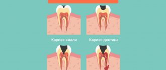
- Pulp hyperemia is a condition caused by the expansion of the lumens of the blood vessels of the pulp. Accompanied by minor inflammation and increased sensitivity to cold irritants. Pulp hyperemia occurs when deep carious cavities appear, during tooth treatment for a crown, or when the technology for installing composite fillings is not followed. Hyperemia is a reversible process, therefore only a biological method is used to treat it.
- Accidental exposure of the pulp can occur during the preparation of a deep carious cavity, as well as as a result of tooth trauma. The conservative method in this case is used if there is no prolonged bleeding from the tooth cavity; if no more than a day has passed since the tooth was injured and the pulp was exposed.
- Acute pulpitis of a limited nature is the initial stage of the process, in which the integrity of the walls of the blood vessels of the pulp is preserved.
Biological pulp treatment is performed in the presence of the following additional conditions:
- The patient's age is not older than 30 years (there are exceptions). This method is often used to treat permanent teeth that have not yet formed roots.
- No changes in the periodontium confirmed by x-ray examination.
- There are no signs of periodontitis development.
Contraindications to biological pulp treatment:
- Significant reduction in electrical excitability of the pulp (indicators of electrical excitability differ for single-rooted and multi-rooted teeth, for different age categories of patients).
- Changes in the periodontium.
- Signs of changes in periapical tissues.
- Using a tooth as a support for a bridge.
- A carious cavity is located in the area of the neck or root of the tooth.
Methodology for biological pulp treatment
1. Indirect pulp capping method - used when there is a thin layer of healthy or partially decalcified dentin, which protects the pulp from exposure and injury. Treatment is carried out in the following sequence:
- Local anesthesia.
- Removal of tissues affected by caries.
- Apply a calcium hydroxide-based medicinal dressing to the remaining layer of dentin. The composition of therapeutic dressings may include antibacterial agents, enzymes, vitamins and glucocorticoids in order to accelerate the elimination of inflammation and stimulate regeneration.
- Placement of a temporary filling.
- A week later - a second visit. If the patient has no complaints, the temporary filling is replaced with a permanent one. If the pain persists, apply a new therapeutic bandage for several days; if the pain does not disappear after that, surgical treatment of pulpitis is indicated. In some cases, the period between the installation of a temporary and permanent filling can be up to 6 months.
2. Direct pulp capping method - used on accidentally exposed pulp.
The medicinal dressing is applied directly to the exposed pulp; under the influence of medications, its plastic function is restored - dentin begins to form. An important point when applying a therapeutic dressing is careful removal of the blood clot formed during bleeding during exposure of the pulp. If this is not done, the clot will prevent contact of the dressing with the pulp, making treatment more difficult.
Third visit to the dentist.
If the canal filling was completed efficiently, then by the third visit to the dentist the patient will have no pain or other unpleasant symptoms. Having checked this, the doctor removes the temporary filling, applies medicine and begins to recreate the tooth. With the help of modern filling agents and restoration methods, the previous shape of the tooth with all functions is restored. Then the fruit of the dentist’s creativity is polished to a shine and its surface is fluoridated with special means. This protects the remaining “living” tissues from bacteria and strengthens them. Devital pulp extirpation method
popular among dentists and patients as effective, reliable and efficient.
Additional methods of conservative treatment of pulpitis
Physiotherapeutic treatment is used as an addition to the biological treatment method. The following procedures are shown:
- Helium-neon laser - its radiation has an analgesic effect, helps reduce swelling, reduce inflammation.
- Electrophoresis - using this method of electrotherapy, anesthetics are injected into the site of inflammation.
- Ultrasound therapy – low-frequency ultrasound stimulates regenerative processes and accelerates the delivery of medicinal substances to tissues.
After treatment of pulpitis with a biological method, patients must undergo electroodontic diagnostics in order to monitor the condition of the pulp. If the electrical excitability of the pulp is not restored after treatment and pulp death is diagnosed, a depulpation procedure (pulp removal) is performed.
Why is it so important to keep the tooth alive?
The thing is that the pulp performs trophic (nourishes the tooth tissue from the inside) and protective functions. After its death, the hard tissues of the tooth become more fragile, in addition, removal of the pulp is tied to the need to remove a sufficiently large volume of hard tissues of the tooth. Also, very often after pulp removal, inflammatory foci appear in the area of the apex of the tooth roots, associated with poor-quality filling of the canals after pulp removal.
Of course, to avoid all this, it is better to try to leave the pulp alive. However, this is also not always possible, because Patients very rarely come at the very beginning of the development of pulpitis, when the use of conservative treatment methods brings good results. More often, patients come with diffuse purulent inflammation of the pulp, when only its complete removal is indicated - followed by filling the root canals.
Read more about the treatment method with complete removal of the pulp in the article: → “Traditional methods of treating pulpitis”
Surgical methods for treating pulpitis
Vital amputation
Another name for the procedure is vital pulpotomy . The technique consists of performing a partial depulpation operation, that is, removing an area of the pulp susceptible to inflammation. The coronal pulp is removed, while the viability of the root pulp is preserved. This method is important in the treatment of pulpitis in multi-rooted teeth with immature roots.
Sequence of pulpotomy:
- Pain relief with injection of local anesthetic.
- Removal of tissues affected by caries.
- The tooth cavity is then opened and exposed to allow access for the next step.
- The dentist performs a pulpotomy - removes the coronal part of the pulp.
- If bleeding from the pulp occurs, it is stopped using tampons or a hemostatic sponge.
- A medicated pad with calcium hydroxide is applied to the root part of the pulp.
- Applying an insulating gasket and installing a temporary filling.
- After 1-4 weeks, replace the temporary filling with a permanent one (after confirming the viability of the pulp using electroodontodiagnostics).
Vital extirpation
Another name for the procedure is vital pulpectomy . As a result of using the technique, the pulp is completely removed. The method is used for all forms of pulpitis, as well as after a pulpotomy, when the inflammatory process continues to develop in the root part of the pulp. Depulpation using the vital extirpation method is carried out in one visit to the dental office.
Sequence of the procedure:
- Local anesthesia. Pulp anesthesia is provided by infiltration, conduction, intraligamentary anesthesia, their combination, or, less commonly, general anesthesia. Sometimes intrapulpal anesthesia is used. The operation to completely remove the pulp lasts 1-1.5 hours, so anesthetics must be guaranteed to provide anesthesia for this period.
- Preparation. Preparation of a carious cavity (removal of pathologically altered tooth tissues - enamel and dentin) is carried out to create access to the root canals. In other words, the dentist performs caries treatment
- Opening of the tooth cavity. When properly opened, the tooth cavity should merge with the carious cavity, without forming overhangs or bends at the border. The opening and development of the cavity is carried out in such a way that its walls smoothly transition into the walls of the tooth cavity;
- Pulpotomy. Amputation of the coronal part of the pulp, access to the mouths of the root canals is prepared;
- Expansion of canal mouths. At this stage, conditions are created for further endodontic treatment.
- Pulpectomy. The root pulp is removed; for this purpose, a pulp extractor is used - an endodontic instrument that is inserted into the root canal; when it is rotated around its axis, it engages with the pulp and is removed along with it. It is important that there are no pulp particles left in the root canal, as this causes infection and the development of residual pulpitis and periodontitis.
- Channel research. It is carried out by probing them with root needles, drills and drills with limiters. The exact parameters of the canal during the study (working length of the canal) are obtained using an apex locator and a visiograph;
- Stop bleeding. If bleeding occurs during pulpectomy, a turunda impregnated with a hemostatic agent is inserted into the root canal. Bleeding is also stopped using diathermocoagulation - a needle is inserted into the root canal, which is an active electrode, and an electric current is applied with high frequency, high strength and low voltage. As a result of this effect, blood clotting occurs, bleeding stops and necrosis of the remaining pulp occurs.
- Mechanical and medicinal treatment of canals. At this stage, the dentist must completely remove remaining pulp and infected tissue from the canals and prepare them for filling by giving them a cone shape. In addition to the mechanical treatment of each canal with special tools, medicinal treatment is carried out: the canal is washed with antiseptic solutions or cotton wool soaked with medicinal substances is inserted into it. Solutions used: sodium hypochlorite, furatsilin, chlorhexidine, hydrogen peroxide.
- Drying of the root canals is carried out with turundas soaked in alcohol and then ether and air;
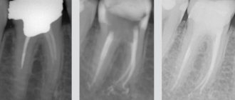
- Filling canals, for example, using three-dimensional obturation using liquid gutta-percha. It is performed using special filling materials, which have many requirements: they must not stain the tooth over time, fit tightly to the canal walls, be radio-opaque, do not shrink, not cause irritation to surrounding tissues, and much more. Filling the root canal is a very important final stage; the success of endodontic treatment depends on the quality of filling the canal. Filling of canals is carried out with gutta-percha - this material best meets the requirements for modern materials for filling root canals, and therefore provides a reliable treatment result. The quality of canal filling is checked using an x-ray image - the canal must be completely sealed, without voids.
- Installation of a temporary filling.
- Installation of a permanent filling. It is carried out a few days after the installation of a temporary filling. Excess canal-filling material is removed from the canal, an insulating gasket and a permanent filling are applied.
Treatment of pulpitis in Dental World is carried out using a dental microscope, which provides an enlarged image of the surgical field, this improves the quality of treatment and canal filling.
Devital extirpation
Previously, patients referred to this method as “putting arsenic.” The technique involves removing the pulp after it has been previously devitalized ( necrotization ) by applying medications. Devital extirpation is carried out if it is impossible to perform depulpation using the vital pulpectomy method. For pulpitis of permanent teeth with unformed roots, this method is not used.
To devitalize the pulp, arsenic acid paste is used. The paste also includes painkillers, antiseptics and anti-inflammatory agents, glucocorticoids and components that slow down the absorption of arsenic into tissues, since the substance is highly toxic to the body.
Arsenic paste is applied after removing softened dentin and opening the pulp. Arsenic paste is applied to single-rooted teeth for 24 hours, and to multi-rooted teeth for 48 hours.
A devitalizing agent with less toxic properties is paraformaldehyde paste. It causes pulp necrosis within a week in single-rooted teeth, and within 10-14 days in multi-rooted teeth.
Depulpation using the devital pulpectomy method is carried out in two visits:
- After treating the carious cavity, a devitalizing paste is applied to the pulp. A temporary filling material is placed.
- The temporary filling is removed, the tooth cavity is opened, and the pulp is removed. Next, the cavity is washed, dried, the root pulp is removed, the canals are treated with medications and filled - the same as with vital pulpectomy, but without the use of anesthesia, since after devitalization and death of the pulp, the tooth becomes insensitive to irritants.
Devital amputation
The devital amputation technique is used for complete obstruction of the canals. After applying the devitalizing composition, the necrotic coronal pulp is removed and the root pulp is impregnated using resorcinol-formalin paste or other mummifying compounds. After 2-3 times impregnation, the root pulp polymerizes and cannot rot.
This method is rarely used in clinical practice and, as a rule, in weakened patients who have suffered a myocardial infarction, stroke, or various severe operations. After removal of the devitalized coronal pulp, mummification of the root pulp is carried out using impregnation of resorcinol-formalin or any of the mummifying pastes.
The development of pulpitis can be prevented if you monitor the condition of the oral cavity and treat caries in a timely manner, since most often pulpitis develops as a consequence of a deep-seated carious process. A preventive examination by a dentist, which should be carried out twice a year, helps to detect caries at an early stage.
First visit to the dentist.
On the first visit, a complete sanitation of the diseased tooth is carried out. After anesthesia, all infected tissues are removed, the tooth is treated with medications and an opening is created to access the pulp. A small amount of arsenic paste is placed there. Which should kill the tissue. In teeth with one root, this process takes a day; for teeth with two roots, it will take 48 hours. If the second visit cannot be scheduled so quickly, then a similar substance is given, only with a slower effect. For a period of one to two weeks. After placing the paste, the hole is closed with a temporary bandage.
Will we treat him or let him live?
Often the fear of doctors forces us to make do with improvised methods. We take painkillers, fast, endure, in the hope that the sharp pain will go away on its own. Is it really possible to “wait out” an exacerbation, or is it better to get professional help in time? Let's figure it out.
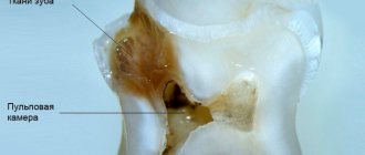
To find the right method of treating a disease, you need to know the origins of its occurrence. The core of the tooth, or pulp, is responsible not only for tooth sensitivity, but also for the production of dentin. The tissue strengthens the tooth from the inside.
The anatomical features of the pulp depend on the type of teeth. In the lateral row, as a rule, there is a chamber with three dental canals. The front row and incisors each have one branch. It is logical that molars are much more difficult to treat than canines or premolars.
Pathological changes in the pulp tissue form swelling, causing unbearable pain. The pain intensifies upon contact with hot food or when pressing on the tooth.
Survey
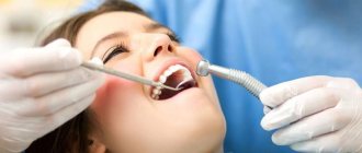
To determine an accurate diagnosis, the dentist conducts an initial examination. Because If pulpitis develops against the background of other diseases, then diagnosis includes 4-5 stages. The procedure begins with communication with the patient. It is important to understand at what stage the inflammation is, which is especially important in chronic pulpitis. Therefore, the doctor asks you to describe in detail the nature of the pain. Then a “manual” examination is carried out using medical instruments (dental mirror, etc.). Additionally, the doctor checks the sensitivity of the affected tooth for temperature changes and exposure to a weak electric current charge.
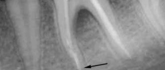
The examination is completed by an X-ray examination to assess the condition of the dental nerve and canals. After collecting information and analyzing the image, the doctor plans the pulpitis treatment process and coordinates it with the patient.
Possible complications
If you delay visiting the dentist, inflammation can spread to the bone tissue of the tooth - periodontium. This is how one of the most common and dangerous complications of this disease begins - periodontitis, which can lead to tooth extraction. Periodontitis also occurs due to an unskilled approach to cleaning root canals.
If you are concerned about an elevated temperature after treatment for pulpitis, urgently contact a more reputable dentist, as the inflammation is progressing. Professional clinics use modern methods of canal treatment using microscopes, binoculars, visiographs, endomotors, and apex locators. These instruments prevent the risk of complications.
Unfortunately, pulpitis remains a common occurrence after caries treatment. The reason for its appearance is the same - the unprofessional actions of a doctor who violated medical protocols and made mistakes when filling. Perhaps he accidentally opened the pulp and gave bacteria access to it.