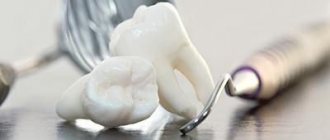
Etiology
The causes of fibrous pulpitis can be different. As already mentioned, it can be the outcome of an acute form of inflammatory processes, in which the crown of the tooth is opened and exudate flows out, or it can occur primarily. The causes of pathological processes are:
- medical errors during caries treatment;
- advanced caries;
- dental injuries.
In our clinic you can get a free dental consultation!
Symptoms of chronic pulpitis
Chronic pulpitis affects the dental pulp tissue and is much more common than acute pulpitis (approximately 75 to 25 percent). It can be either an independent (primary) disease or the next stage of an acute one (on average, the transition is noted after 10–12 weeks from the onset of the acute form). There is chronic pulpitis of permanent and primary teeth, and in childhood, if possible, it is recommended to carry out tooth-preserving manipulations in order to minimize the risk of disruption of the development of permanent teeth.
The disease can be almost asymptomatic, therefore, with chronic pulpitis, patients often have no complaints: pain is dull and occurs quite rarely. In contrast to the acute form, in which the pain is usually sharp and pronounced. However, the symptoms of chronic pulpitis are not limited to pain. Therefore, with due attention to the reactions of your body, noticing the problem is not so difficult.
Complaints with chronic pulpitis
- Painful sensations.
If pain manifests itself, it is almost always dull and uniform (has no peak or decline). Exacerbation of chronic pulpitis is characterized by more severe attacks. - Reaction to stimuli.
In most cases, a reaction to a stimulus (for example, cold or hot) occurs only after some time. - Bleeding while eating.
It appears only if granulation formations appear in the pulp.
Our doctors
18 years of experience
Baghdasaryan
Armen Evgenievich
Chief physician, dentist-orthopedist-therapist
Graduated from VSMA named after. N.N. Burdenko. Internship on the basis of MGMSU named after. A.E. Evdokimov in “General Dentistry”.
Clinical residency at the Moscow State Medical University named after. A.E. Evdokimov in “Orthopedics”.
More about the doctor...
5 years experience
Sadina
Ekaterina Vladislavovna
Dental therapist, surgeon
Penza State University Medical Institute, specialty “Dentistry”.
In 2021, she underwent professional retraining in the specialty “Therapeutic Dentistry” at the Moscow State Medical and Dental University named after A.I. Evdokimov.
More about the doctor...
8 years of experience
Arzumanov
Andranik Arkadievich
Dentist-orthodontist
Graduated from Moscow State Medical University. Internship - Moscow State Medical University at the Department of Orthodontics and Children's Prosthetics.
Residency at Moscow State Medical University at the Department of Orthodontics and Children's Prosthetics. Member of the Professional Society of Orthodontists of Russia since 2010.
More about the doctor...
Clinical manifestations
- Feeling of heaviness in the affected tooth;
- The occurrence of aching pain symptoms in response to exposure to stimuli of a thermal or mechanical nature;
- Unpleasant odor from the mouth;
- Irradiation of pain into the gums and temporal region;
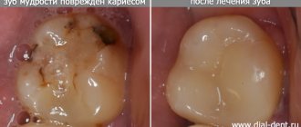
- The presence of a large carious cavity in which the dentist finds a large amount of destroyed dentin.
Diagnostics
It is important to distinguish the fibrous variety from the gangrenous and focal ones, as well as from diseases such as advanced caries. In order to make a correct diagnosis, responsible specialists conduct not only a dental examination, but also electrodiagnostics along with radiography. During the examination, the dentist reveals:
- intense pain symptoms during probing;
- reaction to exposure to high temperatures;
- pulp bleeding.
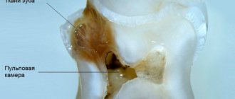
On the x-ray you can see that there is no barrier between the carious and dental cavities, there is an expansion of the periodontal gap.
Diagnosis of chronic pulpitis
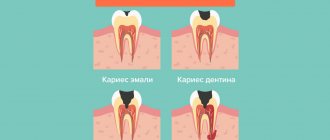
The disease most often occurs as a complication of caries, when bacteria enter the pulp and cause an inflammatory process in it. However, the penetration of microorganisms is possible with dental injuries, gum disease (through periodontal pockets), as well as with sinusitis and osteomyelitis (much less often). Diagnosis of chronic pulpitis is carried out using a number of techniques that make it possible to accurately determine the form and stage of the disease.
- Visual inspection.
With the help of a dental probe and a mirror, you can almost always notice damage to the teeth and the development of the disease (the exception is chronic pulpitis under a filling). - Radiography.
The most common way to diagnose most dental diseases. - EDI (electroodontodiagnosis).
Use of electrical current to assess the viability of pulpal nerve endings. - Thermometry.
Exposure of the pulp to temperatures to assess its condition.
Treatment
Fibrous pulpitis requires surgical treatment, which involves partial or complete removal of the pulp. Treatment tactics are selected based on the results of diagnostic studies. Partial removal is more gentle, i.e. vital extirpation, which is used in patients no older than forty years. It involves removing the pulp in the crown and orifice of the tooth, after which the dentist applies a calcium pad and a temporary filling. If the patient does not experience pain, then a permanent filling is installed. Complete removal of the pulp is called devital extirpation. This technique is used in elderly patients if the patient is allergic to anesthesia or has obstruction of the root canals. It includes 2 visits to the dentist, during which:
- a devitalizing paste is applied and a temporary filling is installed;
- The temporary filling is removed, the pulp is removed, an insulating lining is applied, and the tooth crown is restored.
Anesthesia is used during the procedure, so the treatment is painless.
PATHOGENESIS OF THE DISEASE
Hypertrophic pulpitis is a fairly rare type of disease, most often found in young people. As a rule, it occurs in teeth with an open carious cavity. Under the influence of external irritating factors, the process of proliferation—cell multiplication—begins in the pulp (neurovascular bundle). As a result, the pulp chamber is filled with bleeding granulation tissue.
In the absence of timely medical intervention, the disease becomes chronic - a polyp is formed, tightly fused to the underlying tissue.
Where to contact?
Dentists at the Good Hands clinic have been treating fibrous pulpitis for several years now. You can contact us if you find signs of pulpitis. Our specialists will relieve toothache and provide appropriate treatment. They will make every effort to preserve the functionality of the tooth, however, if this is not possible, the pulp will be removed. Treatment in our clinic is painless and effective. You can schedule a free consultation with us and find out all the necessary information right now by filling out an application on our website or calling us by phone.
Clinical manifestations:
- Mildly manifested pain symptoms arising from exposure to irritants of various natures;
- Pulp bleeding;
- An almost completely destroyed dental crown and a deep carious cavity from which the pulp protrudes;
- Halitosis, which occurs due to the inability to carry out full hygienic procedures for the oral cavity.
In our clinic you can get a free dental consultation!
EXAMINATION OF THE PATIENT
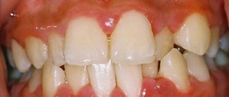
Visual inspection:
The skin of the face and visible part of the neck is of natural color, there is no pronounced asymmetry of the face, there are no scars or sinus tracts on the skin.
Regional lymph nodes (occipital, postauricular, submandibular, submental, facial, cervical, subclavian) are not palpable.
The red border of the lips is bright pink, moisturized, there are no scales, crusts and other lesions, swelling, abrasions, or tears.
Examination of the vestibule of the oral cavity:
The mucous membrane of the vestibule of the oral cavity is pale pink, moist, edema, there are no morphological elements of the lesion.
The depth of the vestibule of the oral cavity is average (7 mm).
The condition and level of attachment of the labial frenulum is within the physiological norm, there are no vestibule cords, there is no blanching or separation of the gums from the necks of the teeth when the lower and upper lips, cheeks, and tongue are abducted.
The bite is orthognathic, there is no crowding or dystopic teeth.
Examination of the oral cavity itself:
The mucous membrane of the hard palate is dark red, moderately moist, and there are no pathological elements. The level of attachment and length of the frenulum of the tongue are within the physiological norm.
| P | |||||||||||||||
| 8 | 7 | 6 | 5 | 4 | 3 | 2 | 1 | 1 | 2 | 3 | 4 | 5 | 6 | 7 | 8 |
| 8 | 7 | 6 | 5 | 4 | 3 | 2 | 1 | 1 | 2 | 3 | 4 | 5 | 6 | 7 | 8 |
| P | P | P | P |
The shape of the dental arches, the shape and size of the teeth correspond to the physiological norm: the position of the teeth in the dentition is not changed, the lateral teeth are pale yellow, the front teeth are dark yellow, there are no changes in the thickness of the enamel, the shine of the enamel is preserved. Mobility of teeth I degree. On the oral surfaces of teeth 12, 11, 21, 22, 34, 33, 32, 31, 41, 42 and 43 there is supragingival and subgingival calculus.
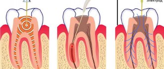
Depulpation in the treatment of a tooth with pulpitis
The depulping process is divided into several successive stages:
Stage 1
Using a drill, the dentist will remove a layer of enamel from the diseased tooth, as well as all dentin (hard dental tissue) damaged and destroyed by caries. In some cases, the doctor has to remove a small percentage of healthy tissue. This is required to gain full access to the dental nerve.
Stage 2
Measures are taken to protect the tooth being treated from moisture. Saliva, which contains large quantities of pathogenic bacteria, should not get into the open cavity and canals of the tooth. To protect the tooth, a thick latex scarf with holes for the teeth is used. This device is called a rubber dam, and it does a much better job of protecting the tooth from moisture than cotton swabs and a saliva ejector.
Stage 3
Using a special tool, a pulp extractor, the specialist will remove the nerve bundle from the pulp chamber. The pulp extractor has sharp serrations that help quickly remove the dental nerve when treating pulpitis.
Stage 4
The length of the tooth canals is measured.
Correct determination of the length of the tooth canals in the treatment of pulpitis is of fundamental importance. If the measurements are carried out with inaccuracies, the canal may be either underfilled or overfilled, which is equally bad, as this can lead to the re-development of inflammation in the treated tooth. To measure the canals, special equipment is used - an apex locator, which helps determine the exact location of the end of the tooth root. To measure the length of the canals, K-files connected to the apex locator are introduced into their cavity and when the file reaches the bottom of the canal, the equipment will generate a signal.
Stage 5
At this stage of treatment of pulpitis, the cavities of the tooth canals are treated, which involves their expansion, cleaning and treatment with antiseptics. When the doctor has processed all the canals, he will put a temporary filling on the tooth and send the patient home. In a few days you will need to come to the clinic again.
Stage 6
An image is taken that shows whether the inflammatory process was stopped or not. If everything is normal and no signs of inflammation are detected, the temporary filling is removed from the tooth, the canals are filled with gutta-percha, and the tooth itself is restored with a photopolymer filling. Each stage of treatment requires maximum care from the doctor, because the slightest mistake in the treatment of dental canals can lead to a relapse of the disease and the inability to save the tooth. Therefore, choose a clinic in which you will treat pulpitis competently: do not chase low prices, choose a well-equipped dentistry with certified and experienced specialists.
Forecast and prevention of fibrous pulpitis
It is very important to start treatment of fibrous pulpitis on time by seeking dental help. Exacerbation of fibrous pulpitis is quite painful and treating its advanced stage becomes increasingly difficult. A competent and timely approach to the treatment of fibrous pulpitis helps to eliminate pain and discomfort, preserve the tooth and prevent the spread of the pathological process to periodontal tissue.
Prevention of fibrous pulpitis consists of following hygienic rules for caring for the oral cavity, both personally and with the help of professionals. It is necessary to undergo regular dental examinations, keeping the condition of your own teeth under control, promptly treating caries and fibrous pulpitis.
Causes of fibrous pulpitis
Chronic fibrous pulpitis manifests itself when the coronal part of the tooth is opened, and the outflow of inflammatory exudate occurs through the carious cavity connecting to the pulp chamber.
Fibrous pulpitis can develop primarily, for example, in some people with low body reactivity. In this case, the disease develops independently in the closed cavity of the tooth.
Another reason for the occurrence of pulpitis is poor-quality treatment of deep caries: non-compliance with the rules of dental treatment, incorrect application of medicinal pads, etc.