Tooth root caries is considered the most dangerous. It is difficult to identify due to its unclear clinical picture. The risk that disease will lead to its removal is very high. It is almost impossible to determine pathology during a visual examination; an x-ray is required. Let's consider the features of tooth root caries: its causes, signs and treatment.
In this article
- Dental root caries: causes
- Symptoms of tooth root caries
- Diagnosis of root caries
- Treatment or tooth root extraction
- Drug treatment of tooth root caries
- Surgical treatment of tooth root caries
- Severe root damage
- When is the best time to remove a tooth?
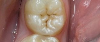
Based on localization, carious lesions are divided into 4 types: crown caries, cervical, basal and root caries. Damage to the root part occurs under the gum; upon examination, it is almost invisible. If the patient has no complaints of pain, the disease is not always detected even during a preventive examination.
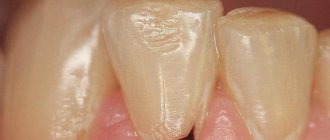
As a rule, pain occurs in cases where root caries has spread to the cervical area. However, at this stage it can already be detected during a visual inspection. The likelihood that the tooth will have to be removed at this stage of the disease is quite high. Moreover, tooth root caries can lead to infection of the gums and jaw, which can lead to various complications.
Often, this form of caries develops in older people, which is associated with atrophic periodontal processes, which are a consequence of age-related changes. Atrophy of the gums leads to exposure of the roots of the teeth, as a result of which they become more vulnerable to cariogenic microorganisms - the main “culprits” of caries. But this pathology can also develop in young people who do not take care of their oral hygiene. Tooth root caries affects 35% of pensioners; among young people it is diagnosed in 1.3% of cases.
Dental root caries: causes
Root caries occurs as a complication of gum disease, in which pathological pockets form between the tooth and gum. They accumulate food debris and pathogenic microbes. They are difficult to remove with a toothbrush and require professional cleaning at the dentist's office.
Without proper oral care, cariogenic bacteria multiply rapidly. They secrete acids that corrode the cement of the tooth root. Gradually, microorganisms penetrate deep into hard tissues down to the pulp, causing pain.
Often this pathological process is caused by osteomyelitis, periodontitis, periodontitis or periostitis, that is, inflammatory diseases of the gums and hard tissues of the oral cavity. Indirect causes of the development of tooth root caries are past infectious diseases and reduced immunity. In addition, the likelihood of carious lesions increases with tobacco smoking, alcohol abuse, poor diet and lack of vitamins and minerals in the body.
If a person rarely brushes his teeth or does it incorrectly, the carious process spreads even faster. As a result, you can lose a tooth without even feeling symptoms of the disease. As a rule, the first signs appear after the pathology has affected most of the tooth root.
How to prevent the development of inflammation
Prevention of periodontal inflammation includes the following recommendations.
- Oral hygiene. Brushing your teeth twice a day and using dental floss will help regularly remove bacterial plaque and tartar deposits from the surface of your teeth and gums.
- Use of toothpastes with fluoride and fluoride compounds, the dosage of which should not exceed the norm (1.5 thousand ppm for adults and 1 thousand ppm for children under 12 years of age).
- Balanced diet. Dentists recommend minimizing the consumption of sugar, sucrose, as well as sugar-containing foods and drinks (especially carbonated sweets). At the same time, the table should have fresh vegetables, seasonal fruits, dairy products without preservatives, protein-rich foods and complex carbohydrates on the table every day.
- Regular removal of tartar at the dental clinic (as needed).
Remember that regular visits to the dentist every 6 months for a routine examination are the key to ensuring that your teeth will always be in excellent shape. As mentioned above, the main reasons leading to the appearance and development of apical periodontitis are untreated advanced forms of caries and pulpitis, and a timely visit to the doctor will help avoid unpleasant consequences.
Symptoms of tooth root caries
Ordinary caries goes through four stages of development: initial, superficial, middle and deep. A person notices the first signs already at the second stage of the disease, when increased sensitivity of the enamel appears after eating sweet foods. However, tooth root caries occurs in a hidden form. Symptoms appear only when the lesion reaches a large size and the dental nerve is exposed. The carious cavity becomes so extensive that it becomes visible to the naked eye. It is localized near the gum and under it.
At this stage, there are many disturbing symptoms:
- Acute pain when cold, hot, sweet or sour food hits the nerve.
- Severe pain when the tooth is exposed to chemical irritants: rinses, medications, toothpastes.
- Gum hypersensitivity. Discomfort while chewing food, bleeding.
- Tooth displacement to the side, loosening, especially after eating.
- Inflammation of nearby soft tissues, dark plaque appears on the lower part of the dentition.
- Bad breath, which is almost impossible to eliminate with the help of pastes, rinses and chewing gum with a refreshing effect.
At first, pain only occurs during eating, but as the pathology develops, the pain becomes constant. Even strong analgesics do not help eliminate it.
Diagnosis of root caries
During a visual examination, dental root caries can be detected only in 13% of cases. If this disease is suspected, additional examination methods are prescribed:
- Probing. The doctor inserts an instrument with a curved tip under the gum and examines the structure of the tooth root, assessing its integrity. During such a study, it is possible to identify chips, roughness, and irregularities that indicate the development of pathology.
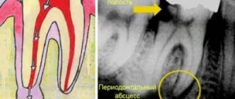
- Radiography. The dentist orders an x-ray of one tooth or the entire jaw. The images show the localization of the pathological area with an accuracy of up to a millimeter. In addition, radiography allows you to assess the condition of neighboring teeth and identify hidden inflammatory processes.
- Thermal diagnostics. The study is carried out using a directed stream of water, which is used to treat the tooth from different sides. After such a procedure, it is possible to determine which areas of the root are susceptible to carious lesions.
- Electroodontometry is a diagnostic of pulp viability, allowing you to find out whether the nerve is damaged and how deep the inflammation has penetrated.
- Visiography. Using a special device called a visiograph, the dentist scans the tooth in different projections. He can then view the root from different angles on a computer.
Before treating the root of a tooth, the dentist suggests the patient undergo ultrasonic cleaning to clean the enamel of plaque. Tartar may obstruct your view during the procedure. Based on the results of the examination, the doctor determines whether the tooth can be saved or whether it needs to be removed.
How is an overgrown tooth removed?
Root extraction is a fairly common dental operation. It is performed under local anesthesia and, as a rule, does not cause complications. An overgrown root cannot be removed using traditional methods, since it is completely covered with gum tissue and there is no access to it. Treatment is carried out in several stages:
- Examination by a dentist, x-ray examination. X-ray allows the doctor to see the size, position, and depth of the root. The dentist assesses the risk of damage to adjacent teeth during surgery.
- The operation is performed under anesthesia. First, the doctor makes an incision in the gum, providing himself with access to the root. After this, the root is removed and sutures are applied.
- During the rehabilitation period, the doctor selects anti-inflammatory drugs and agents that accelerate wound healing.
The process of removing an overgrown root is individual in each case. The complexity of the operation is influenced by many factors:
- Tooth location. Removal of roots on the upper and lower jaws has its own characteristics and is carried out using various dental instruments.
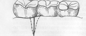
- The degree of damage to the root and nearby tissues, the presence of fragments. Sometimes, for a successful and as painless removal for the patient as possible, the root is destroyed by a drill into several parts. Separation is required in cases where the tooth has several strong, deep roots. As a rule, after destruction, individual parts are easily removed using ordinary tools.
- The presence of pathology in the surrounding bone tissue, inflammation of the gums.
If you have a problem similar to that described in this article, be sure to contact our specialists. Don't diagnose yourself!
Why you should call us now:
- We will answer all your questions in 3 minutes
- Free consultation
- The average work experience of doctors is 12 years
- Convenient location of clinics
Single contact phone number: +7
Make an appointment
If there is no inflammation, the gum is carefully separated from the alveolus and the tooth is removed using suitable dental forceps. The inflammatory process “melts” the surrounding tissue, so for successful removal it is enough to provide access to the root by making an incision in the gum. Application of forceps does not require labor, and there is no need to additionally separate the gingival tissue from the alveoli. In difficult cases, elevators are used.
Treatment or tooth root extraction
If the root of a tooth is more than 50% caries-covered, it is usually removed. Further treatment is aimed at eliminating the inflammatory process and preventing infection of neighboring tissues. In some cases, it is possible to save a tooth by restoring the root with an artificial onlay.
Dental root caries spreads through hard tissues quite quickly. Gradually, the process of destruction covers the gums and spreads to neighboring teeth. There is a risk of damage to the jaw bone. For this reason, doctors recommend coming for an examination every six months in order to detect pathology in time. At the initial stage it is treated with medication, at the next stage - surgically.
Drug treatment of tooth root caries
At the first stage of dental caries, demineralization of the enamel occurs. At the roots, the tissues are protected not by enamel, but by a cement surface. It also consists of minerals. To restore the natural protection of the tooth, the doctor carries out remineralization by applying gels or pastes with a large number of microelements to the root. At this stage it is possible to restore the structure of the cement. In addition, during the medical treatment of root caries, antiseptic tissue treatment is performed to destroy cariogenic microorganisms.
Remineralization is carried out in the dentist's office over several procedures. Your doctor may also prescribe medicated toothpaste for use at home. You must follow all the specialist’s recommendations for the treatment to be successful. If the patient continues to eat sweets and neglect hygiene rules, the therapy will not bring results.
Surgical treatment of tooth root caries
Treatment is often complicated by gum disease. Inflammation of soft tissues is an obstacle to the filling procedure or tooth root removal. In such cases, plastic surgery or gum excision is required. Only after this a temporary filling is installed. When the tissue is completely restored, the doctor performs a permanent filling.
During the treatment of dental roots, a rubber dam is used - a latex plate that allows you to isolate one or more teeth from the area that is affected by the carious process.
In general, the procedure is carried out according to the standard algorithm:
- Application of anesthesia. It is impossible to do without an anesthetic drug during such an operation, because the root and basal areas are most sensitive to external influences.
- Cleaning the carious cavity from necrotic tissue. The doctor uses a drill to excise damaged areas of the tooth to prepare the cavity for filling.
- Treating the tooth with an antiseptic solution. If you apply a filling without disinfection, caries will begin to develop underneath it.
- Filling. Compomer materials that are placed on the root of the tooth are most resistant to the effects of saliva. In addition, they are the most wear-resistant and durable. They contain fluorides, which restore root cement and promote rapid tissue healing.
Tooth root caries develops more often in older people, who, as a rule, have crowns, dentures and other structures in the oral cavity. If they were installed incorrectly or worn out, they must be replaced with new ones. They must exactly match the bite, otherwise the likelihood of recurrent caries increases significantly.
Severe root damage
Modern dental methods make it possible to save a tooth even with extensive damage.
Depending on the specific situation, the doctor may prescribe:
- Resection - removal of the root apex.
- Hemisection - excision of the damaged part along with the crown that is adjacent to it.
- Amputation is the extraction of the affected part of multi-rooted teeth: one of them is removed.
- Separation is the separation of closely spaced teeth if the carious lesion is localized in the area of branching of the root system.
In rare cases, the doctor strengthens and restores the root not with a filling, but by installing a ceramic or metal stump that protects the tooth from fracture and infection. Of course, such treatment methods are expensive and not available to everyone. Most patients have to have their tooth removed.
Content
- Causes of tooth decay
- Signs
- Dental diagnostics
- Methods of dental treatment
Pathological changes in teeth restored with crowns are an extremely unpleasant situation and, moreover, dangerous for the health of the entire body. With the correct use of a well-made and installed crown, the likelihood of such problems occurring is minimized. However, cases of tooth decay under crowns still occur. It is important to recognize the problem in time and seek help from a doctor.
PROMOTION
Zirconium crowns and bridges
RUB 12,500
When is the best time to remove a tooth?
It is more advisable to cure deep root caries, which develops against the background of severe inflammatory processes, by removing the tooth. There are a number of reasons for this:
- A diseased tooth is a source of infection. If caries was on one root, most likely it will develop in another area.
- A person will constantly be accompanied by bad breath, which is impossible to get rid of.
- Pathology makes teeth mobile, which prevents you from chewing food normally. Problems with the gastrointestinal tract may occur.
- There is a risk of infection entering the circulatory system, resulting in the development of general infections, including sepsis.
In each specific case, the doctor selects an individual treatment program based on medical indications, the patient’s age and financial capabilities.
Why does crown destruction occur?
The most common cause of crown destruction is the development of carious lesions. Also, a damaged crown can result from mechanical trauma. Below are the most common factors that lead to this problem:
- advanced caries: the most common cause. Caries begins with a small spot on the enamel, but gradually the inflammatory process goes deeper, first affecting the entire coronal part, and then its nerve,
- absence of a nerve: they are more fragile than living ones, especially if the coronal part has been restored with a filling or inlay.
- excessive load: there are often situations when a tooth breaks right at the root due to the fact that while eating a hard piece was caught - a stone, a bone. True, more fragile teeth are susceptible to this, for example, if the human body lacks certain vitamins,
- the presence of chips and cracks that were not treated: in such situations, the tooth will sooner or later break under load,
- the filling is too large and worn out: over time, composites wear out - they wear off, and the tightness of the seal to the tissue changes. The crown often breaks when there is a very large filling and at the same time thin walls of the tooth itself,
- a previously restored tooth: for example, built on a pin using composite materials.