Gum cancer is included in the group of malignant neoplasms of the oral cavity along with carcinomas of the tongue, floor, cheeks and palate. Malignant processes in different anatomical structures of the oral cavity proceed differently, the reasons for their development and treatment methods are similar.
- Causes of gum cancer
- Stages
- Symptoms of gum cancer
- Risk group
- Diagnosis of gum cancer
- Treatment for gum cancer
- Prevention
- Forecast
- Possible complications of gum cancer
Causes of gum cancer
Statistics know the annual incidence of oral cancer - almost 9,300 Russians, but the number of patients with gum or tongue cancer is unknown. Processes that are close in localization are not similar in prognosis, for example, in terms of aggressiveness, “two big differences”: gum tumor behind the wisdom tooth and under the rest of the dentition, the common thing is the causes leading to malignant transformation.
Until the 2000s, tumors of the oral cavity were considered to be the property of alcoholics and heavy smokers, and cancer processes in this area were called “marginal.” Alcohol and tobacco abuse of all types - smoking or chewing - are still top of the list of risk factors today.
In the 2000s, the population of young and never-smoking intelligent patients leading a sober and healthy lifestyle increased sharply. It was clear that this cohort did not have a generally accepted root cause of cancer, and it was found - infection with human papillomavirus type 16. Unlike cervical cancer, which is also associated with HPV infection, oral carcinomas are not considered markers of promiscuity; a single encounter with HPV-16 is sufficient to initiate the disease.
Modern risk factors also include background processes in the mucous membrane - leukoplakia and erythroplakia, which are initially benign, but with a great potential for proliferation, which can lead to carcinoma in the distant future.
The third cause of gum cancer is chronic trauma to the mucous membrane from dentures, as well as a sluggish inflammatory process maintained by damaged parts of the tooth that are not removed in a timely manner.
Failure to comply with oral hygiene is also included in the causal list, although modern foreign scientists are skeptical about this factor, suggesting that all people brush their teeth twice a day. As targeted surveys and examinations of dentists show, in our country not all social groups have the same level of hygiene, and for Russians this reason is still relevant.
Unlike tumors of all other areas of the oral cavity, which men suffer from several times more often, gum cancer is not gender specific - both sexes are affected equally. The increase in incidence begins after 40 years of life, with each subsequent decade of life marked by a doubling of the number of cases.
Stages
In most cases, gum cancer is not detected at an early stage, which is typical for all oral tumors. People are not used to looking into their own mouth until there is a good reason for this - pain that occurs when the periosteum is involved in the process.
The height of the gum of an adult is small, however, when staging cancer, the tumor is graded in “steps” of 2 centimeters: less than 2 cm, from 2 cm to 4 cm, and more than 4 cm, because malignant processes of the oral cavity are often non-nodular. , and flat infiltrates and ulcers.
The stages of gum cancer are distributed as follows:
- stage 0 or carcinoma in situ - cancer cells have not penetrated into the deep layers of the mucous membrane, as a rule, the disease is visually manifested by leukoplakia, an oncological diagnosis is established only by microscopy;
- Stage 1 - carcinoma is less than 2 cm and there are no signs of damage anywhere else;
- Stage 2 - the primary lesion is more than 2 cm, but less than 4 cm, grows no more than a centimeter deep, there are no metastases in the lymph nodes;
- Stage 3 involves variability in the size of the primary gum lesion, including more than 4 cm, without metastases or with cancer cells in one lymph node no more than 3 cm in diameter;
- Stage 4 is divided into subgroups:
- 4A - extensive tumor without involvement of lymph nodes, or smaller in size with a large lymph node, but not more than 6 cm;
- 4C - the local condition is not important because there are distant metastases.
4B - cancer growing into the jaw and surrounding tissues without metastases, or a smaller lesion with large or multiple tumor lymph nodes on either side of the neck;
Symptoms of gum cancer
Gum tumors are divided into:
- exophytic - growing outward and forming a node;
- endophytic - spreading deep into the gums, in the form of a flat infiltrate or ulcer;
- mixed - a combination of two growth options; as a rule, this is a far advanced or recurrent process.
I detect a knot on the surface of the gum at an early stage, because it gets in the way, the tongue “feels” for it, and it gets injured when brushing your teeth or while eating. Infiltrates grow faster and are found later, when complaints of pain or bleeding appear.
Cancer in the initial stage does not hurt, especially since the oral mucosa is accustomed to irritating, spicy foods and hot foods. Temporary discomfort while eating does not cause everyone anxiety with a desire to see what is happening there. Gum cancer can masquerade as toothache, forcing you to see a dentist. Most often, it is the dentist who discovers the malignant process.
In stages 3-4, when cancer grows into the jaw, destroying the bone, the pain becomes constant, sharply intensifying while eating, and due to basic malnutrition leading to significant loss of body weight.
A large cancerous ulcer is accompanied by inflammation of the surrounding tissues and decay, causing intense pain radiating to the ear and temple. Secondary infection of the cancerous infiltrate is inevitable; it is manifested by a “heavy” putrid odor. The patient cannot open his mouth due to a painful spasm of the masticatory muscles - trismus. The general condition worsens, the temperature rises, and weakness increases.
Citizens who abuse alcohol drown out the pain by increasing the dose of alcohol, and complain of the appearance of a painful and often inflamed metastatic lymph node under the jaw or on the neck. The presence of various microflora in the mouth complicates metastatic conglomerates in the lymph nodes with secondary infection; when the size is more than 3-4 cm, a zone of decay is formed in the center of the lymph node, often with the formation of a fistula. During this period, symptoms of intoxication with high fever appear.
A reflex increase in salivation with impaired swallowing is characteristic. Salivation is very profuse, so it is possible that saliva may be thrown into the respiratory tract at night with the development of aspiration pneumonia. All malignant tumors lead to increased thrombosis and ease of development of bacterial infection in a weakened body. An additional negative factor is malnutrition up to complete starvation with exhaustion and the development of the fatal anorexia-cachexia syndrome.
Distant metastases are not typical for gingival carcinoma, but recurrences are common after treatment. Cancerous destruction of oral tissues with frequent bleeding from vessels destroyed by the tumor against the background of chronic inflammation in metastatic lymph nodes leads to a serious condition and death.
Why does granuloma appear on the root of a tooth?
At the tops of the roots, such formations can appear for several reasons:
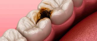
- Poorly performed treatment of pulpitis. If caries is started, the affected cavity gradually becomes quite deep. When microorganisms enter the pulp, its inflammation begins, accompanied by acute pain, which may stop over time, indicating the death of the nerve.
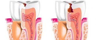
But the development of the disease does not end: bacteria through the root canals extend beyond the boundaries of the affected tooth, as a result of which a focus of inflammation appears near the upper parts of the roots, called periodontitis. The further course of the disease can occur according to several scenarios, one of which is the formation of granuloma.
It is necessary to understand that a tooth with a granuloma does not necessarily have to be affected by caries, since the root can become inflamed near a tooth that has already been treated. If the doctor has not completely removed the tissues affected by caries by placing a filling on top of them, then there is a high risk of developing pulpitis with the subsequent formation of granuloma.
- Filling of root canals performed with violations. A granuloma that appears on the root of a tooth whose canals were filled some time ago indicates the unsatisfactory quality of the procedure performed. Due to inattention, haste or inexperience, the doctor could not fill the canals to the very top. Dentists often refuse to admit their own mistake, because in this case they will have to treat the tooth for free. Such unfortunate specialists can evade until the last minute, pretending that they do not understand the causes of pain, recommending taking a course of antibiotics.
Risk group
Knowledge of risk factors allows you to calculate a likely “candidate” for the development of oral cancer and timely modify behavior towards a healthy lifestyle, eliminating actions that are hazardous to health.
High-risk group for malignant processes in the gum area:
- People who smoke have celebrated their fortieth birthday, because the frequency of neoplasms increases with age and the “control point”, after which the incidence doubles with each decade of subsequent life, is precisely 40 years.
- Young people with a full range of risk factors. Before the age of 40, carcinomas of this localization are rare, but early introduction to tobacco simultaneously with chewing betel or nas, with persistence in the mucosal cells of the human papillomavirus type 16, significantly increases the chances of cancer.
An extremely high-risk group when the prospect of becoming a cancer patient over time is very high:
- Suffering from precancerous processes - leukoplakia and erythroplakia;
- Older people who smoke, use dentures, and the older they are, the more likely malignant transformation is;
- Adults with a hereditary predisposition, which is identified by the presence in the family of patients with oral carcinomas.
All patients at risk should visit the dentist at least twice a year and undergo treatment for precancerous processes.
Signs and symptoms of periodontitis
The main symptoms of tooth root inflammation include:
- pain in the tooth during eating and when closing the jaws;
- increase in body temperature;
- the feeling that the tooth has become “higher”;
- change in gum color (the gums darken or have a bluish tint);
- tooth mobility;
- bad breath;
- accurate determination of the location of the affected tooth by the patient himself;
- discharge of fluid from under a tooth or pus from a fistula;
- irradiation of pain to the temporal or ear region;
- swollen lymph nodes;
- swelling in the area of the root, gums, its redness;
- the appearance of interdental gaps.
It should be borne in mind that sometimes periodontitis occurs without symptoms.
Diagnosis of gum cancer
Oncopathology of the oral cavity is visual, that is, diagnosed during a routine examination without the use of any equipment.
During the initial examination, cells are scraped from the wound for cytological analysis, or, in the absence of an ulcer, the tumor is punctured with a thin needle. If possible, a part of the tumor is “bitten off” with a special instrument - this is a biopsy for histological analysis. The diagnosis of cancer is established solely by the result of a biopsy; cytology allows only to suspect the disease.
After morphological confirmation, an ultrasound of the oral cavity is performed, which explains the relationship of the neoplasm with the surrounding tissues, primarily with the bone and blood vessels, and the CT scan clarifies the condition of the entire anatomical region.
Next is the search for metastases: ultrasound of the neck, CT scan of the lungs or MRI.
Diagnostics: basic methods
It is possible to notice a cyst during a routine examination only at the stage of obvious symptoms (swelling of the gums, tubercle, etc.).
Only an X-ray examination can definitively diagnose a cyst and confirm a specialist’s suspicions. As a rule, the picture is taken in the area where the cyst is located.
In some cases, a panoramic photograph of the jaw or an orthopantomogram is additionally required. OPTG is necessary for the maxillofacial surgeon in case of surgical intervention and if the X-ray image does not allow the full picture of the disease to be seen. Such cases often occur when a dental cyst is caused by infections of the nasopharynx and maxillary sinuses.
Help from other specialists
Cysts caused by gum disease require further consultation and treatment with a periodontist. For a cyst caused by ENT diseases, consultation with an otolaryngologist is necessary.
Treatment for gum cancer
It is optimal to perform the operation at the first stage, if technically possible. The radicality of the surgical intervention ensures a distance of at least 2 centimeters from the tumor border; it is clear that this is possible with a small stage 1 lesion.
At stages 2-3, the possibility of surgery is limited by the depth of penetration of cancer cells deep into the tissues of the jaw; with a large depth of infiltration, the process is considered questionably operable, but a lot is determined by the experience and talent of the oncologist surgeon. Techniques have been developed that make it possible to replace large surgical defects of the jaw with skin flaps, ribs, metal structures or artificial tissues. Plastic surgery may be postponed until antitumor treatment is completed.
If the tumor thickness is more than 4 millimeters, the surgical plan includes removal of the lymph nodes of the neck; if the cancer infiltration is localized in the middle of the oral cavity, the nodes are removed from both sides. Lymphadenectomy has a positive effect on life expectancy, but reduces its quality. It is possible to refuse lymphadenectomy if there are no metastases, which is proven or rejected by a morphological study of a biopsy obtained from a “sentinel” lymph node.
Surgery for cancer above stage 1 is complemented by prophylactic radiation, which should begin one and a half months after surgery.
Initially inoperable stage 3 tumor conglomerates are tried to be reduced by radiation in combination with chemotherapy with platinum derivatives, postponing surgery to the second stage.
Contraindications for surgery: stage 4, proliferation of metastases from the lymph node into the skin or tissue of the neck, severe diseases in the decompensation stage.
If surgery is not possible, 7 weeks of radiation therapy is performed on the primary lesion and lymph nodes of the neck, with the introduction of platinum chemotherapy every 3 weeks. If radiation is contraindicated, chemotherapy is used; this is also done in case of relapse after chemoradiotherapy.
Treatment methods for dental cysts
Treatment of dental cysts is carried out using two methods:
- therapeutic;
- surgical.
If the cyst is small and is not accompanied by serious symptoms such as purulent abscesses and swelling, conservative treatment is possible. In this case, the cyst in diameter should not exceed 8 mm.
Treatment is carried out through the canals of the tooth. After opening them, an antiseptic treatment is performed and a specialist injects a copper-calcium suspension through the canal. Next, the tooth is filled with temporary materials. After a time determined by the dentist, a repeat x-ray is taken to see progress in treatment or lack thereof.
A copper-calcium suspension allows you to restore destroyed bone-jaw tissue.
Additionally, at the discretion of the specialist and in case of severe inflammation, antibiotic treatment may be prescribed. For this disease, this is an auxiliary measure, and in no case is the main treatment.
If there is no progress or if the cyst is large, treatment is carried out through surgery. The operation is performed by a maxillofacial surgeon under local anesthesia. During surgery, the top of the tooth root is cut off, after which the cyst is completely removed.
In some cases, only the anterior wall of the cyst shell may be removed. The recovery period after such an operation is longer.
Among the modern methods of treating cysts are operations using lasers. In this case there is practically no pain. In addition to removing the tumor, during the manipulations the tooth canals and the affected area are disinfected. Wounds with this method heal quickly, complications are extremely rare.
When treating a cyst, specialists try to preserve the tooth. After the suture has healed, the temporary cement filling is removed from the canals, and they are filled with permanent materials. If this is not possible, the tooth itself is removed after surgery.
Forecast
An individual prognosis for the course of the disease is based on the size of the neoplasm and the depth of its penetration deep into the jaw, involvement of vessels and nerves in the conglomerate.
The degree of damage to the lymph nodes affects life expectancy; metastases almost halve the likelihood of cure. Much determines the aggressiveness of cancer cells; low differentiation is not in favor of the patient.
Specific percentages by years of survival are unknown because the incidence is low and statistically gum cancer is not distinguished from the group of oral carcinomas.
Tooth granuloma: symptoms of the disease
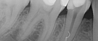
Only a doctor can make a diagnosis based on an x-ray, where a darkening will be visible in the root area. Any darkening in the image is a sign of the presence of a cavity, but if a granuloma occurs, it means that periodontitis, which is chronic in nature, begins to develop.
For a long time, the tooth may not bother a person at all. Sometimes pain may occur when biting or when eating hot food: these are the first signs of the development of chronic periodontitis. An exacerbation of the disease is observed during periods of weakening of the body's immunity: then the pain intensifies significantly, which is especially noticeable when biting. During such periods, as a rule, the gums become swollen, and in the area of inflammation it becomes painful to touch.
Possible complications of gum cancer
First of all, patients are concerned about the cosmetic consequences of treatment; in this regard, chemoradiation treatment is preferable, but is fraught with a higher frequency of relapses and is inferior to surgery in terms of survival.
The list of dangerous complications of carcinoma includes deterioration in nutrition with exhaustion and the development of fatal anorexia-cachexia syndrome. Long-term cancer-preventive therapy worsens nutritional status and worsens underweight. From the first days of treatment, the patient necessarily needs nutritional correction and nutritional support with special protein mixtures. In some periods, the patient may require tube and parenteral nutrition, since swallowing is seriously impaired and spasm of the facial muscles develops, up to the inability to open the mouth due to trismus.
Malnutrition and tooth loss after irradiation worsen the course of concomitant diseases and contribute to the development of infections against the background of a natural decrease in immunity. A change in the swallowing reflex due to too intense production of saliva, literally flowing in a stream, leads to the night-time reflux of saliva with food debris and bacterial flora into the respiratory tract and the occurrence of severe aspiration pneumonia.
Blockage of lymph outflow and proliferation of neck vessels by metastatic lymph nodes is complicated by constant headaches and loss of consciousness.
The disintegration of a cancerous ulcer is fraught with bleeding, which can only be stopped by ligation of the artery. Tumor masses growing into the bones cause intense pain, aggravated by secondary infection. The most severe suffering for the patient's family is caused by the disintegration of a cancerous ulcer with the addition of a secondary infection and a foul odor. It is absolutely impossible to clear a progressive tumor of necrosis on your own; this is a very difficult task even for specialists.
Treatment of such patients is a great oncological art, which is available to the specialists of our clinic. We are ready to help any patient, with or without complications, who has been treated for a long time and is just beginning to fight for life.
Book a consultation 24 hours a day
+7+7+78
Bibliography:
- Alieva S.B., Alymov Yu.V., Kropotov M.A. etc. /Cancer of the oral mucosa. Oncology. Clinical recommendations // Ed. Davydova M.I.; M.: Publishing group RONC, 2015.
- Gelfand I.M., Romanov I.S., Udintsov D.B. /Tactics for the treatment of localized forms of cancer of the oral mucosa// Tumors of the head and neck; 2016; 6.
- Romanov I.S., Yakovleva L.P. /Issues of treatment of oral cancer// Farmateka; 2013; 8.
- Kurokawa H., Yamashita Y., Takeda S. et al. /Risk factors for late cervical lymph node metastases in patients with stage I or II carcinoma of the tongue// Head Neck; 2002; 24.
- Little M., Schipper M., Feng FY et al. /Reducing xerostomia after chemo-IMRT for head-and-neck cancer: beyond sparing the parotid glands// Int J Radiat Oncol Biol Phys; 2012; 83.
- Midgley AW, Lowe D, Levy AR et al. /Exercise program design considerations for head and neck cancer survivors// Eur Arch Otorhinolaryngol; 2018; 275(1).
What to do with a dental cyst?
Since it is difficult to identify a cyst under a tooth, since it forms slowly and without pronounced symptoms, patients often turn to the doctor in the later stages of the disease - when pain, malaise and other signs of inflammation are felt.
Previously, in such a situation, the only way out was to remove the affected tooth. Today, there are different methods for treating cysts without losing a tooth. Depending on the degree of the disease, you can get rid of it therapeutically or surgically.
Therapeutic treatment includes cleaning out the cyst from the canals, treating them and filling them. Surgical treatment involves removing the damaged part of the tooth root while preserving the tooth itself. In this case, the cyst is eliminated and the missing tissue is replaced with a special material.
Today, indications for tooth extraction due to a cyst are special cases that threaten complications, as well as a wisdom tooth cyst.
Is it possible not to treat a dental cyst? Only if it appeared in a child before the change of baby teeth, over time such a tooth cyst will burst on its own, due to the friction of the gums against each other.