Indications for treatment of periodontitis
There are two main ways to treat periodontitis: conservative and surgical. Each of them has its own indications and contraindications.
According to modern dental standards, a doctor should give preference to conservative methods. They are indicated for both acute and chronic periodontitis, including the appearance of cysts and granulomas, loose teeth, and increasing inflammation.
However, orthograde treatment cannot be used in all cases. Indications for surgical intervention are:
- obstruction of the tooth root canals;
- the presence of a stump tab or pin that cannot be removed without damaging the roots;
- multiple perihilar cysts or cysts growing into the maxillary sinus;
- wide affected area (over 10 millimeters);
- perforation of the tooth cavity or root wall;
- ineffectiveness of conservative treatment methods.
Important!
When we talk about periodontitis, we often mean apical (also known as periapical or apical) periodontitis - that is, inflammation at the apex of the tooth root. The cause of this disease is endodontic problems. Another type of periodontitis, marginal, affects the gums in the cervical area of the tooth, but it already belongs to the field of periodontology. This material is devoted to the treatment of apical periodontitis only.
Surgical methods
The arsenal of a dental surgeon in the treatment of periodontitis includes the following manipulations:
- Opening the gums - used to facilitate the drainage of pus that has accumulated at the apex of the root and caused the formation of an abscess on the gum.
- Hemisection is an operation to remove the root of a multi-rooted tooth, at the apex of which a focus of inflammation is found, while at the same time the crown part of the tooth adjacent to the root is removed. The canals of the remaining roots must be thoroughly treated before surgery. The hole remaining after hemisection is filled with osteoplastic material, and the mucous membrane is sutured. The tooth is later covered with a crown.
- Root amputation is an operation to remove the root of a multi-rooted tooth, but preserving the crown part.
- Root apex resection – the root apex is removed through a hole formed on the outer surface of the jaw in the area of the projection of the root apex of the diseased tooth. The operation is often performed in combination with cystectomy - removal of a granuloma or cyst. The cavity formed after their removal is filled with osteoplastic material. Also, resection is indicated if it is not possible to unseal the canal (provided that it is thoroughly sealed throughout its entire length, except for the apical part).
- Tooth replantation is a complex operation that involves removing a tooth and then returning it to the socket after curettage of the alveoli, filling of the canals and resection of the apex (sometimes resection is not performed). The method is used on multi-rooted teeth, the healing of the replanted tooth can last up to 10 weeks, during which time the tooth must be kept at rest using splints or trays. Even with successful engraftment, a replanted tooth must be removed on average after five years due to root resorption and loss of stability.
- Tooth extraction – if it is impossible to use other methods of treating periodontitis, they resort to tooth extraction. Subsequently, it is recommended to carry out implantation - implantation of an artificial root (implant) with further installation of a crown. Thanks to implantation, full chewing function is preserved, and the appearance of the artificial tooth does not differ from the natural one.
Modern methods of treating periodontitis allow, in most cases, to save the tooth, but if the patient seeks dental care late, it may be lost. To avoid unpleasant consequences, you need to monitor your dental health, conduct timely preventive examinations of the oral cavity and treat identified diseases.
Treatment methods for dental periodontitis
| Conservative treatment | Surgery |
Therapeutic:
Physiotherapeutic:
Conservative treatment of periodontitis is accompanied by the use of antibiotics. | Surgical treatment:
|
Important!
The probability of successful conservative treatment of periodontitis is 70 - 90%
What is a periodontal tooth?
The periodontium is the complex of tissues surrounding each tooth. This includes the bone around each root and the dental ligaments that occupy the space between the cementum of the dental root and the bone socket. Thus, the tooth is provided with some mobility, which is very important for the correct distribution of the chewing load. During active chewing, each tooth “springs” inside the socket; Thanks to this property, hard tissues are protected from excessive abrasion.
The vital activity of the periodontium is supported by an extensive network of blood and lymphatic vessels, as well as nerve fibers. It is interesting that periodontal tissue has a fairly high rate of renewal, which gradually decreases with age (at the same time, thinning of the periodontal fibers occurs), as a result of which some elderly people may experience severe mobility, even loss, especially with incorrect positioning and excessive load.
The most important functions of periodontium can be considered:
- anatomical (mechanostatic) – thanks to the periodontal ligaments, the tooth is securely held in the bone socket or alveolus;
- distributive-regulating - thanks to the main substance of the periodontium with fibers, the load on the tooth is evenly distributed during chewing;
- trophic - thanks to a developed network of vessels and nerve fibers;
- protective creation of an antimicrobial barrier and maintaining healthy tissue balance;
- plastic – thanks to the ability of the periodontium to restore its own and surrounding tissues;
- sensory - thanks to an extensive network of nerve fibers and endings.
Stages of periodontitis treatment
The number of visits to the clinic for the treatment of periodontitis depends on the stage of the disease (acute periodontitis, chronic, chronic in the acute stage) and the chosen technique. Often, therapy is carried out in several stages and requires at least 2 - 3 visits to the attending physician, since it is not recommended to install a permanent filling until the inflammation is completely removed.
- Preparation for treatment: diagnosis using an x-ray, anesthesia injection.
- Drilling a tooth to access canals, removing a nerve, or removing an old filling.
- If necessary, expand channels.
- Antiseptic treatment of canals, application of medications, physiotherapeutic procedures.
- Installation of a temporary filling.
- Removal of the temporary filling, antiseptic treatment of the canals (this stage is repeated until the source of inflammation is completely eliminated; sometimes this may take several months).
- Installation of a permanent filling, control x-ray.
In parallel, the patient is prescribed antibacterial and anti-inflammatory therapy, as well as home rinses with disinfectant solutions.
DIAGNOSIS OF GRANULATING PERIODONTITIS
The main symptom of granulating periodontitis is subcutaneous granulomas or fistulas, but the disease can occur without visible changes. Instrumentally, pathology can be detected using a probe and assessing electrical sensitivity. There is a change in the threshold of excitability and slight painful sensations in the area of vertical percussion.
In response to pressure with the probe, the gum mucosa first turns pale, then turns intensely red. You can distinguish the disease from pulpitis by the shade of the crown - it becomes grayish, and with pulpitis it retains its normal color. To differentiate the disease from the granulomatous form of periodontitis, an x-ray examination is prescribed.
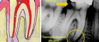
DIFFERENTIAL DIAGNOSTICS AND RADIOGRAMS
Radiography helps clarify the diagnosis, establish the causes of the pathology and develop treatment tactics. The image clearly visualizes the deformation of the gap between the root cement and the alveolus. In the area of the root apex, areas of destruction or rarefaction of bone tissue are visible, the edges of the plate lose clarity of contours, the affected area is characterized by an irregular “eaten away” shape without clear boundaries. In the granulomatous form of the disease, the image shows the regular round shape of the focus of bone destruction with clear boundary lines.
Features of the treatment of periodontitis with fistula
Odontogenic fistula is one of the complications of periodontitis, mainly granulating. It consists of holes in the mucous membrane, which are formed due to the proliferation of granulations and destruction of the tissues surrounding the tooth. In severe cases, a fistula can appear not only in the gum, but also in the cheek, and even on the skin of the face. Purulent contents are released through the hole, which appears due to the inflammatory process in the periodontium.
On the one hand, the formation of a fistula facilitates the course of the disease, since inflammatory products are eliminated through it (which means that the patient most likely will not suffer from severe pain). On the other hand, non-intervention over time can lead to tooth loss.
You can get rid of a fistula only by eliminating its cause - damage to periodontal tissue. Treatment follows a standard scheme: mechanical treatment of the canals, disinfection and thorough filling. Due to the outflow of pus through the fistulous tract, treatment is most often successful and takes less time. After creating suitable conditions, the fistula goes away on its own, but in severe cases, surgical removal of overgrown granulations may be necessary.
Causes of the disease
The main reason for the development of the disease is a chronic inflammatory process due to an advanced form of pulpitis or carious lesions. Other reasons may be injuries to the dentition due to mechanical impact, for example, when biting hard foods or during a fall or impact.
Also, the body’s reaction to incorrectly formulated drug treatment and incorrectly selected dental braces can also be expressed by inflammation in the gum area. An exacerbation of the process occurs in the presence of endocrine diseases, as well as a lack of vitamins and minerals in the body, reduced immunity.
Treatment of chronic forms of periodontitis
There are three types of chronic periodontitis: fibrous, granulating and granulomatous.
- In fibrous periodontitis, the tissues surrounding the apex of the tooth are replaced by fibrous tissue. The patient usually does not feel pain, and the disease can only be determined by an x-ray.
- Granulating periodontitis is characterized by the growth of granulation tissue: the process of bone resorption (resorption) starts, fistulous tracts are formed, through which inflammatory products are separated. As the granulations expand, the patient begins to experience periodic aching pain.
- Granulomatous periodontitis is accompanied by the appearance of a granuloma - a neoplasm at the root apex. It is a chamber of connective tissue filled with granulations. If the disease is not treated, the growth of granuloma can even lead to a jaw fracture.
Treatment of chronic periodontitis is often carried out using conservative treatment methods. According to modern standards, doctors, as a rule, do not carry out separate treatment for granulomas, cysts and fistula tracts: if the canals are disinfected and properly sealed, the neoplasms will disappear on their own. In advanced cases, surgical intervention is permissible.
Types of granulomatous periodontitis
Classification of the stages of the disease allows us to divide the disease into 3 types:
- The periodontal tissue thickens, the connective tissue grows significantly, and granulomas form. Dimensions do not exceed 5 mm.
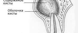
- Development of cystogranuloma. The tumor can reach 1 cm in diameter.
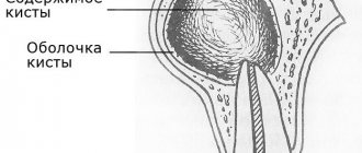
- Cyst formation. (a cavity formation that has an external connective tissue capsule, lined with mucous tissue on the inside). The fluid produced by the mucous layer puts pressure on the adjacent bone tissue, deforming and destroying them.
Features of the treatment of periodontitis in the acute stage
Exacerbation of periodontitis goes through two phases: intoxication and exudation (appearance of discharge). As the disease progresses, the patient first experiences aching and episodic pain, and then constant throbbing and tearing pain, so treatment cannot be delayed.
Acute periodontitis can be serous or purulent. In the second case, purulent exudate accumulates in the apical part of the tooth root, and the main task for the doctor is to remove it. Sometimes this is enough to clean the tooth cavity and treat the canals, but in severe cases it may be necessary to cut the periosteum for drainage.
CAUSES AND DESCRIPTION OF PATHOLOGY
The disease is caused by pathogenic bacteria that attack the periodontium, a fibrous tissue with a dense network of capillaries and nerve endings located between the root cement and the alveoli. Infection occurs as a complication of deep caries, when pathological microorganisms reach first the pulp and then the periodontium. In areas of bone tissue destruction and constant inflammation, pathological granulation tissue begins to form and grow rapidly.
Another reason may be unskilled filling of root canals, penetration of aggressive materials and pastes into the periodontal tissue that stimulate the growth of colonies of anaerobic bacteria.
Treatment of periodontitis at home
Periodontitis cannot be cured at home, since the disease is caused by bacteria that colonize the dental canals. The only way to get rid of them is to carry out antiseptic treatment and sealing of the canals, and this can only be done by a doctor, but by waiting for a visit to the clinic, you can alleviate the symptoms and reduce pain.
Disinfectants that do not irritate the mucous membranes can be used for rinsing 4 - 5 times a day. Doctors also recommend rinsing with a solution of salt and soda, including after treatment, to relieve swelling and reduce inflammation. Non-steroidal anti-inflammatory drugs are suitable for pain relief. All this will help relieve symptoms, but is not a cure.
You may experience pain after periodontitis treatment. Normally, they last 3–5 days and gradually fade away. If the pain does not subside or returns with renewed vigor, re-therapy is necessary.
Prevention
Exacerbation of chronic granulating periodontitis is an extremely painful phenomenon, so it is important not to start the disease and carry out timely prevention, which includes:
- Oral hygiene. Daily brushing of teeth should be thorough, but not traumatic to the gums, otherwise the path for bacteria will be open.
- Regular removal of plaque and tartar in the dentist’s office is an effective measure to prevent the development of the disease, and therefore the need to treat granulating periodontitis.
- Restoration of dentition. If a tooth is missing, you should not leave an empty space on the gum, otherwise the remaining teeth will become more vulnerable to inflammation.
- Early diagnosis and treatment of granulating periodontitis and other dental diseases. For this purpose, you need to visit a doctor at least twice a year.
Still have questions?