From this article you will learn:
- what is a cyst on the gum - photos, symptoms,
- what it looks like in a child.
- how to treat a cyst on the gum.
The article was written by a dental surgeon with more than 19 years of experience.
A cyst on the gum is an inflammatory formation filled with pus that forms on the gum in the projection of the source of inflammation at the root of the tooth. In other words, in appearance such a cyst resembles a purulent sac on the gum, and it looks the same in both children and adults (Fig. 1-3). It should be noted that in dentistry such a term does not exist, and therefore the expression “cyst on the gum” is nothing more than an everyday colloquial expression.
As you already understand, such a purulent sac forms on the gum only if there is inflammation at the root of the tooth, which in dentistry is referred to as apical periodontitis. The formation of a cyst always occurs in the projection of the root of the causative tooth, and on the tooth itself you can always see either caries, a filling or a crown. Well, in children it can also form as a result of a tooth injury (due to an impact or a fall), in which case a chip may be found on the tooth, or the crown part of the tooth will be painted bluish.
Cyst on the gum: photo
In photo 1 you can see a cyst filled with pus, which formed in the projection of the roots of the destroyed 6th lower tooth. What does a cyst on a child’s gum look like – photos 2-3 (and in these photographs we see that in one case the causative tooth has a carious lesion, and in the other there is a traumatic fracture of the crown). Depending on whether it is a baby tooth or a permanent one, on the amount of destruction of the tooth crown and on the size of the inflammatory focus, both conservative therapy for dental periodontitis and tooth extraction can be carried out.
The latter especially concerns children, because approach to the treatment of baby teeth with inflammatory foci in the root area (especially in the presence of a fistula or purulent sac on the gum) absolutely does not involve saving the tooth. This is due to 1) the peculiarity of the structure of the root canals in baby teeth, 2) the risk of death of the rudiments of permanent teeth (the latter are almost closely adjacent to the roots of baby teeth). Read more about this at the link below.
→ Treatment of an abscess on the gum of a child
What is a dental cyst?
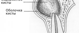
Tooth cyst - what is it? An odontogenic cyst is a pathological neoplasm that occurs in the upper part of the tooth root. The internal cavity of the cyst is filled with liquid or mushy purulent contents; it is enveloped by a dense layer of epithelium.
The size of the cyst starts from a few millimeters, with rapid development reaching several centimeters in circumference. Most often, the pathological process affects the upper jaw, since the roots of its teeth have a more porous structure.
In order to understand what a dental cyst is and how to treat it, you need to know why such a phenomenon occurs. The formation of cysts occurs as a result of inflammation, thus the body restricts healthy tissue from the affected areas, clogging them along with bacteria into bubbles.
How is surgery to remove a cyst (cystectomy) performed?
Removal of the cyst is carried out by a dental surgeon in a surgical office, on an outpatient basis, the operation usually lasts about half an hour, an hour after the manipulation the patient can go home. Improvement in the patient's condition should occur 6-8 hours after the operation. A sick leave certificate is issued for the recovery period - until the stitches are removed.
To relieve all pain, the patient is given local conduction or infiltration anesthesia; we also numb the injection site with an application gel. After surgery, painkillers in tablets are prescribed for the first time.
I Preparatory stage
- The patient is diagnosed by a surgeon, and a consultation with the attending dentist-therapist is required.
- It is advisable, if the operation is not urgent, to provide the patient with professional hygiene and sanitation of the oral cavity before surgery - this reduces the presence of infection in the oral cavity and healing proceeds faster.
II Carrying out the operation
- The patient is given anesthesia.
- The surgical field is treated with an antiseptic.
- The dentist-surgeon separates the mucoperiosteal flap from the bone.
- Access to the affected area is provided, the cyst along with the affected part of the apex of the tooth is removed.
- The cavity is thoroughly cleaned and washed with an antiseptic.
- An antiseptic is injected into the cavity and the wound is sutured.
- During the recovery process, the cavity heals on its own and the bone tissue is restored.
III Postoperative period
- After the operation, you should not eat for 4 hours, then the food should not be hot, liquid and gentle.
- The patient is prescribed antibiotics to fight the infection and painkillers.
- Hygiene can be carried out according to the standard scheme one day after surgery, according to the recommendations of your doctor.
- Antiseptic rinses and healing gels are prescribed.
- Swelling of the gums and face may occur for several days.
- NOT RECOMMENDED:
- Warm the surgical site, because re-development of the infection is possible.
- Take medications without a doctor's prescription.
Causes
There are several reasons why a dental cyst develops. The main reason is the activity of pathogenic microorganisms in a closed dental space; the following risk factors contribute to this:
- severe pathology, lack of timely treatment and incorrect treatment of dental diseases - caries, periodontitis, pulpitis;
- infectious complications after tooth filling, implantation procedures - in such cases, the doctor removes not only the cyst, but also the crown or implant, this avoids relapse;
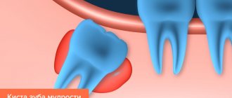
- complications during teething, especially when wisdom teeth erupt - dental tissues injure the gums, bacteria get into microcracks,
- microorganisms also enter wounds that form due to mechanical damage to teeth;
- Nasopharyngeal diseases – infections in the nose and throat can spread to the oral cavity.
To provide adequate treatment, it is necessary to accurately determine the cause of the development of a dental cyst; based on it, the dentist will prescribe suitable therapy. So, in cases of injury, treatment consists of removing the cyst and tissue regeneration, but if the cyst is a complication of another disease, then in addition to removing the vesicle, the patient will be prescribed treatment for the underlying disease.
Why does it occur
Among the main causes of pathology:
- advanced caries;
- jaw injuries;
- illiterate root canal treatment;
- overload of individual units as a result of poorly performed prosthetics;
- congenital anomalies of the upper/lower jaw;
- infections of the nasopharynx and oral cavity.
Regardless of the characteristics of the provoking factor, a gum cyst should be treated under the supervision of a dentist. Self-medication for this diagnosis is unacceptable.
Types of dental cysts
Tooth cysts have different classifications, each of which is formed according to certain characteristic parameters of the pathology.
According to the nature of the disease, they are distinguished:
- residual cysts – occur after tooth resection (removal) surgery; this is the most common type of cyst;
- retromolar – formed during severe eruption of wisdom teeth;
- radicular - cysts are located on or near the tooth root;
- follicular – at the heart of such cysts is the germ of a permanent tooth; follicular neoplasms arise as a result of poor quality care of baby teeth.
Classification of neoplasms according to their origin:
- odontogenic – arise as a result of the transition of the inflammatory process from other dental diseases;
- non-odontogenic - the causes of the development of such cysts include problems not related to the teeth and oral cavity.
Locations of cystic formation:
- anterior teeth;
- teeth that are adjacent to the nasal sinuses with their roots;
- wisdom teeth.
Summary:
You should not think that if a purulent cyst on the gum spontaneously opens, then you don’t need to go to the dentist and not treat the causative tooth. The source of inflammation at the root tip will still be in place, and it will slowly increase. Over time, this can lead to the formation of a large root cyst at the apex of the tooth root, and even tooth extraction. Therefore, treatment should not be delayed. We hope that our article: Cyst is not a gum treatment - was useful to you.
Sources:
1. Higher prof. the author’s education in therapeutic and surgical dentistry, 2. Based on personal experience as a dentist, 3. National Library of Medicine (USA), 4. “Outpatient surgical dentistry” (Bezrukov V.), 5. “Therapeutic dentistry: Textbook” (Borovsky E.).
Symptoms
The danger of a dental cyst lies in the fact that signs of pathology appear only when the neoplasm reaches a relatively large size. In the early stages, small cysts do not manifest themselves in any way, meanwhile the infectious process covers an increasingly larger area of healthy tissue. In the initial development of pathology, cysts are discovered by chance during routine examinations or treatment of other diseases.
The duration of the formation of a dental cyst takes only 1-2 days; as it develops, the following symptoms may occur:
- unpleasant and even painful sensations in the tooth, which intensify when chewing solid food;
- protrusion of the gum of a tooth, in the area of the root of which a cyst forms, the growth of the gum becomes larger over time, redness is observed;
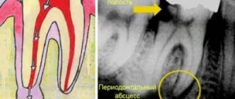
- the formation of a fistula in the area above the root of the tooth, the release of serous or purulent accumulations from it;
- general weakness and malaise;
- increase in body temperature.
Note! When a dental cyst occurs, the symptoms are not immediately visible; they appear in the later stages of development. The pain when a tumor appears is aching in nature, but it is less pronounced than the pain caused by caries and pulpitis.
If a clinical picture occurs and you suspect a pathological process, be sure to consult a doctor. Under no circumstances should you resort to self-treatment - the dental cyst must be removed. In addition, taking the wrong medications can worsen the patient’s overall well-being.
Sometimes there is no pain in the oral cavity; instead, the basis of the clinical picture is intense headaches. The cause of this phenomenon may be a cystic formation in the maxillary sinus.
Classification
All cysts that occur in the human oral cavity can be divided into the following types:
- root;
- residual;
- follicular;
- teething;
- primordial;
- lateral periodontal;
- calcifying odontogenic.
Let's talk about each of them in more detail.
Root
It is a consequence of granuloma that appears due to periapical inflammation and necrosis. Located in the area of the apical part of the root. The boundaries of such a structure are easily determined. Its size can be from 2 mm to 3 cm. But even with a large diameter, root formation does not lead to a change in bone volume or displacement of the incisor. Only if a secondary inflammatory process is involved, the disease makes itself known through pain and swelling.
When identifying a disease, you need to understand what led to its appearance and take measures to eliminate the provoking factors. Usually the problem can be resolved with endodontic therapy.
Residual
It is also called residual. Appears after removal right in the place where the roots of the torn out unit were previously located. X-ray diagnostics helps determine the presence of a residual structure.
Follicular
It is formed from the tissues of the follicular sac that covers the rudiments. Easily visible in the photo. Often leads to serious damage to premolars and molars.
As a rule, follicular formations quickly increase in size and can spread to adjacent teeth, changing their location and angle of inclination. Traditionally, their treatment involves surgical curettage.
Teething
Appears during the period of eruption of permanent units. Covers the emerging rudiment. May contain blood. Visually resembles edema, swelling. Has a bluish tint.
Sometimes it ruptures spontaneously, spontaneously. Then the person simply notices that the “ball” has disappeared, the liquid has flowed out of it. In rare cases, surgery may have to be performed. During this procedure, the blood sac is opened, and the structures that interfere with further eruption of the molar are excised.
Primordial
Formed on the basis of the tooth germ. Most often diagnosed in the area of the posterior units. This type of formation is prone to recurrence. To cure the primordial structure, it is necessary to curettage the altered tissues.
Lateral periodontal
Rarely encountered in dental practice. It has modest dimensions. Clearly visible on X-ray images. This cavity is called a lateral cavity, since it is fixed to the side of the tooth root.
The tumor can only be removed by curettage. The material obtained during the operation is necessarily sent for histological analysis to ensure that the disease present in the person is benign.
Calcifying odontogenic
Another rare type of cyst. Observed in the lower part of the supporting surface of the jaw. Well visualized on x-rays. There is cloudy contents inside. Its color depends on the degree of calcification.
Immediately after diagnosis, surgical curettage is performed.
Consequences
Without adequate treatment, the dental cyst continues to grow and develop; in advanced stages, large neoplasms destroy the bone tissue of the skull, as a result it is replaced by connective tissue formations, which leads to the development of the following complications:
- dissolution of the jaw bone, which depends on the growth of the cyst;
- the formation of pus in the cyst, further purulent inflammation can lead to the development of an abscess;
- inflammatory process of lymph nodes located near the source of infection;
- development of osteomyelitis or periostitis;
- development of chronic sinusitis when the cyst grows in the maxillary sinus;
- pathological fracture of the jaw bones when the cyst reaches a large size;
- development of phlegmon due to a long-term purulent inflammatory process in the cyst;
- sepsis – blood poisoning;
- degeneration of a cyst into a malignant tumor without timely treatment.
Many patients are interested in why a dental cyst appears in the maxillary sinus, how dangerous it is and its symptoms. The formation of a cyst of this type occurs as a result of untreated inflammation of the tooth root in the upper jaw. A granuloma forms at the root of the tooth, which increases in size and becomes a peri-radicular cyst, then takes a position in the maxillary sinus. The volume of such a cyst can reach 9-12 cubic centimeters.
The symptomatic picture includes painful sensations, the nature of which is similar to trigeminal neuralgia, pain in the occipital, temporal and parietal regions of the head. Externally, a dental cyst can be identified by the asymmetry of the face. Tooth cyst - photo shows a cyst in the maxillary sinus.
What will happen if left untreated?
Ignoring the symptoms of the disease leads to dangerous consequences for health. Thus, the patient may encounter:
- destruction of roots, their strong loosening;
- jaw fracture;
- tumor formation;
- purulent abscess;
- periostat;
- osteomyelitis.
The disease can cause the development of sepsis. This is a very serious condition in which the infection enters the blood and quickly spreads throughout the body.
Diagnosis of tooth root cyst
To make a diagnosis and carry out appropriate treatment, the dentist collects and analyzes the medical history. During the initial diagnosis, many patients report the fact of endodontic treatment performed to eliminate periodontitis or pulpitis. Some patients indicate an exacerbation of the disease after intraoral dissection.
Radiography is used as the main diagnostic method. Below is a photo and x-ray of a dental cyst.
To obtain an x-ray, several methods are used, the first method is based on contact intraoral x-ray, the advantages of this technique:
- determining the degree of destruction of the jaw bones;
- assessment of the condition of the tooth root and tooth canal;
- assessment of the quality of canal filling;
- identifying the presence of perforations and fragments of instruments and materials in the tooth canal;
- determination of the relationship between the cyst and the roots located near the teeth.
The second method of performing radiography is an orthopantogram; the procedure is a panoramic photograph of both jaws and the maxillary sinuses of the upper jaw.
Another method of the procedure is a survey X-ray in the nasomental projection; the X-ray covers the bones of the skull from the nose to the chin; using the image, the doctor assesses the condition of the maxillary sinuses and detects cysts that have grown into the nasal cavity.
In addition to radiography, to detect a tumor, the patient may be prescribed an electroodontic diagnostic procedure. This technique helps to assess the degree of such an indicator as the electrical excitability of the teeth that are located next to the cystic tooth. If the value exceeds 60 microamps, the dentist prescribes endodontic treatment to the patient.
For diagnostic purposes, histological and cytological studies are used to determine whether the neoplasm is benign or malignant.
Diagnosing a dental cyst is not difficult, but only qualified dentists can carry it out in a hospital setting; under no circumstances try to independently determine the presence of a cyst and do not take therapeutic measures; strictly follow the doctor’s recommendations.
Get a consultation
We will answer all your questions before visiting the clinic!
+7
Online registration
Removal of a dental cyst by resection
Removal of a dental cyst by resection of the root apex is a tooth-saving operation, i.e., making it possible not to part with the tooth. It is carried out by excision of the root and removal of the affected fragments. Then, to avoid the spread of infection to adjacent areas of hard and soft tissue, the canal is sealed. As a result, this is a good way to cure a dental cyst at the initial stage without removing the entire unit. If a fragment of the tooth remains, it will be replaced with prosthetics in the future. Tooth-preserving methods are valued by specialists because they help preserve the integrity of the dentition. Therefore, resection of the root apex is also used in other cases:
- Inflammation of the tooth under the crown. The obvious solution is to remove the prosthesis, remove the abscess, and install a new design. However, these manipulations are more costly and time-consuming than root resection.
- Root canal abscess, often irregularly shaped, due to unscrupulous filling or the use of low-quality materials.
- With osteomyelitis. There is a need to cut out the affected areas of bone tissue at the border with the roots. This helps prevent deep infection.
- When foreign objects are detected in the channels - boron particles, files. This happens when using low-quality equipment. Our clinic uses the latest instruments based on titanium alloy.
However, such an effective operation has contraindications. Removal of a dental cyst by resection is not carried out if: diseased and healthy roots are located critically close; in the presence of extensive chips of the crown; high tooth mobility; in the stage of acute periodontitis. Not recommended for people with diabetes, mental and nervous disorders, chronic heart and vascular diseases, pathologies of the lungs and thyroid gland.
Treatment
Treatment of dental cysts is carried out through surgery, laser treatment and conservative therapy. The latter has a positive effect only in the initial stages of the disease; overgrown cysts must be removed.
Surgery
To eliminate the pathology, it is not necessary to remove the entire tooth; only the tooth root on which the cyst is located is subject to resection. After removing the affected area, the dentist seals the remaining root, treats the surgical canal through which he removed the bladder with its contents, and stitches it up.
After a few days, the doctor removes the stitches and monitors the wound healing process. It is important to make sure that there are no cyst particles left in the dental canal; to achieve this goal, repeat radiography is performed.
Note! Sometimes it is impossible to remove the root along with the cyst; in these cases, the doctor completely removes the tooth. Indications for complete tooth resection are a difficult-to-reach location of the cyst and a severe course of the disease.
After surgical removal of a cyst, the patient must regularly visit the dentist and follow the recommendations prescribed by the doctor.
Conservative therapy
Tooth cyst - treatment of the disease with conservative methods is possible only in the early stages of its development. In order to eliminate the tumor, the patient is prescribed injections and rinses.
During therapeutic treatment, the dentist opens the dental canal, which leads to a cystic neoplasm, and pumps out exudate from it. The doctor does not fill the canal for ten days; during this period, the patient uses antiseptic solutions and tinctures to rinse the mouth.
Upon completion of the treatment course, the dentist treats the dental canal with medications and then fills the tooth.
Laser removal
Laser treatment is a modern method of treating dental cysts. When performing the method, the doctor opens the dental canal and uses laser irradiation to treat the area where the cystic tumor is located. The laser destroys not only the epithelium of the cyst, but also hundreds of thousands of bacteria that are inside the bladder.
The advantages of laser removal are rapid tissue healing and no risk of secondary infection in the oral cavity and dental canal.
Treatment with antibiotics
In some cases, dental cysts are treated with antibiotics. Taking antibacterial drugs is an auxiliary measure to destroy an expanded infection or the main method of treatment if a dental cyst develops against the background of a primary infectious disease.
Antibacterial drugs can only be prescribed by the attending physician; the following drugs are most often used:
- amoxicillin – has a high antibacterial effect, greatly facilitates the treatment of cysts with other methods;
- Cifroploxacin is a broad-spectrum antibiotic that actively destroys bacteria and relieves inflammation;
- tetracycline - this drug is prescribed more often than others, it actively relieves the inflammatory process, pain syndrome, and facilitates other methods of treating dental cysts.
Sometimes a doctor can prescribe topical antibacterial agents to a patient, but taking such medications is not always advisable - local drugs - antibiotics are quite difficult to distribute evenly over the diseased area.
Note! Antibacterial drugs are potent drugs that also affect beneficial bacteria in the body. You can take such medications only as prescribed by a doctor, without increasing the number of doses or dosage.
Treatment at home
Treatment of dental cysts at home is possible only as an auxiliary therapy. A cyst should not be confused with a granuloma; the latter can resolve on its own, but the cystic formation must be radically removed. Home treatment is not used to remove the cyst, but to eliminate the inflammatory process and destroy harmful bacteria.
The main goal of therapy at home is to provide an antiseptic effect. Propolis tincture, calendula tincture, eucalyptus tincture have an antiseptic effect. Tinctures are used as follows: a small amount of medicine is applied to a cotton swab and applied to the affected area, held for 5-10 minutes.
Medicines with an antiseptic effect can be used before surgery to remove a cyst and after tooth root removal. The antiseptic effect allows these medications to be used in the treatment of caries and other infectious diseases of the oral cavity.
Prevention
It is always easier and faster to prevent any disease than to cure it, so one should not forget about simple preventive rules that will help avoid the development of a dental cyst. The basic rules for preventing dental cysts are based on compliance with the rules of oral care.
How to prevent the formation of pathology:
- do not trigger the course of dental diseases such as caries, periodontitis, pulpitis; if infections occur, consult a doctor immediately;
- Brush your teeth daily and prevent the appearance of plaque, which can later transform into tartar;
- monitor the condition of the teeth and oral cavity after operations and mechanical injuries;
- visit your dentist regularly;
- monitor the condition of filled teeth and dental implants;
Patients who have had their teeth filled or have dental crowns or implants placed are advised to periodically have dental x-rays taken. This will allow timely detection of pathological changes and increase the chances of successful recovery without serious consequences.
Note! All diseases must be treated in a timely manner; inflammatory processes reduce immunity, as a result of which infections move freely from one organ to another; in addition, secondary infections can be added to already developed pathologies. It is important to monitor your health and strengthen local and general immunity.
To strengthen your immune system, strengthen your body, include fresh fruits and vegetables in your diet, play sports and walk outdoors more often. It is more difficult for any infection to get into a hardened body than into a weakened body.
How to treat
Today, doctors are trying to eliminate the disease without resorting to tooth extraction. The unit is pulled out only if it is completely contained in the inflammatory capsule and its tissue is significantly destroyed.
In other situations, therapy may be:
- surgical;
- conservative.
Surgery
Cystectomy is a procedure aimed at removing the cyst and the changed part of the root apex. The technique is considered highly effective. Its only drawback is its difficulty.
Less commonly, for cystic changes, hemisection is performed. The operation is indicated for complete destruction of the tooth root. During treatment, the dental surgeon removes the capsule with its contents, the altered root and part of the crown. This leaves a serious hollow defect. It is closed by laying special composite materials and installing a crown (if necessary).
Conservative therapy
Does not involve surgery. The doctor does not make an incision to access the capsule. Instead, he drills out the roots and treats them. The fact is that the tip of the tooth root connects to the cyst, so after drilling, the contents of the cavity immediately flow out.
When this happens, a medicinal composition and a special anti-inflammatory paste are placed into the canals. Afterwards the filling is carried out. The disease is considered to have resolved if after six months the treated area is not visible on the image.
Conservative treatment helps most patients, so the main emphasis is placed on it when fighting the described disease.
Depophoresis - an innovative method of conservative treatment
The most modern conservative treatment method is depophoresis. It does not require drilling of channels. The doctor exposes only the mouth of the canal, after which he inserts a thin electrode into it. The second electrode is pressed into the patient's cheek. Then it delivers a weak current discharge, along with which copper/calcium hydroxide passes through the channel. It penetrates even the most inaccessible areas, instantly kills pathogenic microorganisms, and destroys necrotic cells. Depophoresis is repeated three times. This is enough to clean the fabrics. Afterwards the tooth is filled. This technique is very effective. It allows you to get rid of the problem forever in 99% of cases.