Caries is a common dental disease that affects children and adults. There are several types of tooth decay, and some of them are difficult to detect initially. Pathology is often detected already in an advanced stage. Let's find out what common clinical methods exist for diagnosing dental caries.
In this article
- What are the methods for diagnosing caries?
- The main examination method for identifying caries: visual inspection with a mirror
- Probing
- Percussion
- Silk thread method
- Additional methods for diagnosing caries
- Vital coloring
- Thermometric examination for caries
- X-ray examination
- Electroodontodiagnosis (EDD)
- Transillumination
- Luminescent diagnostics
- Laser fluorescence spectroscopy
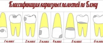
Despite all the advances of medicine in the prevention of dental diseases, caries still remains a pressing problem. It is diagnosed in almost 90% of people in the world. And this is largely due to the fact that carious changes can be difficult to detect in the early stages of the disease. Here's how caries is classified in dentistry:
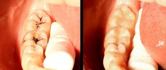
- Fissure - localized in natural depressions on the surfaces of chewing teeth.
- Approximal - occurs on the contact surfaces between teeth.
- Cervical - carious changes are localized in the area adjacent to the edge of the gums.
Some types of pathology are difficult to diagnose at the initial stage, even during examination. For example, approximal caries, which is also called interdental or contact, occurs at the junctions between teeth, where even the dentist may not notice them at first during a routine visual examination of the patient’s oral cavity.
It has been proven that caries is a process consisting of several stages, and the transition from one to another is very smooth, it is almost impossible to track it. The formation of a carious cavity requires a combination of time and risk factors. In this case, significant dentin destruction may already occur inside the tooth, a large carious cavity may form, and on the outside this will only manifest itself in the form of a slight darkening of the enamel.
What are the methods for diagnosing caries?
The objectives of modern dentistry are to maximize the preservation of the patient’s teeth and prevent the development of the pathological process at the initial stage of development. For the purpose of early detection of caries, there are various effective diagnostic methods. The main ones include:
- survey (the doctor finds out whether the patient has any general diseases, for example, endocrine diseases, finds out lifestyle, etc.);
- visual examination;
- probing of teeth;
- percussion, etc.
In addition to the basic ones, there are additional methods for diagnosing dental caries. These include radiography, transillumination, electroodontodiagnosis (EDD), laser fluorescence spectroscopy, etc. Hardware diagnostic methods make it possible to identify carious lesions at the earliest stages, which is the key to successful and timely treatment of caries.
As a comparison of basic and additional methods for diagnosing dental caries shows, there is no single ideal research method for identifying different types of caries. An optimal understanding of the course of the disease and the processes occurring inside the teeth is provided by an integrated approach that allows one to obtain a complete clinical picture.
Prevention of caries in the spot stage
Avoiding the development of a disease is always easier than treating it later. Dental caries in the staining stage can always be prevented. To do this, you need to follow several rules:
- Brush your teeth at least twice a day, choosing high-quality toothpaste and a brush that is not too hard. It is better to ask your dentist to select hygiene products for you.
- Use dental floss and a special mouthwash after each meal.
- Rinse and dry your toothbrush well to prevent the growth of pathogenic bacteria in its bristles.
- Eat nutritiously, do not overindulge in frequent snacks, drink enough water.
- Quit smoking, as it causes the thinning of tooth enamel.
- Visit your dentist regularly for a preventive examination and removal of dental plaque.
Enamel caries in the white spot stage is dangerous because it can quickly progress, developing into superficial, and then medium and deep caries. The sooner a patient begins to take care of oral hygiene, the less likely he is to lose a tooth in the foreseeable future.
The main examination method for identifying caries: visual inspection with a mirror
This is the very first and main method used in clinical practice. Conclusions about the condition of a patient's teeth were previously based solely on visual examination data. But this method of diagnosing caries has significant drawbacks: low insufficient information content, as a rule, almost complete impossibility of identifying fissure or proximal caries.
The quality of the visual inspection depends on subjective factors. Mandatory testing conditions include preliminary cleaning and drying of the teeth being examined, and good lighting. This is a suitable method for diagnosing superficial caries, as well as medium or deep caries - at the initial stage of the disease, a visual examination does not reveal defects. Approximal (contact) caries can sometimes be identified by changes in the color of the enamel (it darkens, becomes grayish or chalky), floss gets stuck between the teeth, and the interdental papilla is inflamed.
Probing
The doctor uses a periodontal or button probe to determine the condition of the teeth. By examining different areas of the tooth with a probe, he finds out how sensitive the reaction of the dental tissues is.
Probing also makes it possible to identify an inflamed pulp - in this case, the patient feels a sharp pain from the touch of the probe. At an advanced stage of the disease or an acute course, the enamel may be loose and even break off when touched by a probe.
Silk thread method
Despite the fact that modern dentistry has high-precision computer diagnostic methods in its arsenal, the silk thread method is still used by doctors during the initial examination, mainly to identify approximal caries. To do this, floss is passed between the teeth using sawing movements (it used to be a silk thread, hence the name).
If there are carious lesions between the teeth, their edges are uneven, and the thread begins to cling. This method is used after cleaning the enamel and between the teeth that do not border the filled ones. The thread may touch tartar or filling, which reduces the reliability of the method.
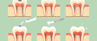
Vital coloring
To perform this diagnostic procedure, the teeth are first thoroughly cleaned, dried and isolated from saliva. Then a swab with a dye solution is applied to them. After 2-3 minutes, the swab is removed, excess dye is removed, and the mouth is thoroughly rinsed with water.
If the tooth enamel is not demineralized, as happens with caries, then there will be no traces of coloring matter left on its surface. If there are carious lesions, then dye residues of varying intensity are visible on the teeth, it all depends on the degree of damage. The more stained the tooth tissue is after removal of the tampon, the more global the degree of demineralization. This method is one of the most accessible and suitable for all categories of patients, allowing to detect the presence of the disease in the early stages.
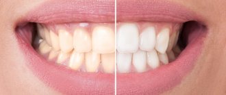
Clinic of caries in the spot stage
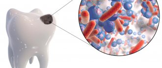
The cause of caries is microorganisms that live in the oral cavity. Due to poor hygiene, they actively multiply and secrete substances that gradually destroy tooth enamel. In addition, due to improper nutrition, which does not cover a person’s needs for minerals, a process of demineralization occurs: the surface of the teeth becomes loose and porous, loses its shine, and whitish spots appear on it. This disease is called white spot caries.
Diagnosing this condition is not easy due to the similarity in the natural shade of the enamel and the affected areas. White spots of caries are often localized in the cervical and interdental areas, so they are difficult to see. However, in the smile area this is still possible if the enamel is first cleaned and dried. Self-diagnosis is also made difficult by the fact that white spots on teeth can easily be confused with other causes of enamel damage - for example, fluorosis (excess fluoride in the body) or pigmented plaque. However, spot caries is usually single and can affect any teeth, while fluorous areas are multiple and are characteristic of incisors and canines.
Thermometric examination for caries
This diagnostic method is based on exposure of dental tissue to hot and cold irritants. With its help, you can make a differential diagnosis between caries and pulpitis, pulpitis and periodontitis, checking the viability of the pulp.
To carry out the procedure, the teeth are dried, then a hot or cold cotton swab is applied. Previously, ether or water melted after ice, as well as hot gutta-percha, were used for cooling. Currently, for this additional method of caries research, a special refrigerant is more often used in the form of a spray, spraying it onto the tooth being tested. Painful sensations that arise from exposure to hot or cold, but then quickly pass within 2-3 seconds - a normal reaction of a healthy pulp. If the discomfort lasts 10 seconds or more, this indicates that inflammation is occurring. Quite sensitive and prolonged pain is a clear sign that the process is irreversible. Usually in such cases, pulp removal is prescribed.
Teeth with a healthy pulp are sensitive to temperatures below +5-10°C and above +55-60°C. A complete lack of response to thermal effects indicates that the pulp tissue has already died.
Other drugs for remineralization at home -
Below we have listed a few more decent medications that you can use to remineralize weakened tooth enamel.
- Remineralizing gel ROCS Medical Minerals –
This Russian-made gel contains increased dosages of easily digestible calcium, phosphorus and magnesium. After its use, a thin film is formed on the surface of the teeth, from which microelements gradually penetrate into the tooth enamel. The gel should be used immediately after brushing your teeth (an important point - to brush your teeth before directly using the gel, you need to use fluoride-free toothpastes).
ROCS Medical medicated toothpaste – this Russian toothpaste contains a combination of amino fluoride and sodium fluoride; total fluoride content – 5000 ppm. You can apply it to your teeth using applications, or brush your teeth with it (for 3-4 minutes).- Elmex gel (Germany) – highly effective fluoride gel for teeth cleaning, containing a combination of amino fluoride and sodium fluoride; total fluoride content 12,500 ppm. To consolidate the effect of the ROCS remineralizing gel, you just need to use Elmex-gel only 2 times a week, brushing your teeth with it before going to bed for 3 minutes.
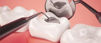
X-ray examination
Modern dentistry cannot do without this method of diagnosing dental caries. Along with the traditional method, compact devices are now successfully used in practice, making it possible to carry out various types of research. For example, microprocessor-controlled orthopantomographs make it possible to perform literally any diagnostic examination in the field of dentistry. Radiography helps to identify hidden carious cavities on the contact surfaces of teeth, under an artificial crown. Using a radiovisiograph, you can carry out diagnostics without leaving the office. In this case, the patient himself has the opportunity to observe on the screen what exactly is done in the oral cavity during the treatment process.
Clinical researches
Laboratory studies have proven that regular use of professional toothpaste ASEPTA REMINERALIZATION after 4 weeks improved the condition of the enamel by 64% and reduced tooth sensitivity by 66%.
Sources:
- Report on the determination/confirmation of the preventive properties of personal oral hygiene products “ASEPTA PLUS” Remineralization doctor-researcher A.A. Leontyev, head Department of Preventive Dentistry, Doctor of Medical Sciences, Professor S.B. Ulitovsky First St. Petersburg State Medical University named after. acad. I.P. Pavlova, Department of Preventive Dentistry
- Clinical experience in using the Asepta series of products Fuchs Elena Ivanovna Assistant of the Department of Therapeutic and Pediatric Dentistry State Budgetary Educational Institution of Higher Professional Education Ryazan State Medical University named after Academician I.P. Pavlova of the Ministry of Health and Social Development of the Russian Federation (GBOU VPO RyazSMU Ministry of Health and Social Development of Russia)
- Clinical studies of antisensitive toothpaste “Asepta Sensitive” (A.A. Leontyev, O.V. Kalinina, S.B. Ulitovsky) A.A. LEONTIEV, dentist O.V. KALININA, dentist S.B. ULITOVSKY, Doctor of Medical Sciences, Prof. Department of Therapeutic Dentistry, St. Petersburg State Medical University named after. acad. I.P. Pavlova
Electroodontodiagnosis (EDD)
This clinical method is used in dentistry most often for the diagnosis of deep caries. Its essence is as follows: the tooth pulp is exposed to an electric current of minimal voltage until slight pain appears. For a healthy pulp, the values are in the range of 2-6 µA, for a pulp affected by caries - no more than 8 µA. This method also has disadvantages: it does not detect initial caries and does not allow one to determine the depth and extent of the process. The EDI method is not prescribed for children.
When is the outcome of treatment of initial caries considered successful?
The initial stage of caries is the easiest to correct, but incomplete or poor-quality treatment almost always leads to the transition of the disease to the next, more serious, stage. The outcome of treatment of initial caries is considered successful when:
- the carious stain disappeared, the tooth color became uniform;
- repeated diagnostics confirms the absence of traces of enamel demineralization;
- there is no reaction to temperature stimuli and the feeling of soreness, which sometimes appears at the stage of initial caries, disappears.
Transillumination
The method is intended to identify various types of caries in the initial stage. Based on the heterogeneous light-absorbing ability of dental structures. Transillumination is carried out by shining a light from inside the mouth. Hidden carious cavities are revealed in the rays of light passing through the hard tissues of teeth.
The doctor makes conclusions about the degree and type of the disease based on the visible results. Fissure caries is characterized by a dark, fuzzy shadow, the intensity of which depends on the degree of damage to the fissures: the deeper they are, the darker the shadow. If the caries is approximal, then the carious lesions look like brown hemispheres, delimited from healthy tissue. Transillumination can also detect mineral deposits (tartar).
Is it necessary to treat enamel caries at the spot stage?
Now we know how to treat caries in the spot stage, but is this necessary? Indeed, there are cases when the enamel is naturally saturated with calcium due to the composition of saliva, and the problem disappears. However, this does not happen too often due to insufficient hygiene and monotonous nutrition, which does not meet the body’s need for minerals. Therefore, it is better not to rely on chance and consult a doctor, otherwise early stage caries will develop into a full-fledged defect.
Luminescent diagnostics
A research method for identifying superficial caries, based on the ability of enamel to change its color under the influence of UV rays. A beam of UV rays is directed at the teeth from a distance of 20-30 cm. Their hard tissues begin to glow. Normally, dentin and enamel emit a bright blue glow. With carious lesions of internal structures or enamel, the intensity of luminescence changes significantly: for example, in areas with chalk spots it is noticeably extinguished. This method of diagnosing dental caries allows you to detect changes inside them at the earliest stages.
Why do stains appear on teeth: the main reasons
The initial form of caries can be provoked by various factors, but most often this happens due to the formation of an acid-base imbalance in the oral cavity. A large number of harmful microorganisms constantly live in the mouth, which actively participate in the process of food decomposition. Food particles tend to remain between the teeth and on their surface, which leads to the destruction of tooth enamel.
Other reasons leading to the destruction of the structure of the enamel layer on the teeth include:
- incomplete or poor oral hygiene. After some time, failure to comply with hygiene rules leads to the appearance of white plaque on the teeth, which acts as an excellent environment for the accumulation of pathogenic bacteria. As a result, microorganisms produce harmful acids, which negatively affects the condition of tooth enamel. This process can be prevented by regular oral hygiene procedures. If, nevertheless, destruction of the enamel is observed, then treatment of caries at the spot stage will help prevent complications;
- hereditary predisposition. Even at the moment of conception and during intrauterine development, the structure and strength of tooth enamel is laid in the baby. When the mother’s body during this period lacks fluoride, calcium, vital vitamins and minerals, the risks that the child’s teeth will be excessively fragile and prone to the formation of carious processes are maximum;
- poor nutrition. Tooth enamel will be hard and durable if the daily diet contains foods high in calcium, fluorine and phosphorus (fish, cheese, cottage cheese, nuts, milk). But foods containing a large amount of carbohydrates (sweets, baked goods) must be kept to a minimum. This will protect the enamel from premature destruction;
- changes in the composition of saliva and its increased viscosity. Along with the fact that saliva is involved in the process of softening the food consumed, it also cleanses the surface of the teeth from food debris. When, due to changes in the structure of saliva, its acidity increases, a constant destructive effect on the teeth is formed. This process leads to the development of carious pathologies.
Often the initial form of caries appears in the area of the neck of the tooth, which is difficult to clean with a toothbrush. Regular examination of the condition of the teeth by a dentist and, if necessary, professional cleaning will help prevent the development of carious processes.
Laser fluorescence spectroscopy
When irradiated with a laser, hard dental tissues acquire the ability to fluoresce. The device operates at a specific wavelength - 655 nanometers. The pulsed flow directed at the tooth penetrates approximately 1 mm inside it and is partially reflected. Carious tissues and bacteria, when exposed to light rays, begin to fluoresce, that is, emit waves of a different length - 680 nm. These changes are recorded and analyzed by a laser device.
The effectiveness and sensitivity of the method increases significantly after applying a fluorescent composition to the teeth - in this case, it is possible to detect demineralized tooth tissue at a depth of 5-6 microns.
By the shade of the glow, the doctor can determine the degree of caries and carry out differential diagnosis. The method makes it possible to identify hidden fissure and proximal caries and the dynamics of foci of enamel demineralization.
In the article we have listed various basic and additional methods for diagnosing caries, which are widely used today in modern dentistry. Of course, they are not prescribed all at once for one patient—the doctor chooses one or two methods at his own discretion. Most often, this is thermal exposure, radiography, probing, less commonly, vital staining and electroodontic diagnostics are prescribed. But there may be other methods depending on the condition of the dentition. Together, diagnostic procedures allow us to establish a complete picture of the disease and select the appropriate method of therapy.
Fluorescence of teeth (Conclusions)
Light is a simple and safe method for making a clear diagnosis. Fluorescence of teeth helps to quickly establish contact between the doctor and the patient. Using laser technologies, we can detect early and hidden caries, monitor its development, and control treatment.
The translation was made specifically for the OHI-S.COM website. Please, when copying material, do not forget to provide a link to the current page.
Literature
- Abdelaziz M, Krejci I. DIAGNOcam - a Near Infrared Digital Imaging Transillumination (NIDIT) technology. Int J Esthet Dent. 2015 Spring;10(1):158-65.
- Yu JL, Tang RT, Feng L, Dong YM. Digital imaging fiber optic transillumination (DIFOTI) method for determining the depth of cavity. Beijing Da Xue Xue Bao. 2021 Feb 18;49(1):81-85
- Young DA, Featherstone JD. Digital imaging fiber-optic trans-illumination, F-speed radiographic film and depth of approximal lesions. J Am Dent Assoc. 2005 Dec;136(12):1682-7.
- Astvaldsdóttir A, Ahlund K, Holbrook WP, de Verdier B, Tranæus S. Approximal Caries Detection by DIFOTI: In Vitro Comparison of Diagnostic Accuracy/Efficacy with Film and Digital Radiography. Int J Dent. 2012;2012:326401. doi: 10.1155/2012/326401. Epub 2012 Nov 4.
- Yang J, Dutra V. Utility of radiology, laser fluorescence, and transillumination. Dent Clin North Am. 2005 Oct;49(4):739-52, vi. Review.
- Söchtig F, Hickel R, Kühnisch J. Caries detection and diagnostics with near-infrared light transillumination: clinical experiences. Quintessence Int. 2014 Jun;45(6):531-8
https://styleitaliano.org/