Dental caries is usually classified according to different indicators. One of the most widespread is Vinogradova’s classification, according to which there are compensated, subcompensated and decompensated forms of dental caries. We will tell you more about the symptoms and treatment features of different forms of the disease in this article.
In this article
- How does dental caries occur?
- Three forms of dental caries
- Compensated form of caries
- How is compensated caries treated?
- What is subcompensated caries and what are its features?
- Features of decompensated caries
- How is decompensated caries treated?
- Pulpitis as a complication of decompensated caries
- Periodontitis with decompensated caries
- Is it possible to prevent tooth decay?
- Conclusion
What happens if you ignore a toothache or the appearance of a small spot on a tooth?
Already at the very first stage, caries slowly approaches the tooth enamel and tends to destroy it. Further, harmful acids penetrate deeper into the dentin, making their way to the roots of the tooth.
At the very beginning, as a rule, there is no pain. This is where the danger lies. Caries develops, and we continue to live, unaware.
The doctor can detect affected carious teeth:
- at preventive examinations
- when undergoing professional oral hygiene.
Caries has several stages of development
What is subcompensated caries and what are its features?
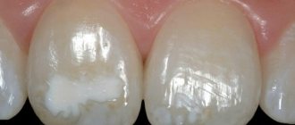
Subcompensated caries occurs in approximately 25% of children with dental caries. This form is characterized by an average rate of spread of the pathological process. Characteristic features are dull enamel and superficial lesions without deep tissue involvement. Most often, subcompensated caries does not cause discomfort or pain, so it can only be detected during an examination by a dentist.
People with this form of caries are recommended to have scheduled visits to the dentist two to three times a year, as well as oral sanitation and preventive measures.
Treatment of superficial caries
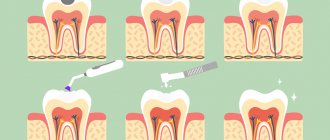
| Examination in a chair and carrying out diagnostic procedures - an x-ray to suggest the volume of the carious cavity; |
| Anesthesia, pain relief for the working field. For painless removal of carious tissues; |
| Selecting the color of the filling; |
| Installation of rubber dam. Complete isolation of the tooth from saliva. Saliva is the richest environment in microbes and can cause poor adhesion of the filling and the development of recurrent caries; |
| Removal of carious tissues using a drill. Drilling, which in most cases causes fear in the patient, in this case will not last at all, because superficial caries usually affects small areas of enamel. Formation of a cavity for filling; |
| Drug treatment; |
| Drying the surface; |
| Restoring the natural shape of a tooth by filling. Installation of a light-curing filling; |
| Grinding and polishing the filling. |
| Grinding and polishing the filling is not just mandatory, but an important step. The surface must be smooth, because a rough surface is a favorable environment for the accumulation of bacteria. Anatomically, the filling, completely replicating the tooth, should not cause any discomfort. |
How is compensated caries treated?
The method of treating compensated caries depends on the stage at which it is detected and, accordingly, on the degree of tooth destruction.
At the initial stage of a carious stain on the enamel, non-invasive methods that do not require drilling a dental crown can be effective.
- Remotherapy (remineralization).
Using special pastes, gels, and solutions, tooth enamel is saturated with calcium and fluoride ions - the main components of its mineral composition. They normalize the structure of the enamel, increase its protective properties and resistance to the corrosive effects of acid.
- Deep fluoridation.
It is similar to the remineralization procedure with the difference that the composition of medicinal preparations necessarily includes fluoride. It penetrates deep into the enamel, makes it stronger, has an antiseptic effect, and prevents the teeth from losing calcium and other minerals.
- Fissure sealing.
Fissures are natural grooves on the surface of chewing teeth. They can be of different shapes and depths, and due to their anatomical features, food debris easily accumulates in them. In addition, the enamel in the fissure area is thinner compared to other areas of the dental crown. The sealing method is often used to prevent caries in the chewing teeth of children and adults with deep grooves on their teeth. The essence of the method is that the dentist seals the natural recesses of the dental crown with a special sealant, so that food debris no longer accumulates in them and bacteria cannot multiply.
- Filling.
This method treats a compensated form of caries in the later stages, when cavities have formed in the dentin of the tooth. Most often, the dentin treatment process is painful, so the procedure is performed under local anesthesia. After the anesthetic injection takes effect, the doctor prepares the affected tooth with a drill, removes carious tissue, and then covers the cured tooth with high-quality filling material. The best treatment option is without drilling or filling. Methods of remineralization and deep fluoridation are safe, painless, and do not cause discomfort in the patient. But they are effective only at the initial stages of the carious process.
Therefore, it is important to regularly check the condition of your teeth with a dentist (for compensated forms of caries, a visit once a year is recommended) in order to notice the beginning of caries as early as possible.
Average caries
This is not a death sentence for the tooth, but it is the first sign that an infection has entered inside. The upper layers of dentin are subject to carious lesions.
Dentin is a hard tissue that makes up the main part of the tooth, but its structure is still softer than that of enamel. However, despite the fact that enamel is considered the hardest fabric, it is quite fragile and can crack. Through these microcracks, bacteria penetrate into the dentin, where in more fertile soil they multiply much faster, which will lead to the formation of a carious cavity. On the surface, the diameter of the lesion is small. But even a tiny dark point in depth can extend to the pulp.
This stage is not the easiest, but still, with timely contact with a specialist and proper treatment, you can avoid the worst and keep the tooth alive.
How does dental caries occur?
During caries in hard dental tissues, under the influence of organic acids, first areas of enamel demineralization are formed, and then carious cavities. Acids that destroy teeth are produced by cariogenic bacteria. This occurs during the fermentation process of carbohydrates that we consume in food. According to statistics, caries is the most common dental disease; it affects the teeth of people of any age, including the development of baby teeth immediately after teething.
There are different classifications of caries, in particular the Vinogradova classification. According to this division, there are compensated, decompensated and subcompensated caries.
There are two manifestations of average caries:
- Chronic May last a long time and not manifest itself in any way. Layers of dental tissue are gradually affected. Pathology can develop over several years. This is fraught with transition to a more advanced form, deep caries, and even threatens tooth depulpation and canal filling.
- Acute Rapidly developing stage. Perhaps an acute reaction to various irritants, which quickly passes after their removal. This form is characterized by an abundance of softened dentin on the walls and bottom of the tooth cavity with sharp and fragile edges
The chronic form of caries can transform into pulpitis.
Three forms of dental caries
Vinogradova’s classification is based on the degree of activity of dental caries:
- The compensated course of caries is characterized by the fact that the pathological process develops slowly, carious cavities are hidden under dense dentin, they are rare, and the disease progresses relatively slowly.
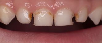
- The subcompensated form of the disease develops slightly faster than the compensated form. It is characterized by the appearance of dark areas on the enamel and horizontal spread without penetrating deep into the tooth.
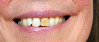
- Decompensated caries is often multiple and spreads very quickly. With such caries, all teeth located near the cheeks are often affected.
The decompensated form is considered the most severe, in most cases accompanied by complications in the form of pulpitis or periodontitis.
Let's talk about all forms of caries in more detail.
Deep caries
It is at this stage that the tooth begins to react and sends us SOS signals - sharp aching pain. However, they are temporary; unfortunately, patients can endure these moments or help themselves with painkillers and delay going to the doctor. It’s not worth the effort to guess; this has never ended in anything positive. Medium caries that is not treated in time slowly and quite difficultly turns into deep caries. The rate of damage varies from compensated to decompensated, which in turn is more dangerous, because in a short period of time it can cover even two teeth. It is important to prevent infection from entering the pulp chamber and to prevent complications - pulpitis and periodontitis.
Pulpitis as a complication of decompensated caries
The course of caries in a decompensated form can be complicated by inflammation of the neurovascular bundle - the pulp. The inflammatory process in this area is called pulpitis. It occurs when the infection spreads beyond the tooth
The pulp contains blood vessels, nerves, and nourishes dental tissue. If it becomes inflamed, there is a risk of tooth loss due to loss of vitality. With pulpitis, the patient complains of severe, prolonged pain, which is often pulsating in nature and radiates to the temples and ears. Painful attacks are repeated with a certain frequency, last for five minutes, intensify in a supine position.
Depending on the severity and neglect of the process, pulpitis is treated with a conservative or surgical method. At an early stage, the doctor, if possible, preserves the pulp and root of the tooth, eliminating the inflammatory process with the help of medications. If the inflammation is too strong, there is a risk of it going beyond the pulp, and it is not possible to stop the pathological process with medications, in this case the doctor chooses a surgical treatment tactic, partially or completely removing the pulp. This option is less preferable, because without a neurovascular bundle, the tooth becomes virtually “dead” and serves much less time.
The complications of caries should be taken seriously and at the first symptoms, contact your dentist. If you neglect timely diagnosis and treatment, the infection can spread to other organs and tissues, which can lead to serious disruptions in the functioning of the entire body. In particular, untreated pulpitis is dangerous for the development of gumboil, periodontitis, pulp necrosis and even sepsis.
Periodontitis with decompensated caries
In addition to pulpitis, with decompensated caries another serious complication often develops - inflammation of the tissues surrounding the tooth root. This disease is called periodontitis.
The periodontium is a ligamentous apparatus that holds the tooth in the jaw and provides shock absorption. If an inflammatory process develops in the periodontal tissues, this can lead to loosening and tooth loss. Against the background of periodontitis, ulcers can form, the removal of which will require surgical opening of the gums.
Periodontal inflammation can be acute or chronic. In the first case, inflammation develops rapidly, accompanied by darkening of the tooth, acute pain, swelling of the affected tissues, and inflammation of the cheek. Chronic periodontitis is characterized by more smoothed symptoms, pain syndrome may be absent. Treatment for the chronic form is long and difficult and can take several months.
Periodontitis greatly weakens the immune system and creates a source of chronic infection in the body.
Classification according to the degree of tooth damage
- Enamel caries
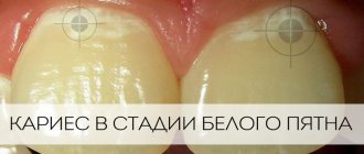
This species is often called the “spot stage.” Some areas of tooth enamel become thinner due to a lack of minerals. A dark or white spot appears at this place.
- Dentin caries
It is also called internal, since microorganisms penetrate deep into the main tissue of the tooth - dentin. This type of caries is diagnosed in most people.
- Cementum (root) caries
The carious cavity is located directly below the gum level. It often affects the cervical part of the tooth. Root caries progresses rapidly and almost immediately leads to pulpitis. The reason is the formation of a periodontal pocket and exposure of the neck of the tooth. Most often, cement caries occurs in older people. Unfortunately, in most cases the disease is discovered when severe pain occurs.
- Odontoplasia
This is a pathology of the root system that is observed in most young children. Caries in primary teeth must be treated immediately. If this is not done, the carious process will affect permanent teeth. In addition, the enamel and dentin layers of children's teeth are very thin, and the carious process reaches the pulp in a matter of months.
- Suspended caries
If you take care of your health and the body’s defenses are high enough, then caries can seem to “freeze” at a certain stage, and the tooth does not further decay. However, this does not mean that you can postpone your visit to the doctor.
Classification according to the location of the outbreak
- Fissure caries
Fissures are called tubercles and depressions on the chewing part of the tooth. They are an ideal place for food debris to stick and bacteria to grow, so cavities are not uncommon there. Fissure caries is always located strictly in the central part of the tooth and never affects its neck and roots. Neglected cavities are usually very deep and painful. If there are no complications, the tooth is filled.
- Contact caries
If the teeth grow crowded, and it is not possible to regularly clean the interdental spaces with floss from stuck pieces of food, then contact caries develops. It got its name due to the fact that the lesion occurs on the proximal part of the tooth. These sides are where the teeth touch each other. The main symptom is the appearance of a dark carious cavity. During treatment, the doctor first restores the side wall and then fills the tooth.
- Cervical caries
The carious process affects the neck of the tooth, it is located near the gingival margin. Here the enamel-dentin layer is very thin, so tartar destroys its integrity in a short time. First, a characteristic white or dark spot appears in the lesion, then the tooth begins to react painfully to sour and sweet foods, as well as changes in temperature. Treatment of cervical caries cannot be delayed, because You can lose a tooth in a short time. Most often, cervical caries affects the front teeth.
- Circular caries
This is a form of cervical caries. They talk about it if the pathological process covers the diseased tooth in a dark ring. Circular caries mainly occurs on baby teeth or is an occupational disease in adults - for example, in musicians who play wind instruments.
Complications
Expected consequences of the current:
- pulpitis – inflammation of the dental nerve;
- periodontitis – inflammation of the periodontium (periodontal tissues);
- fracture of the crown part of the tooth;
- tooth decay.
In pregnant women, caries worsens the condition of the body and negatively affects the health of the baby. Also keep in mind that cariogenic bacteria enter the gastrointestinal tract, disrupting the functioning of the digestive system.
Types of disease
Acute caries has two forms:
- medium spicy;
- deep spicy.
The difference between medium and deep acute forms lies in the size of the carious cavity. With moderate acute caries, there is no need to remove the nerve, and the tooth can be treated and restored. In case of deep acute caries, depulpation is usually required, and in case of severe tooth decay it is necessary to remove it.
Since in the acute form of the pathology the destruction of tooth tissue occurs very quickly, and children are most susceptible to the disease, in pediatric dentistry the following grouping is accepted:
- Compensated form (I group);
- Subcompensated (group II);
- Decompensated (group III).
Groups have been created to carry out clinical observation.
Pediatric dentist T. F. Vinogradova identified several dispensary groups:
- practically healthy teeth;
- compensated form;
- subcompensated form;
- decompensated form.
Children with a compensated form are examined once a year, with an undercompensated form - 2 times, with a decompensated form - 3 times. Planned sanitation reduces the risk of complications in the development of caries, the number of fillings and extracted teeth decreases. The need for treatment is also reduced by almost half, and the number of annual scheduled examinations is reduced.
Monitoring risk groups allows you to keep records based on a number of criteria:
- general prevalence of caries;
- anamnesis of life;
- health status;
- severity of the disease.
Planned sanitation and timely diagnosis of caries in adults and children make it possible not only to cure it in the initial stages, but also to prevent the development of blooming caries.
Among the pressing problems of modern practical dentistry, dental caries occupies one of the leading places. Its medical and biological significance is determined not only by the huge prevalence of various forms of caries and its immediate complications (acute forms of pulpitis), but also by the negative impact on the body as a whole.
The high incidence of pulp damage, according to various statistics, is about 30% of all dental patients, and is largely due to the characteristics of both structure and function, constant contact with the external environment, the presence of microflora, a variety of types of load, etc.
Experience gained in recent years shows that it is possible to stop the increase in pulp pathology with therapeutic measures. In this regard, it is necessary to develop and widely implement measures to prevent diseases such as dental caries and its complications.
Dental caries (Caries dentis) is a pathological process that manifests itself after teething, during which demineralization and softening of the hard tissues of the teeth occurs, followed by the formation of a defect in the form of a cavity.
Dental caries is a key problem in dentistry, very interesting theoretically and extremely important in practical terms.
Etiology
About 400 theories have been proposed to explain the etiology and pathogenesis of dental caries. Without dwelling on all available theories, we will present those that, at least to some extent, provide an explanation of the origin of the most common pathological process - caries.
Modern concept of the etiology of caries.
The generally accepted mechanism for the occurrence of caries is the progressive demineralization of hard dental tissues under the influence of organic acids, the formation of which is associated with the activity of microorganisms.
Many etiological factors take part in the occurrence of the carious process, which allows us to consider caries a polyetiological (multi-causal) disease. The main etiological factors are:
1) microflora of the oral cavity;
2) nature and diet, fluoride content in water;
3) quantity and quality of salivation;
4) general condition of the body;
5) extreme effects on the body.
All of the above factors were called cariogenic and divided into general and local, which play an important role in the occurrence of caries.
General factors:
1) Poor diet and drinking water; 2) Somatic diseases, a shift in the functional state of organs and systems during the period of formation and maturation of dental tissues. 3) Extreme effects on the body; 4) Heredity, which determines the usefulness of the structure and chemical composition of tooth tissue. Unfavorable genetic code.
Local factors:
1) Dental plaque and plaque, isolating microorganisms; 2) Violation of the composition and properties of oral fluid; 3) Carbohydrate sticky food residues from the oral cavity; 4) Resistance of dental tissues, due to the complete structure and chemical composition of the hard tissues of the tooth; 5) Deviations in the biochemical composition of hard dental tissues and defective structure of dental tissues; 6) Condition of the dental pulp; 7) The state of the dental system during the period of formation, development and eruption of permanent teeth.
A cariogenic situation is created when any cariogenic factor or group of factors, acting on a tooth, makes it susceptible to the effects of acids. Of course, the trigger is the microflora of the oral cavity with the obligatory presence of carbohydrates and the contact of two factors with tooth tissue.
In conditions of reduced resistance of dental tissues, the cariogenic situation develops more easily and quickly.
Clinically, a cariogenic situation in the oral cavity is manifested by the following symptoms:
1) poor oral hygiene;
2) abundant dental plaque;
3) tartar;
4) crowding of teeth and malocclusions;
5) bleeding gums.
Dental resistance to caries or caries resistance is ensured by:
1) the chemical composition and structure of enamel and other tooth tissues;
2) the presence of a pellicle;
3) the optimal chemical composition of saliva and its mineralizing activity;
4) a sufficient amount of oral fluid;
5) low level of permeability of tooth enamel;
6) good chewing load and self-cleaning of the tooth surface;
7) properties of dental plaque;
 good oral hygiene;
9) diet features;
10) correct formation of rudiments and development of dental tissues;
11) timely and complete maturation of enamel after tooth eruption;
12) specific and nonspecific factors for protecting the oral cavity.
The susceptibility of teeth to caries or caries susceptibility contributes to:
1) defective enamel maturation;
2) a diet deficient in proteins, macro- and microelements, and excess carbohydrates;
3) water with insufficient fluoride;
4) absence of pellicle;
5) composition of oral fluid, its concentration, viscosity, quantity and flow rate;
6) the biochemical composition of the hard tissues of the tooth, which determines the course of caries, since a dense structure with minimal spaces in the crystal lattice slows down the course of caries and vice versa;
7) state of the neurovascular bundle;
functional state of organs and systems of the body during the formation and maturation of dental tissues;
9) improper development of the tooth due to common somatic diseases.
Pathogenesis
As a result of frequent consumption of carbohydrates and insufficient oral care, cariogenic microorganisms become tightly attached to the pellicle, forming dental plaque. When consuming sticky food, its remains harden in the retention points of the teeth (fissures, pits, contact surfaces, fillings, dentures) and undergo fermentation and rotting. The formation of dental plaque is influenced by:
1) the anatomical structure of the tooth and its relationship with surrounding tissues;
2) tooth surface structure;
3) food intake and chewing intensity;
4) saliva and gingival fluid;
5) oral hygiene;
6) the presence of fillings and dentures in the oral cavity;
7) dental anomalies.
Soft plaque has a porous structure, which allows liquid food components to penetrate into the saliva. This is a soft amorphous substance that adheres tightly to the surface of the tooth. The accumulation of end products of microorganisms and mineral salts in plaque slows down this diffusion, as porosity disappears. And this is a new substance - dental plaque, which can only be removed by force, but even then not completely. Under the dental plaque there is an accumulation of organic acids - lactic, pyruvic, formic, butyric, propionic, etc. The latter are products of fermentation of sugars by most bacteria during their growth. It is these acids that play the main role in the appearance of a demineralized area in a limited area of enamel. Neutralization of these acids does not occur, since there is a restriction of diffusion both into and out of dental plaque.
Dental plaque contains streptococci, in particular Str. mutans, Str. Sanguis, Str. salivarius, which are characterized by anaerobic fermentation. In this process, the substrate for bacteria is mainly carbohydrates, and for certain strains of bacteria - amino acids. Sucrose plays a leading role in the occurrence of caries. It is this that causes the fastest decrease in pH from 6 to 4 in a few minutes. The process of glycolysis occurs especially intensively during hyposalivation, xerostomia, and during sleep. And the activity of the fermentation process depends on the amount of carbohydrates involved. It has been established that during the period of consumption of excess sugars, the amount of plaque increases significantly.
The formation of plaque is influenced by the composition of food and its consistency. It has been observed that soft foods accelerate its formation as well as high sugar content. It has been proven that dental plaque forms faster during sleep than during eating, since salivation and mechanical stress help slow down the formation of dental plaque.
Plaque microorganisms are capable of fixing and growing on the hard tissues of the tooth, metal, and plastic.
In persons with multiple caries, there is an increase in the biochemical activity of streptococci and lactobacilli located on the tooth surface. High enzymatic activity of microorganisms is regarded as caries susceptibility.
Classifications of the carious process.
1. Topographical.
a) caries in the spot stage (white, pigmented); b) superficial caries; c) average caries; d) deep caries.
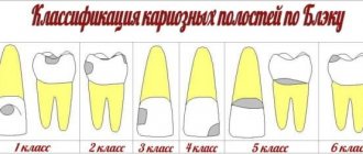
2. By localization:
1st class - carious cavities in the area of natural fissures of molars and premolars, as well as in the blind fossae of incisors and molars; Class 2 – carious cavities located on the contact surfaces of molars and premolars; 3rd class - cavities located on the contact surfaces of the incisors and canines without violating the integrity of the cutting edge; 4th class - cavities located on the contact surfaces of the incisors and canines with a violation of the integrity of the angle and cutting edge of the crown; 5th class – cavities located in the cervical areas of all groups of teeth; Class 6 is atypical, that is, where caries usually does not occur, these are the immune-resistant zones of the crown of the tooth.
Clinical picture.
Initial caries (spot stage)
With initial caries, there may be complaints of a feeling of sore throat. The affected tooth does not respond to a cold stimulus, as well as to the action of chemical agents (sour, sweet). Demineralization of enamel upon examination is manifested by a change in its normal color in a limited area and the appearance of matte, white, light brown, dark brown spots with a black tint. The process begins with the loss of enamel shine in a limited area. This usually occurs at the neck of the tooth near the gum. The surface of the spot is smooth, the tip of the probe slides over it.
Superficial caries.
For superficial caries, the occurrence of short-term pain from chemical irritants (sweet, salty, sour) is the main complaint. It is also possible that short-term pain may appear from exposure to temperature stimuli, more often when the defect is localized at the neck of the tooth, in the area with the thinnest layer of enamel, as well as when brushing the teeth with a hard brush.
Average caries.
With average caries, patients may not complain, but sometimes pain occurs from exposure to mechanical, chemical, thermal irritants, which quickly pass after the irritant is eliminated.
Deep caries.
Patients complain of short-term pain from mechanical, thermal, and chemical irritants, which quickly passes after the irritant is eliminated.