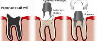
48642
Crooked, unhealthy teeth can ruin the appearance of an otherwise attractive person. Oral health is important not only for our attractiveness, because the main function of teeth is to grind food. The condition of the stomach and intestines depends on this, which directly affects health and life expectancy .
Scheme
Human teeth consist of three elements:
- Crown . The uppermost visible part, which fully or partially protrudes above the alveolus after eruption;
- Cervix . A narrower area located in the gum between the main crown and the root.
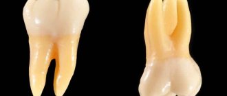
- Root . The lowest part, located in the alveolus. The pulp of the root contains intertwining nerves and blood vessels. With the help of the periosteum, the roots are tightly fixed in the alveolar socket. Depending on the functions performed by the tooth and the anatomical features of the person, the number of roots can vary from 1 to 4 units.
The main substance in the structure of a tooth is dentin, which makes up most of its mass. From a chemical point of view, dentin is collagen impregnated with various salts, phosphorus and other minerals.
Photo: diagram of the structure of human teeth and jaw
The crown is covered with enamel on top. Due to the fact that the crown is based on inorganic compounds, its strength is close to that of diamond . Metabolic processes take place only in a thin skin that tightly envelops the surface of intact enamel.
The tooth is fixed using “special cement” covering the root. In its structure, cement is very close to the structure of bone tissue. Blood flow occurs through the branches of the external carotid artery, tightly intertwined. The outflow of venous blood occurs through vessels directly connected to the blood circulation of the brain.
Such blood circulation, in turn, carries a danger: if the initial infection is localized in the oral cavity, through these vessels it can enter the dura mater of the brain and cause a number of serious diseases.
In an adult, the row consists of two arches, each of which contains from fourteen to sixteen teeth. For children under twelve years of age, the row looks a little different - they, as a rule, only have twenty dairy products.
The external similarity of the structure of the upper and lower jaws does not indicate their identity, so you should familiarize yourself with their structure and distinctive features.
In the following video you can clearly see all of the above:
Anatomical structure of teeth with pictures
External anatomical structure of a human tooth:
- The crown is the visible area covered with enamel, the hardest part of the tooth. The surface layer performs a protective function and is designed to preserve dentin.
- The root part is a complex of nerve fibers, arteries and veins that provide nutrition and innervation to the tooth. The second function of the root is retaining.
- The neck is the area between the root and the crown, which performs a connecting function. Differs in a narrowed shape.
Diagram: external structure of a human tooth
How does a human tooth work at the tissue level?
All human teeth have a similar internal structure. The diagram below shows all the dental layers - enamel, dentin, cement, pulp.
The figure shows the histological structure of the tooth
Enamel covers the teeth like a cover and makes up 25% of the total mass of all dental tissues. The hardness and strength of this tooth layer is ensured by a high mineral content of 397 kg/mm.
The composition of the enamel layer includes:
- minerals – 96%;
- organic substances – 1.2%;
- liquid, water – 2.3%.
The enamel is covered by an outer shell, the cuticle, which covers the chewing surface of the tooth. Most of the tooth is occupied by dentin, located immediately under the enamel. It has less strength, its hardness is 58.9 kg/mm.
Cement – covers the root part and in most respects is similar to bone tissue. Depending on the type of structure, there are two types of cement:
- acellular or primary - consists of collagen fibers;
- cellular or secondary - the composition includes cemenoblasts.
The internal dental cavity is occupied by the pulp - soft tissue, including:
- numerous nerves;
- blood vessels;
- connective tissue.
Sectional structure of a tooth
Despite the differences in the purpose and structure of the root system, all teeth have the same internal structure:
- Enamel. A very hard surface layer that protects teeth from environmental influences. It is designed as a structure of glued prisms. Thanks to the large amount of mineral salts in its composition, enamel is the strongest tissue in the human body. Its main task is to preserve the dentin of the crown. The presence of a pellicle ensured the tissue's resistance to the effects of acids.
- Dentine. It consists of coarse fibrous tissue, the basis of which is collagen fiber. The structure is similar to porous bone. There are two types of dentin: superficial and internal. The first has a higher density and prevents infection from entering the dental cavity.
- Cement. Consists of collagen fibers saturated with limestone salts. Layer thickness - from 50 to 150 microns, depending on location. There are two types of cement: cellular and acellular.
- Cavity. It has a shape identical to the crown and is located under the dentin. The entire area of the cavity is filled with pulp, a type of tissue responsible for nourishing the tooth and performing a connecting function. The presence of blood and lymphatic vessels ensures high sensitivity of the pulp.
Photo: this is what a tooth looks like in cross-section
Upper jaw
The central incisor is characterized by the presence of a flat shape, a beveled cutting edge and a single root. The front part of the incisor is convex and contains three small tubercles.
The appearance of the lateral incisor is identical to the central one. But due to the fact that the central tubercle is large and stands out much more strongly, the cutting edge itself takes on a convex, streamlined shape.
Find out the reasons for the formation of a yellow coating on the tongue, and ways to prevent this disease.
In this article you will find photos with symptoms of oral leukoplakia.
Here: https://www.vash-dentist.ru/lechenie/zubyi/kakie-simptomyi-vospaleniya-troynichnogo-nerva.html - you can find out why the trigeminal nerve becomes inflamed and how it is treated.
Fang is an element inherited by a person from predatory representatives of the fauna. There is only one voluminous tubercle on the canine crown. With the help of a groove running along the inside, the fang is divided into two parts.
Small molars (called premolars ). Unlike the frontal ones, premolars are characterized by a more square shape. The roots, although flattened, are already beginning to bifurcate.
Large molars (aka molars) are the largest in the entire row and are responsible for the direct grinding of food. The first molar has a rectangular shape with four cusps, which allows you to chew food as efficiently as possible. The second molar is somewhat smaller in size, but in terms of functionality and root structure it is practically no different from its predecessor.
The third molar, also called the wisdom tooth, grows much later than the others. Sometimes it may not erupt at all, which is not very scary, since it does not perform any important functions and is largely a vestigial organ.
Lower jaw
The name and number of teeth in the upper and lower jaws are the same, but they have differences in structure and functional features.
The front incisors are significantly smaller than their counterparts on top. The outer surface has two edges: sharp and blunt. The roots are shallow and not large.
The lower fangs are practically no different from those located above, they just have narrower edges.
The molars and premolars of the lower jaw have a different number of tubercles for chewing food, as well as roots and canals in them. Unlike upper molars, lower molars have one less root.
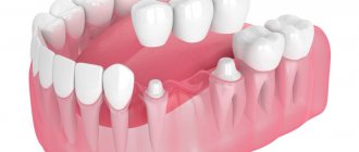
Photos of dental implantation before and after
Photo of dental implantation on a beam
All on 4 implantation photo
All on four implantation photo
Installation of 4 implants of the Superline Dentium system using All on 4 technology and installation of a fixed denture. Orthopedist Shagin L.V., surgeon-implantologist Kurbatov V.O.
All on Four implantation photo
Photo of one-stage dental implantation
Tooth extraction and photo of one-stage implantation
Simultaneous removal and implantation in the area of 25 and 26 teeth. Without bone grafting, but using a centrifuge.
Anatomy of molars and premolars
Molars in dentistry are divided into large ones - molars, and small ones - premolars. And their structure in humans is very different from the anterior ones.
Premolars
A person has two small molars on the left and right sides. In the first premolar, the central part of the chewing surface has a long shape, while the distal part is shorter and larger.
The second premolar retains all the features of the first, but it is more massive. The upper premolar is slightly smaller in size than its lower counterpart.
Molars
Depending on individual anatomical features, the number of molars in a person can vary from eight to twelve. Due to the structural features of the jaw, the molars gradually become smaller from the center to the edges.
The crowns of molars are large, with a pronounced square or even triangular closure surface. From three to five chewing tubercles are located on top, allowing the molars to fully perform their functional duties - the primary processing of food.
Upper molars are characterized by the presence of three roots, two of which are directed to the cheek, and one to the tongue. The lower molars have only two roots: posterior and anterior. In the outer molars, the roots sometimes grow together. Third molars also have a very unpredictable crown shape, which depends on the structure of the skull and jaw.
We'll tell you what antibiotics are usually prescribed for gumboils and how to take them correctly.
How malocclusion is corrected with braces - see our “Before” and “After” photos.
Here: https://www.vash-dentist.ru/lechenie/desnyi/parodontoz/sposobyi-narodnyimi-sredstvami.html - you will learn about the causes of periodontal disease and traditional methods of treating it.
The importance of proper bite for a person
Having understood a little what an optimal bite looks like from a physiological point of view, we can move on to the question of the importance of its presence for the full functioning of a person.
With an incorrect bite, you can experience 2 types of problems that affect the state of the body and behavior. Bite defects often affect the psychological state. People who experience this are likely to feel discomfort while building friendships, business and intimate relationships.
This state of affairs provokes the emergence of complexes and can interfere with adequately assessing oneself and the attitude of others, which will definitely affect the quality of achievement of assigned tasks in all areas of life. On the other hand, an incorrect bite can cause negative consequences for your well-being, disrupting the natural course of physiological processes in the body.
Now we will highlight the most common pathologies and diseases caused by malocclusion: displacement of the upper cervical vertebrae, narrowing of the airway gap, impaired cerebral circulation, difficulty breathing during sleep, digestive disorders due to poorly chewed food entering the stomach by improperly closing jaws.
Uneven pressure on teeth in an incorrect bite leads to their abrasion, damage and even premature loss. Improper closing of the jaws creates a large load on the temporal joint, and the jaw muscles do not relax. This can cause regular headaches.
Incisors and canines
Dentists divide human front teeth into canines and incisors.
Incisors
The incisors include two teeth located in the upper and lower jaw arches. The crown has a narrow, flattened shape with a sharp edge, as it is intended for cutting pieces of food, which are subsequently chewed by molars and premolars.
The incisors of the upper jaw are much wider and more massive, while the lower ones are almost half as large. The roots are single and flat, especially for the incisors located below. The upper part of the roots deviates to the side.
Fangs
The canines are located directly behind the incisors in the upper and lower jaw arches. Their distinctive feature is that both cutting edges converge at an angle at one point, forming such a recognizable shape. The canines have one long root with grooves on the side.
The upper canine is larger and more massive, while the lower one is less pronounced. The fangs located below have a shorter and smoother cutting edge and narrow longitudinal ridges. The roots are noticeably shorter than the upper ones and have pronounced grooves.
Wisdom teeth
a — vestibular surface; b — mesial surface; c - lingual surface; d — figure-of-eight cut; d — mesiodistal section; 1, 2, 3 - cross section: in the plane of the crown, in the middle and upper part of the root
Wisdom teeth, or third molars as they are correctly called, can erupt at any age, and not necessarily all of them. But at the same time, even if they never appeared, remaining in their infancy, this is not a deviation from the norm.
Third molars are among the most problematic teeth in humans. They are located at the end of the row on both sides, and there are four of them in total. The structure of the third molars is no different from the structure of the other large molars. But at the same time, it also has its own characteristics:
- the wisdom tooth is located last in the row and is not sandwiched between its neighbors;
- at the location of the third molar, children do not have milk teeth that prepare the ground for its eruption, which makes this process more unpleasant and painful;
- the roots of third molars often grow together into one large one, which may have an irregular cone shape;
- the crown does not necessarily erupt completely and has different shapes.
Typically, third molars grow in between the ages of eighteen and twenty-five. But sometimes they can appear much later, or even not appear at all. Such unerupted teeth are called impacted or semi-impacted if the crown has only partially appeared.
Problems with the growth of wisdom teeth are caused by evolutionary changes in the skull. In the jaw of a modern person, they are a rudimentary organ, and often there is simply no space left for their normal development.
Human dental formula
Many people have heard that dentists call teeth by different numbers. Thus, each unit is assigned a specific number, which guarantees maximum accuracy in records of dental conditions or treatment procedures. The count is made starting from the middle of the dentition to the right and left sides. The anterior units - incisors - are No. 1 and 2, 3 - canines, 4-5 - premolars, 6, 7 and 8 - molars, the last of which is the wisdom tooth.
To determine exactly where the tooth is located, the jaw is figuratively divided into segments. The beginning of the numbering is the right side of the upper jaw of the teeth. Units are written as two-digit numbers, where the first determines the serial number, and the other determines the so-called segment. The upper right half of the teeth is designated by numbers 11-18, the left - 21-28, and the lower left segment - 31-38 and 41-48. Regarding primary teeth, the count is made starting from the front units on the left in a clockwise direction. They are assigned the numbers 50, 60, 70 and 80.
READ ALSO: structure of the human upper jaw and functions of its processes
There are several types of numbering that dentists use:
- With the square-numeric Zsigmondy-Palmer method, Arabic numerals are used for radical units, and Roman numerals are used for dairy units. Often used by orthodontists and surgeons.
- Haderup's method is based on the symbols "-" and "+", where the first denotes the lower and the other the opposite arc. Units are written in Arabic numerals, and in the case of dairy units, 0 is added.
- The Viola system uses Arabic numerals 1-8 for units and two-digit numbers for segments. This type is very convenient, which is why it is widely used by dentists around the world.
- The last system is universal. Each group of units is assigned a letter, number and segment number.
Baby teeth
Their formation in a child begins to occur in the womb at the twelfth week . As a rule, the first to appear in a child are the incisors and canines, and only at the very end the molars.
The timing of this process is purely individual and can vary, but in most cases, the formation of the primary bite begins to occur at the age of seven months and ends at three to four years. By this time, the child should have twenty baby teeth.
Compared to permanent teeth, milk teeth have their own characteristics:
- smaller sizes;
- fewer chewing tubercles;
- the roots spread out to the sides.
Despite this, primary and permanent teeth have the same number of roots.
The deciduous row in the jaw consists of ten teeth: four molars, four incisors and two canines. At the age of six or seven years, baby teeth begin to fall out and are replaced by permanent ones.
First of all, the large molar is replaced, and the final formation of the row ends by the age of twelve to fourteen, with the exception of the third molar.
If you find an error, please select a piece of text and press Ctrl+Enter.
Tags: tooth structure
Did you like the article? stay tuned
Previous article
Photos of the smiles of stars and ordinary patients BEFORE and AFTER installation of veneers
Next article
What to do if a wisdom tooth grows into the cheek?
Bite correction
It is better to correct the correct bite, or rather to form it, in childhood, but adults often turn to orthodontists for whom the problem of a malocclusion prevents them from leading a full life.
Malocclusion can be corrected at any age, however, the more advanced its severity, the more difficult it is to work with it, but for modern medicine this is a solvable problem. For example, during dental prosthetics, special attention is paid to the problem of bite, so it is better to solve this problem in advance.
Nowadays, various technical means are used to create the correct bite: LM activators, retaining caps with a system of elastic bands or tourniquets, orthodontic plates, trainers and brace systems.
When the pathology is particularly complex and cannot be corrected using the above means, doctors perform surgery to eliminate the malocclusion. Alternative methods of influencing bite correction include myotherapy and wearing special aligners. Let us dwell in more detail on the main types of orthopedic structures that help to create a correct bite.
LM activator
LM activators stimulate the development of the jaw arch, chewing muscles, facial expressions of the child, and promote normal breathing.
LM activators are also suitable for getting rid of bad childhood habits - for example, if a baby sucks fingers, bites lips, bites pencils or nails. Due to its effect on the gums and tongue, the device fixes the rudiments of teeth in the desired position and prevents the development of maxillofacial pathologies in the future.
Correcting a child's bite at 3 years old requires minimal costs. At this age, the dental system is still very plastic, and treatment, as a rule, is limited to the use of a cap to correct the bite in children. It is worn only at night to straighten baby teeth and avoid problems in the future when they are replaced by molars.
The cap is a bandage made of rubber straps that are fixed on the jaw. The device is also used to correct mesial occlusion - the design restrains the development of the lower jaw and allows the upper jaw to form correctly.
To correct the bite of a child from the age of 4, plates are used - thanks to them, you can straighten the teeth or prevent them from moving, correct the shape of the palate, and slightly widen the jaw. However, more complex violations cannot be corrected using this device. Orthodontic plates are made from individual impressions and secured in the oral cavity with metal brackets on the outer teeth.
Trainers for correcting malocclusion in children are also called silicone trainers. They are quite soft but durable mouthguards, and are used to correct the bite of a child after 5 years. At this age, the teeth will already be replaced with permanent ones.
These devices are elastic, which are used for children under 8 years of age, and hard, recommended after reaching this age. These are standard designs that are produced without taking into account the individual characteristics of the patient. There are no contraindications for their use, and the effectiveness of mouthguards allows you to finally solve this problem and form the correct bite.
The general name for devices for correcting malocclusion in children is removable orthodontic structures. They are suitable for children up to 12 years old and correct minor defects.
For older children, the orthodontist will most likely recommend braces. They allow you to correct almost any pathology of the dentition, as they have a strong effect on the entire maxillofacial apparatus. For example, to correct distal occlusion in children, systems with two arches are used that move the teeth in the desired direction.
Facebow braces are effective in correcting deep bites in children because they inhibit the development of the upper jaw to encourage growth of the lower jaw. If your baby, on the contrary, has the upper jaw moved back and the lower jaw forward, correcting the child’s malocclusion will take a little longer, but braces will also cope with this task. In addition, the doctor may prescribe certain surgical procedures, such as lingual frenuloplasty.