Language, which emerged in the process of phylogenesis as an organ of mechanical action and an organ of touch, acquired several additional functions and properties in the process of evolution. And it is not surprising, because this is an organ that, in addition to the mucous membrane, muscles, nerves and blood vessels, has glands and taste buds. The tongue is a marker of the physical condition of the body, and is often a great help for diagnosis. In this article we will look at the disease glossitis, its clinical picture, diagnosis and treatment.
Glossitis is a condition of the tongue in which all its tissues are in the stage of inflammation. Most often, glossitis is a manifestation of some general disease of the human body, less often - as an independent disease. The term "glossitis" is used to refer to any pathological condition of the tongue.
Classification of glossitis
Classification of glossitis according to clinical course:
- Spicy;
- Chronic;
Professors Borovsky E.V. and Danilevsky N.F. The following forms of glossitis were identified:
- Desquamative glossitis (geographic tongue);
- Chronic hyperplasia of nontivid tongue papillae (black hairy tongue);
- Diamond-shaped glossitis;
- Folded tongue;
Classification of tongue diseases according to ICD - 10:
- K14.0 Glossitis: tongue abscess; traumatic tongue protrusion;
- K14.1 Geographic tongue: benign migratory glossitis; exfoliative glossitis;
- K14.2 Median rhomboid glossitis;
- K14.3 Hypertrophy of the papillae of the tongue: glossophytia; coated tongue; hypertrophy of leaf-shaped papillae;
- K14.4 Atrophy of the papillae of the tongue;
- K14.5 Folded tongue;
- K14.6 Glossodynia: burning tongue; glossalgia;
- K14.6 Other diseases of the tongue;
- K.14 8 Diseases of the tongue, unspecified
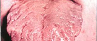
Desquamative glossitis
Desquamative glossitis is a dystrophic-inflammatory disease of the tongue. Synonyms for desquamative glossitis are “geographical tongue”, “escfoliative glossitis”, “benign migratory glossitis”.
Etiology of desquamative glossitis
The etiology of desquamative glossitis is not fully understood and unclear. However, scientists associate the occurrence of desquamative glossitis with many diseases. Thus, desquamative glossitis most often (primarily) is a manifestation of diseases of the gastrointestinal tract. Now the appearance of desquamative glossitis is associated with endocrine disorders, diseases of the hematopoietic system, and neurodystrophic processes. Most often, desquamative glossitis is diagnosed in women and children.
Complaints with desquamative glossitis
Complaints with desquamative glossitis are most often absent. Patients may complain only of slight tingling in the tongue area, irritation when eating hot or spicy foods, especially on the tip of the tongue or its lateral surfaces.
Clinical picture of desquamative glossitis
The onset of desquamative glossitis is associated with the appearance of a whitish-gray coating on the mucous membrane of the tongue. That is, the process of rejection of the epithelium on the surface of the tongue begins. Over time, when the rejection process is more developed, an erosive surface appears. Most often this process is observed in the area of filiform papillae. In the central zone, the filiform papillae are completely atrophied. The fungiform papillae are preserved. The rejection process begins to spread to other surfaces of the tongue. But parallel to this, there is also a process of new formation of the tongue mucosa, that is, a regeneration process. And the alternation of these two early-directed processes creates a picture like a geographical map. Hence the synonymous name for desquamative glossitis – “geographic tongue”. It should be noted that these processes occur very quickly. And new epithelium is formed within 3 days after rejection.
There are three forms of desquamative epithelium:
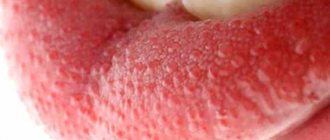
- Surface form. The superficial form of desquamative glossitis is characterized by the presence of clearly defined areas of de-epithelialization. These areas are limited by the halo/circle of redness and normal mucosa. When the epithelium is desquamated, the tongue becomes smooth and bleating. Patients feel itching and burning.
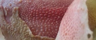
- Hyperplastic form. In the hyperplastic form of desquamative gingivitis, the filiform papillae thicken. And in areas of this compaction/hypertrophy there are areas of white, yellow and gray color.
- Lichenoid form. In the licheoid form, there are areas of de-epithelialization of different shapes and sizes. Fungal papillae are preserved. Most often, the licheoid form of glossitis is observed in patients who have increased sensitivity to metal.
The course of desquamative glossitis is chronic. Repeated inflammation (remissions) occurs spontaneously and, as a rule, does not last long. Changes in the mucous membrane are reversible.
Treatment of desquamative glossitis
Treatment of desquamative glossitis is complex, including both general and local treatment:
- General treatment of desquamative glossitis:
- Identification and treatment of the main pathology of internal organs;
- Sanitation of the oral cavity, professional hygiene;
- For cancerophobia - psychotherapy;
- Sedatives;
- Desensitization therapy;
- Calcium pantothenate (0.5 – 3 g) 3 times a day for a month;
- Multivitamins with microelements;
- Vascular drugs;
- Drugs such as dalargin, which have epithelial and analgesic effects.
- Local treatment:
- In case of severe pain, use drugs with an analgesic effect locally;
- If a burning sensation occurs, use irrigation or oral baths with citral solution;
- Application with vitamin A, rosehip oil, carotoline, solcoseryl;
- Eikonol – concentrated fish oil;
- Physiotherapy;
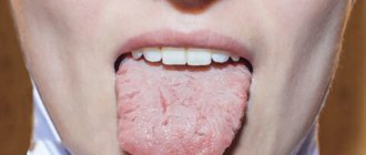
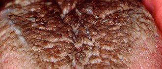
CHRONIC HYPERPLASIA OF FILIVID PAPILLAS
Chronic hyperplasia of filiform papillae is characterized by growth in area and increase in size of filiform papillae. That is, hypertrophy of the filiform papillae occurs.
Etiology of chronic hyperplasia of filiform papillae
The etiology of chronic hyperplasia of filiform papillae is not fully understood. Microbiological research was important, but no specific pathogens were found. Commonly encountered leptotrichia and saprophytes were found.
Scientists have identified predisposing factors to the occurrence of chronic hyperplasia of filiform papillae:
- Violation of metabolic processes;
- Alcohol;
- Smoking;
- Taking medications;
- Changes in the acid-base balance of oral fluid;
- Gastrointestinal diseases;
- Infections;
- Hypo-, vitamin deficiencies.
- Males;
- Middle or old age.
Complaints in chronic hyperplasia of filiform papillae
Complaints in chronic hyperplasia of filiform papillae may be absent. Only subjectively can the patient note the sensation of a foreign body on the back of the tongue, an unusual appearance of the tongue, the appearance of a gag reflex when chewing, swallowing food, or when talking. Itching and burning, spreading to the palate.
Clinic of chronic hyperplasia of filiform papillae
The clinic of chronic hyperplasia of filiform papillae is characterized by elongation and thickening of the filiform papillae on the back of the tongue (up to 2-3 mm); the keratinizing cells covering the back of the tongue have a color from dark brown to black. The area with hyperplasia most often has a triangular shape. The lateral and anterior surfaces of the tongue are free from hyperplasia. It was assumed that such a dark color is formed from iron compounds coming from food and the work of microflora.
Treatment of chronic hyperplasia of filiform papillae
Treatment of chronic hyperplasia of filiform papillae is complex. Consists of both general and local events.
- General treatment of chronic hyperplasia of filiform papillae:
- Treatment of autonomic pathology;
- Sanitation of the oral cavity, professional hygiene;
- Tranquilizers, sedatives, psychotherapy as needed;
- Calcium pantothenate;
- Multivitamins with microelements;
- Desensitization therapy;
- If candidiasis is present, antifungal therapy can be prescribed;
- Local treatment of chronic hyperplasia of filiform papillae involves the use of keratoplasty, strict twice-daily oral hygiene and complete cessation of smoking.
Diagnostics
Diagnostic measures begin with examination of the oral cavity and collection of medical history. After listening to the patient's complaints, palpation is performed along the midline of the tongue. With this pathology, compacted tissue will be palpated, and atrophic areas will be visually observed, the configuration resembling a rhombus or an oval.
To clarify the diagnosis, bacteriological and cytological examinations are prescribed. This allows for differential diagnosis with such pathological processes as:
- syphilis or tuberculosis;
- candidiasis or lichen planus;
- papillomatosis or malignant formation.
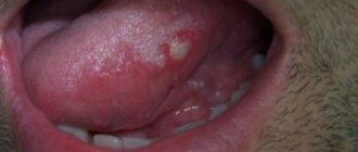
RHOMBID GLOSSISTIS
Diamond-shaped glossitis is characterized by a COMPLETE absence of papillae on the surface of the tongue.
Etiology of rhomboid glossitis
The etiology of rhomboid glossitis includes the following factors:
- Smoking;
- Candidiasis; hypo-, avitaminosis of vitamin C;
- Gastrointestinal diseases.
Complaints with rhomboid glossitis
There may be no complaints with rhomboid glossitis. Sometimes patients note a burning sensation, tingling on the surface of the tongue. These symptoms may worsen when eating and spread to the surface of the hard and soft palate.
Clinical picture of rhomboid glossitis
There are three forms of rhomboid glossitis:
- Smooth or flat form of rhomboid glossitis. With this form of rhomboid glossitis, the area has a limited character. There are no papillae in the area and the tongue is red or pink. The area does not rise above the mucous membrane of the tongue. On palpation it is dense and painless. The submandibular lymph nodes are not enlarged.
- Lumpy or tuberculate shape. The mucous membrane of the tongue has dense tubercles that are separated from each other by grooves. There is a complete absence of papillae on the mucous membrane of these tubercles. These tubercles are either diamond-shaped or triangular in shape. Red with a cyanotic tint. If you look at the mucous membrane of the tongue from above, an association with cobblestones is created.
- Hyperplastic form. In the hyperplastic form, papillomotous growths with a flexible base and a sharp apex appear on the mucous membrane of the tongue. These formations are pale pink in color and rise above the surface of the tongue. Patients experience a sensation of a foreign body in the mouth.
The course of any form of rhomboid glossitis is benign. And only with a hyperplastic form or a lumpy form with constant irritation or poor-quality and untimely treatment there is a risk of malignancy.
Treatment of rhomboid glossitis
Treatment of rhomboid glossitis is complex.
- General treatment for rhomboid glossitis:
- Treatment of general pathology;
- Sanitation of the oral cavity, professional hygiene;
- Calcium pantothenate;
- Sedatives, tranquilizers, psychotherapy as necessary;
- Smoking ban.
- There is no local treatment for rhomboid glossitis. In case of severe growth of papillomas - cryodestruction.
Forms of the disease
In dentistry, there are three types of disease:
- flat
- smooth epithelium without roughness and papillae, clear boundaries of the lesion, bright red-pink color, dense tissue, a tingling sensation appears when pressed; - tuberous
- there are clearly defined tubercles connected by folds, but the tissues of the formations themselves are smooth and have a dark red tint; - papillomatous
- growths in the form of spines protrude strongly above the surface of the tongue, the tissues are loose and rough, covered with a milky pinkish coating.
FOLDED TONGUE
Most often, a folded tongue is a sign of an abnormal development of the tongue, and this form of glossitis manifests itself in childhood. The synonymous name for the folded tongue is “scrotal tongue”, this is due to the similarity of the tongue to the skin of the scrotum. Most often, a folded tongue is accompanied by an enlargement of the tongue - macroglossia, which makes it not only large, but also prominent. With a pronounced longitudinal groove, the tongue is called “slit-like”. Because of this anatomy (the presence of deep grooves), excellent conditions are created for the development of pathogenic microflora, especially for fungi of the genus Candida. Therefore, in such patients the risk of candidiasis increases. Treatment for a folded tongue is not required. Only the patient should be well motivated to perform thorough oral hygiene and professional hygiene at the dentist.
BE CAREFUL NOT ONLY OF YOUR PATIENTS, BUT ALSO OF YOURSELF. LOVE TO ALL! WITH:
THANK YOU FOR READING ^_^
The article was written by N. Shidlovskaya specifically for the OHI-S.COM website. Please, when copying material, do not forget to provide a link to the current page.
Where to run?
At the first signs of illness, you should contact your dentist. He will determine the cause of the disease and prescribe appropriate treatment. For example, in the case of a mechanical injury, it consists of grinding down the sharp edge of a chipped tooth or adjusting the prosthesis. One of the important procedures is rinsing the mouth with antiseptic solutions.
Article on the topic
Diseases of the lips and tongue. Why are cheilitis and glossitis dangerous?
If the disease is caused by pathogenic microbes, antibacterial and anti-inflammatory drugs are used. To reduce pain while eating, a gentle diet is recommended, including, in particular, pureed soups, cereals, and various purees.