The development of technology has relatively recently introduced this type of preliminary examination of the body, such as computed tomography, which you can do here in Moscow. Today it is used very widely for diagnosing diseases of various organs and systems of the body.
Previously, before the computer tomograph came into practice, diagnostics of the oral cavity in dentistry was carried out using a targeted image of one tooth or a detailed panoramic image - an orthopantomogram (abbreviated as OPTG). Having assessed the diagnostic capabilities of jaw CT, our dentists have significantly improved the quality of services provided, obtaining reliable and long-term results from treatment, prosthetics and dental implantation. Computed tomography of teeth is a three-dimensional 3D image that gives a specialist the opportunity to see, study in detail the clinical picture of each element, and objectively see all the individual structural features of a particular segment.
What does the doctor see in a CT scan?
- Presence and volume of carious processes.
- If there was an injury, then its consequences are clearly visible.
- If there are anomalies in the development of the dentofacial apparatus.
- Volumes and structure (dense or loose) of bone tissue.
- Presence of a tumor.
- How well or not the root canals of the teeth are filled.
- Perforation of root canals.
- Fragments of tools in the channels.
- Formation of fistulas, cysts, granulomas.
- Inflammatory processes in periodontal pockets.
- Condition of installed implants.
- Position of impacted teeth.
- Pathological diseases of the palate.
- Condition of the maxillary sinuses.
A specialist can enlarge, examine, measure by millimeters in a 1x1 ratio any of the elements of the dental system reflected in the picture. The program of our tomograph also allows you to carry out virtual implantation visually in front of the patient, where you can see how possible implantation is without additional preparation for bone tissue augmentation. In a CT image, you can “immerse” the implant into the bone, rotate it from all sides and objectively see down to a millimeter how much the implant will be surrounded by bone.
Diagnocat artificial intelligence: fast CBCT analysis, accurate diagnosis
Only in our clinic, Diagnocat artificial intelligence is used for CBCT analysis! The program uses 3D images to determine the condition of the teeth, finds problems and suggests how to treat them. In just a few minutes, Diagnocat will examine your CT scan, identify problem areas, and generate a report for each tooth. And the doctor will make an accurate diagnosis! More about Diagnocat here.
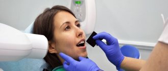
How is a dental CT scan done?
The CT scan procedure does not take much time. Before starting the examination, the person being examined needs to remove all metal jewelry and glasses. If there are metal-containing crowns in your mouth, you need to warn your doctor about this so that he can change the settings to obtain a high-quality image.
Next, a lead apron is put on the patient. Despite the fact that with a computed tomography the level of radiation is several times less than with a conventional x-ray, an apron is required in order to minimize the radiation dose to the entire body.
The picture is taken in a sitting or standing position. The chin is fixed on a special backing. While the device is operating, while it is rotating and scanning, you must remain completely motionless; this takes approximately 1-2 minutes.
After the device stops its movement, you need to wait for the doctor’s signal that you can leave the device area.
Next, the image data is loaded into the computer using a special program. The patient will not be able to understand and decipher the CT image data on his own. But the specialist sees all the subtleties on it and can show it to the patient, while giving appropriate explanatory comments.
Areas of dentistry in which it is important to conduct a CT examination of the jaw.
Therapy.
Often, a targeted or panoramic image is enough to detect caries. But sometimes this type of x-ray diagnosis is not enough. The carious process can develop hidden and sometimes only a CT scan helps to assess the extent of tooth damage. In this image you can clearly see the full volume of affected tooth tissue and can already see in advance how thin the line is between healthy tooth tissue and pulp. Thus, the doctor can predict the treatment plan in advance. Borderline conditions of very deep caries cannot always be eliminated while preserving the nerve of the tooth. Often the margin of remaining healthy tissue is so thin that the tooth has to be depulped.
Endodontics.
To assess the quality of root canal treatment, computed tomography is the best assistant for a specialist. When applying for retreatment of dental canals from which the nerve has already been removed, the specialist sees in the CT image:
— the canal is sealed efficiently, tightly, up to the top, or there are voids and the canal is not filled;
— it is impossible to miss the perforation of the canal, formations on the roots;
— fragments of instruments from previous treatment;
- branches and additional channels of the tooth.
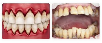
When diagnosing, treating and monitoring the dynamics of treatment of a disease such as periodontitis, CT is indispensable. When the inflammatory process from the tooth passes into the periodontal tissue, an inflammatory “sac”, a cyst, or cystogranuloma is formed at the apex of the tooth root. OPTG makes it possible to see this formation, but only in one plane, and sometimes a misleading perception is created about the real size of the inflammatory process. The 3D image makes it possible to see in all planes and make a decision on the advisability of saving the tooth. Treatment of periodontitis is not a quick process, sometimes it takes more than one year. But CT also helps to observe the dynamics of improvements or their absence.
Periodontology.
For a more accurate and correct diagnosis of periodontal diseases, a computed tomograph is an invaluable assistant.
When diagnosing periodontal disease, a specialist also needs CT examination data of the jaw. This 3D volumetric image allows you to see a problem hidden from view, without distortion and in full, the possibility of a diagnostic error of the disease is reduced to zero. Computed tomography gives a complete picture and vision of periodontal pockets. It is also important for the periodontist to see the condition of the jaw bone tissue. All these points allow the doctor to make the only correct diagnosis and severity of the disease in order to conduct a high-quality course of treatment.
Prosthetics.
When the question of dental prosthetics arises, a qualified specialist will first assess the full picture of the condition of the oral cavity.
This includes a visual inspection, determining the correct jaw relationship, and assessing the correct functioning of the TMJ. In addition to manual and visual examination, the orthopedist needs to know and understand what is happening in the area hidden from view. A panoramic image of the OPTG will not provide an objective understanding of the condition of the bone, the presence or absence of inflammatory processes. Only a computed tomography scan of the jaw will allow you to create an objective and correct course of treatment.
Implantation.
This area of dental treatment using CT of the jaw deserves separate and more detailed attention.
The process of restoring teeth with implants can vary greatly in complexity. From one missing tooth to rehabilitation for complete tooth loss.
Whether it is one-stage, one-stage, two-stage implantation, fixed or conditionally removable prosthetics on implants, the first stage of the patient is a computed tomography diagnosis. It is important for the orthopedist to evaluate the number, location and angle of the implants.
The implantologist needs to see the condition of the bone tissue in the planned installation area. How dense is it, is there enough volume? It is also necessary to exclude inflammatory processes in the planned area of dental implantation.
Regarding the volume of bone tissue, the OPTG image gives a visible representation only in height. Only a CT scan of the teeth will show how wide the ridge is enough to install an implant. Specialists who are focused on the long-term result of treatment for the patient, and not on their own immediate benefit, will never perform such a serious procedure as implantation without an objectively necessary CT scan procedure.
Regarding the installation of implants in the upper jaw in the area of the maxillary sinuses, the fundamentally important point here is the presence of the required volume of bone up to the mucous membrane of the maxillary sinus. If the bone tissue height is insufficient, a sinus lift operation is performed before tooth implantation, or alternative options for tooth restoration are considered.
After osteoplasty (bone tissue augmentation) or sinus lift surgery, the only diagnostic method that objectively helps to understand the dynamics of bone grafting is CT.
After installing implants, it is necessary to regularly monitor their condition in the bone. The frequency of diagnosis is determined by the attending physician; it depends on the individual characteristics of the body, the age of the patient, and the initial clinical picture before implantation. This is done by examining a 3D computer image.
Before the dental implantation procedure, the question of whether a 3D computed tomography should be done or not is not discussed with qualified specialists. The answer is clear: yes. It is not even possible to conduct a full and objective consultation, not to mention the procedure of implantation and prosthetics on implants.
Do not self-medicate, consult a doctor!
Don't wait for your condition to worsen!
Sign up
Is it possible to take a photo in advance?
When planning to go for a dental implant consultation, do not rush to take a CT scan yourself. If the clinic is equipped with a CT scanner, it is better to take the picture directly in the clinic. Subsequently, during treatment, doctors will always have the opportunity to track the dynamics, always having primary initial clinical data at hand. If dentistry does not have the opportunity to conduct a computed tomography scan of the jaw, then, most likely, a specialist will send you to a certain place where this procedure is performed efficiently and safely using modern equipment. Because there are often cases when old equipment is used, pictures are taken that are cheap in comparison, but they are of no use, because there is no way to clearly see and evaluate the survey data.
Sequence
There is no need to prepare for a CT scan. You come to the clinic, remove all metal objects (metal can distort the results of the tomograph). An apron with a lead layer is placed around your neck to protect your body from radiation. After this, your head is fixed on the frontal, temporal, and chin support to eliminate involuntary movements (which can also ruin the picture). You secure a special contact segment with your teeth, and your hands on the handrails.
In seconds, the study ends, and within five to ten minutes you receive the results.
Cost of CT scan of teeth.
Computed tomography of the jaw can vary in price in different clinics, depending on the level of the medical institution, the availability of more modern and safe equipment (computed tomograph) or old equipment that provides uninformative, unclear research data.
It is not always necessary to do a CT scan of the entire jaw; sometimes you need to see only a fragment, or an area of one row of teeth, upper or lower. In this case, at the Es-Dent Center, you can do a CT scan of the entire jaw, or a separate computed tomography scan of the lower or upper jaw; it is also possible to film a minimal fragment, covering an area of up to 3 teeth. Accordingly, such dental CT images are cheaper in price compared to a full examination.
3D computed tomography of teeth at the German Es-Dent Center for Aesthetic Dentistry can be done at an attractive price in Moscow.
Advantages
The advantages of the computed tomography method include:
- lack of special training;
- suitable for children;
- painlessness (the device only makes a quiet sound, no discomfort, pain, or unpleasant sensations);
- minimal radiation (the dose is 40 - 60 microsieverts, so up to three procedures can be done per day);
- the possibility of obtaining a voluminous, high-quality model.
The advantages also include a short list of contraindications:
- first trimester of pregnancy;
- lactation period.
Some patients consider the price of the procedure to be a disadvantage of dental computed tomography.