What is transmitted genetically
Most diseases of the dental system are not 100% genetically dependent. However, there are signs that are passed on from generation to generation and can ultimately affect the condition of the teeth. These include:
- composition, thickness and shade of enamel;
- shape of dental units;
- bite geometry;
- timing of the eruption of primary and permanent teeth;
- composition of saliva and oral microflora.
Along with these characteristics, the child also inherits a predisposition to certain pathologies. But there is no fatality in this. With a reasonable, competent attitude to oral care, you can avoid the development of one or another disease, the likelihood of which is genetically predetermined.
Publications in the media
Dental developmental disorders - structural anomalies and malformations of teeth - can appear in isolation or in combination with structural anomalies and malformations of organs and systems of the entire child's body.
Etiology. Dental tissue defects are caused by hereditarily fixed changes in the genetic code, but can be caused by environmental factors. Pathogenesis. Teeth during the period of their follicular development, i.e. Before eruption, several stages pass: laying, formation and calcification of crown enamel, formation and calcification of dentin. At each stage of tooth development, conditions can be created that lead to disruption of the formation of its shape, mineralization and color.
Classification • All disorders are divided into: •• anomalies of the structure of dental tissues, transmitted by inheritance •• anomalies in the number, size and shape of teeth, due to the hereditary transmission of the sample •• anomalies of the structure and malformations of dental tissues, which arose as patterns of the pathogenesis of systemic pathology in the child’s body (hereditary, congenital, acquired) •• structural anomalies and malformations of dental tissues caused by the influence of external factors. • Each group has individual clinical and radiological features, determined by the cause of occurrence and the mechanism of development.
Hereditary disorders of the development of hard dental tissues. Men and women suffer equally often. Both temporary and permanent teeth can be affected. • Enamel defects (most often dysplasia) are an isolated symptom (130900, Â). Combinations with cataracts and stenosis of the Sylvian aqueduct (600907, r), nail defects and hypohidrosis (ameloonychohypohidrotic syndrome, *104570, Â), also a sign of a number of hereditary diseases, for example: •• Renal tubular acidosis type II •• Albright’s disease •• Lacrimal duct defect (149700) •• Citrate transport protein defect (190315) •• Dysosteosclerosis (224300) •• Cranioectodermal dysplasia (218330) •• Tumor calcification (114120) •• Mucopolysaccharidosis, type IVB (253010) •• Neurosensory hearing loss, hypoplasia enamel, nail defect (234580) •• Goltz-Gorlin syndrome (305600) •• Menkes syndrome (261900) •• Naegeli syndrome (161000) •• Polyglandular autoimmune syndrome, type I (*240300) •• Stimmler syndrome (202900) • • Tuberous sclerosis (191092) •• Epidermolysis bullosa (226440). Oculodental-digital dysplasia (164200, genes ODDD, SDTY3, ODOD, 6q22 q24) •• Amelogenesis imperfecta (impairment of the process of enamel formation) is the most common pathology. Usually manifested by grooved (wrinkled) enamel or its sharp thinning. Erupting teeth are small, cylindrical, yellowish or brown in color. Amelogenesis imperfecta is inherited independently and is part of a number of hereditary syndromes ••• Amelogenesis imperfecta, type 1 (301200, genes AMELX, AMG, AIH1, AMGX, Xp22.3 p22.1, À) ••• type 2 (104500, gene AIH2 , 4q11 q21 À) ••• type 3 (301201, AIH3 gene, Xq22 q28 À) • with taurodontia (104510, À) ••• with nephrocalcinosis (204690, r) •• Enamel abnormalities are also observed in the following phenotypes: •• • platyspondylia with amelogenesis imperfecta (601216) ••• Kohlschutter syndrome (*226750, r): epilepsy, dementia and amelogenesis imperfecta ••• photoreceptor dystrophy and amelogenesis imperfecta (217080, r).
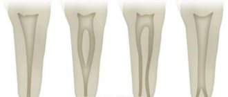
• Defects in dentin formation are numerous ; traditionally, the groups of dentinogenesis imperfecta and dentin dysplasia are distinguished. In addition, dentin defects occur in a variety of other osteogenesis disorder syndromes •• Dentinogenesis imperfecta. The structure of the enamel is not changed, but due to the imperfection of dentin, its connection with the enamel is fragile, which leads to enamel chipping ••• type I (*125490, 4q13–q21, DGI1 gene, Â). Synonyms: opalescent dentin, opalescent teeth without osteogenesis imperfecta ••• dentinogenesis imperfecta, type II, Capdepont teeth ••• type III (125500, Â). Clinically: Rapidly abraded dental crowns, smooth amber-colored dentin, open bite, very large pulp chambers and root canals of primary teeth, small or absent pulp chambers of permanent teeth •• Dentin dysplasia is a dental anomaly in which the teeth are clinically, morphologically and color-wise normal , but x-ray examination reveals short roots, obliteration of the pulp cavity and canals, increased mobility and premature tooth loss ••• type I (*125400, Â) •• type II (*125420, Â) - occlusion of the pulp cavity, the structure of the pulp in the form straws ••• Dentin dysplasia and cortical osteosclerosis (*125440) - compacted long bones, compaction of the alveoli of the teeth, narrow or sometimes closed medullary canals, thickening of the cortical layer.
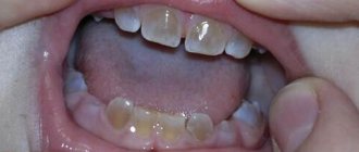
• Mesoectodermal odontopathy (Stainton–Capdepont disease; simultaneous disruption of the formation of enamel, dentin and pulp). The teeth erupt at the usual time, have a normal size and shape, but their color is changed (from gray to blue-gray and yellowish-brown). The length and shape of the roots are changed. The tooth cavity and root canals are obliterated. Characterized by rapid wear of teeth (especially temporary ones). Their surface is shiny, flat and smooth (“polished”). At the same time, teeth practically do not react to mechanical, chemical and temperature stimuli.
• Oculodental-digital dysplasia (ODDD gene, 6q22–6q24, Â - *164200, r -*257850) - microphthalmos, coloboma or iris abnormalities in combination with congenital dental defects and finger abnormalities. Clinically: microphthalmos, microcornea, glaucoma, dental anomalies (enamel hypoplasia, small teeth), camptodactyly of the little fingers, syndactyly of the fourth and fifth fingers, absence of the middle phalanges of the toes, hypotrichosis, small nose, hypoplastic wings of the nose, hyperostosis of the skull bones, orbital (bone) hypotelorism, jaw overgrowth, spinal cord compression, spastic tetraparesis, progressive spastic paraplegia, the effect of paternal age on the frequency, pathology of the white matter of the brain on MRI, calcification of the basal ganglia. Synonyms: oculo-dental-digital syndrome, ODD (oculo-dento-digitalis) syndrome, Meyer-Schwickerath and Weyers syndrome. Note: a mutation at this same locus is the cause of syndactyly type III.
• ADULT syndrome (from Acro-Dermato-Ungual-Lacrimal-Tooth, 103285, Â). Clinically: hypodontia, early eruption of permanent teeth, ectrodactyly, obstruction of the lacrimal ducts, onychodysplasia, freckles, hypoplasia of the mammary glands. • Treatment depends on the degree of tooth tissue loss due to increased abrasion. In case of minor loss, it is recommended to regularly (2 times a year) conduct courses of remineralization therapy; in case of severe loss, orthopedic treatment is recommended. Enamel hyperplasia is the excessive formation of tooth enamel during its development. Typically, enamel hyperplasia is observed in the neck of a permanent tooth in the form of drop-shaped formations. After teething, these formations do not progress and are not affected by caries; do not cause any unpleasant sensations. Treatment is indicated only for noticeable cosmetic defects; Grinding is carried out followed by remineralization.
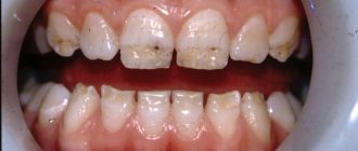
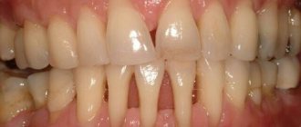
Enamel hypoplasia is a developmental defect that manifests itself in a violation of the structure and mineralization of tooth tissue during the period of their formation. Usually, only the formation of enamel is disrupted, and in severe cases, dentin as well. Most often, enamel hypoplasia is observed on permanent teeth, very rarely on temporary teeth, which is explained by their formation in the prenatal period. The localization of areas of hypoplasia depends on the age at which the child suffered from a general disease, and its severity depends on the severity of the disease.
• Causes: severe infectious diseases, hypo- and avitaminosis, disorders of the gastrointestinal tract (dyspepsia), endocrine organs (especially the parathyroid glands), hemolytic jaundice, hereditary syphilis, etc. • Classification : •• systemic hypoplasia - a violation of the structure of the tissues of all or a group of teeth formed in the same period of time •• focal hypoplasia - a malformation of the tissues of one or more teeth that formed in the focus of inflammation of the tissues surrounding the follicle, in the area of tumor growth or exposed to trauma. • Manifestations. Hypoplasia of dental enamel manifests itself in the form of whitish spots or depigmentation of various sizes with a smooth shiny surface. They can be combined with point or cup-shaped depressions, grooves, and constrictions of the enamel of the crowns of the teeth. In these lesions, the enamel loses its inherent color, but retains its density. Sometimes a complete absence of enamel is observed in a limited area of the tooth, which looks like a cup-shaped depression. Rarely, enamel is absent on almost the entire surface of the tooth crown and then the tooth takes on a bizarre, ugly shape (Turner's, Hutchinson's, etc. teeth).
• Treatment and prevention . Treatment of dental enamel hypoplasia depends on the form and severity of the disease. For spotted hypoplasia, bleaching with medications or grinding is carried out. To prevent the occurrence of caries, remineralizing therapy is carried out. Small enamel defects are filled after preparation. Teeth significantly affected by hypoplasia are covered with artificial crowns. Prevention of dental enamel hypoplasia - harmonious physical development of the child, balanced nutrition, timely treatment of diseases of primary teeth. Fluorosis is an acquired malformation of dental tissues caused by excessive intake of fluoride during tooth formation (a kind of enamel hypoplasia). Most often it develops with an excessive fluoride content in drinking water (more than 1.5 mg/l), but it is also possible with an optimal fluoride content (0.8–1.2 mg/l) in children with a reduced immune status.
• Pathogenesis. Fluoride salts have a toxic effect on ameloblasts, which leads to impaired calcification of tooth tissue. Subsequently, the content of fluorine and calcium in the surface layers of the enamel increases, and, accordingly, the acid solubility of the enamel decreases. The severity of fluorosis depends on the dose of fluoride entering the body during amelogenesis. These teeth are not affected by caries.
• Manifestations: •• chalky spots and stripes on the vestibular surface of the teeth •• chalky and pigmented (to brown and black) spots •• erosive lesions of the enamel •• disturbances in the shape of the crowns of the teeth in combination with stains, erosions and fragility of the enamel (fractures of areas of enamel during mechanical influence).
• Treatment is prescribed taking into account the severity of the process. If the form is spotty, teeth are whitened. Enamel defects are filled or orthopedic treatment is performed (crowns, pinned teeth). • Prevention •• Public prevention: reducing the intake of fluoride from drinking water into the child’s body during the period of teeth formation (up to replacing the water source) •• Individual prevention: partial defluoridation of drinking water by boiling, settling •• Additionally, milk and dairy products are recommended throughout the year , careful hygienic care of the oral cavity.
ICD-10 • K00 Disorders of development and eruption of teeth •• K03.7 Discoloration of hard tissues of teeth after eruption
Structure of teeth
In the presence of a thin layer of enamel on the teeth and deep fissures (slit-like depressions on the chewing surface), the risk of early carious tooth destruction increases. But even with hereditary weakness of the enamel, it will be reliably protected:
- careful hygienic care;
- carrying out remineralization of enamel (saturation with calcium and fluoride salts);
- fissure sealing (sealing with a composite material immediately after teeth erupt).
At the Shifa clinic, children can always get help in strengthening hard dental tissues.
Other factors affecting dental health
Heredity is just one factor. The influence on the human body in general and teeth in particular is exerted by:
- ecological situation;
- food intake and diet;
- Lifestyle;
- regular anxiety and repetitive stress;
- smoking, drinking alcohol, other bad habits;
- woman's behavior during pregnancy;
- infant habits of sucking fingers, pacifiers and other objects.
Even if there are relatives with an “ideal” tooth structure and an even bite, the lifestyle of the expectant mother will affect the formation of tooth buds in the fetus.
During pregnancy, people think about the health of the baby’s body and teeth. That's why:
- treat emerging diseases in a timely manner;
- regularly observe the rules of oral hygiene;
- do not use medications without the consent of your doctor - some of them can affect the color and thickness of enamel, for example, tetracycline;
- balance your diet, eat healthy foods and avoid unhealthy ones;
- normalize your sleep schedule, do not overexert yourself physically.
Composition of saliva
When parents complain about bad teeth, they often do not realize that the cause may be the composition of saliva, which determines heredity. It may vary in the ratio of mineral and organic components. And this has a direct impact on dental health.
With a reduced content of oligosaccharides in saliva, the population of microorganisms increases, which settle on the surface of the enamel, creating a predisposition to caries. To avoid this disease, you need to brush your teeth at least twice a day and use special mouthwashes.
An increase in the concentration of class A immunoglobulins (IgA) in saliva, which have an antibacterial effect, protects the enamel from cariogenic bacteria.
The specialists of the Shifa clinic will help you choose toothpaste and other hygiene products to create the right environment in the oral cavity. Dentists will also give recommendations on diets that reduce the negative effects of saliva.
How is dental fluorosis treated?
First of all, it is important to promptly stop using water with a high fluoride content for drinking. The development of modern technologies allows people to drink clean, healthy water. Drink only purified water, buy quality water in large bottles. During the treatment of fluorosis, you can use a special defluoridation filter to purify water.
It is worth stopping for a while from eating fluoride-containing foods (fish), eating less spinach, and not drinking tea. When choosing toothpaste, buy one that does not contain fluoride. The above measures will help stop the further development of enamel fluorosis. How can you restore your teeth to a beautiful appearance?
Bite
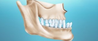
Abnormal bite often causes:
- increased load on individual teeth, which contributes to their rapid destruction or damage to the ligamentous apparatus;
- crowding of teeth, leading to gum inflammation and dental caries.
- The hereditary factor is detected in no more than 50% of cases of abnormal structure of the dental system. The remaining 50% is due to the following circumstances:
- artificial feeding;
- formation of bad habits at an early age (sucking pacifiers, fingers, lips, holding pencils or pens in the mouth);
- the habit of sleeping in one position, especially with your hand under your cheek;
- incorrect posture;
- lack of calcium and other trace elements in the body.
In preventing the development of pathological occlusion, the role of parents is great, who should pay attention to the early identification and elimination of provoking factors.
How to prevent pathology
prevent your child from developing crooked teeth by regularly visiting the orthodontist's office. Even if your baby has a genetic predisposition to certain diseases, dealing with them at an early stage is much easier.
Adults can avoid the appearance of upper and lower crooked teeth if they do not delay the installation of implants after the loss of one of the molars, and also remove their wisdom teeth in a timely manner. In addition, it is necessary to get rid of the habit of gnawing on durable objects, not to knock your teeth against each other and not to clench your jaw during emotional stress.
The importance of prevention
The child subconsciously adopts the behavior and habits of his parents. If older family members do not brush their teeth, eat improperly, or do not exercise, then dental diseases lie in wait for both adults and children. At the same time, the incorrect conclusion is often made that “bad teeth are inherited.”
Mineralization of dental tissue continues after teething. Any disease, weakened immune system, or unbalanced diet have a much greater impact on the hard tissues of teeth than hereditary predisposition.
Parents should monitor the health of their children by providing them with proper care and disease prevention. This will help avoid dental caries and the development of enamel hypoplasia, and reduce the risk of abnormal bite.
In case of poor heredity, timely prevention plays a key role. Regular dental care will reduce the risk of developing dental and periodontal disease in children. The specialists of the Shifa clinic are always ready to help. Our dentists regularly improve their professionalism, conduct diagnostics and treatment using modern equipment. This allows you to identify possible risks and prevent them in a timely manner. Contact professionals to ensure your child’s teeth are always strong and healthy!
Diagnosis and treatment of dental abnormalities
Diagnosing abnormalities in tooth shape is not particularly difficult. During a visual examination, a dentist can easily determine an abnormality in the shape of the teeth. To compile a complete etiological picture of the disease, doctors of other specializations may be involved. To determine deviations in the roots of the teeth, an x-ray can be taken.
Treatment for abnormalities in tooth shape depends on the type of anomaly and its severity. If the anomaly is minor, the bite is not disturbed, and the patient has no complaints about the appearance of the teeth, then it is not at all necessary to correct this pathology. If necessary, the main method of treatment in this case will be to restore the correct shape of the teeth using phytopolymer composites, as well as through prosthetics with crowns, veneers, etc. By performing such procedures, you can restore the functionality and aesthetics of your teeth. In some cases, it may be necessary to remove one or more teeth and replace them with removable or fixed dentures.