16.11.2019
Complications after tooth extraction occur regardless of the complexity of the procedure and the qualifications of the doctor. One of them is a lump on the gum, which signals pathological changes in the tissues. Its appearance cannot be ignored, since it is a precursor to severe pathologies of the mucous membrane and gum diseases. The lesion will not resolve on its own, but timely dental care will help cope with the problem.
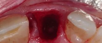
After tooth extraction surgery, swelling with liquid contents may form. If you don't touch them, they will dissolve on their own. In some cases, the cheek swells, which is a consequence of an allergic reaction to anesthesia, infection of the wound (with deep roots), opening of an abscess during tooth extraction, or increased blood pressure during surgery.
Whatever the reasons why the compaction formed, it should not be ignored. The dentist will examine, order an x-ray, determine the nature of the lesion and prescribe the correct treatment.
Content:
- In what cases are molars removed?
- Reasons for the formation of a lump after wisdom tooth removal
- Dangerous symptoms
- How to treat a lump after tooth extraction
- What happens if you don’t treat a lump after a tooth extraction?
Most patients at dental clinics believe that pulling out a diseased tooth will relieve them of painful symptoms.
They treat the extraction procedure as a life-saving one. But, unfortunately, it is not always possible to solve the problem completely surgically. It happens that a small tumor soon forms in the vacant space.
A seal on the gum after tooth extraction indicates the need to urgently visit a doctor. Below we will tell you why it occurs and how doctors fight it.
Types of seals and the probable cause of their appearance
Most often, a lump on the gum occurs due to diseases of the oral cavity. Usually it is localized on the upper jaw, causing discomfort and pain, sharp and aching, which does not give rest. In some cases, the problem remains hidden for a long time. The main factors that provoke growth include:
- soft tissue infection;
- trauma to the mucous membrane as a result of taking spicy, sour foods, alcohol, and a number of medications.
Hard white bumps on the gums are an alarming symptom and a reason to see a doctor as soon as possible. After removal, hematomas are fluid-filled tumors.
They have a reddish tint and are not particularly dangerous. However, they need to be shown to a doctor and never pierced or disturbed. In this case, they gradually dissolve. The following types of pathological changes occur:
- Dense cone of white color. Evidence of an infectious process. There is pain when you press it.
- A bloody lump (hematoma) at the site of an extracted tooth. A consequence of soft tissue injuries during surgical procedures or inflammation.
- Hard lump. The cause may be a piece of bone tissue that was not removed. It does not necessarily cause pain, but it disturbs the patient.
hard lump
If a hard lump appears, it is important to show it to the dentist. The abscess should not be heated; to alleviate the condition, you can apply ice to the cheek on the problematic side. During diagnosis, the doctor may use palpation, periapical x-rays and tomography. In case of inflammation, the surgeon opens the lesion and clears the cavity of pus. The wound is washed with an antiseptic solution and a paste is applied. At home, antibiotics and anti-inflammatory therapy, rinses, and special ointments are prescribed. Monitoring is necessary until the gums heal completely.
Soft red ball
If tooth extraction was difficult and lengthy, a soft red ball may appear on the gum. Inside there is a bloody fluid - a good environment for the spread of infection and suppuration in the future. You cannot pierce the abscess or touch it with your hands. You should also not assume that the lump will go away on its own. The most correct thing is to consult a doctor who will do everything necessary to eliminate it and, if necessary, open it.
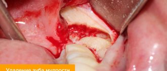
In what cases are molars removed?
Medical indications for extraction are:
- Progressive odontogenic osteomyelitis of the jaw. It is a purulent infectious-inflammatory lesion that affects not only the root of the molar, but also the tissue adjacent to it. Leads to increased body temperature, weakness, and abscess. For osteomyelitis of this type, tearing out the “causal” unit should be done in line order. Under no circumstances should surgery be postponed. Otherwise, the pathological contents of the abnormal focus will be evacuated into the jaw bone. There is a possibility of it entering the systemic circulation.
- Purulent periostitis, phlegmon, abscess, lymphadenitis, sinusitis of the maxillary sinus , if it is impossible to ensure a high-quality outflow of purulent masses from the infectious focus in the jaw.
- Lack of effect after conservative therapy for any purulent-inflammatory lesions of the oral cavity.
- Complete destruction of the crown and unsuitability of the remaining roots for prosthetics. Damaged root canals are a chronic source of odontogenic infection, so they have to be disposed of.
- The presence of teeth in the area of fracture of the alveolar process or jaw bone. Such units do not allow the correct composition of bone fragments and are regarded as conduits for infection.
- Impacted units, during the eruption of which a strong infectious-inflammatory reaction , tumor or cyst developed.
- Fangs, incisors and molars, due to which the mucous membranes of the mouth are constantly injured. They can be pulled out if standard sanding does not stabilize the situation.
- Progressive periodontitis , if loosening has reached the third or fourth stage.
- of a malignant tumor in the tissues of the periodontium or alveolar process
- Severe malocclusions , when “neighbors” do not allow the aligned units to take the correct position.
- Supernumerary teeth , causing curvature of the row, disrupting the aesthetics of the face.
Treatment
Before starting treatment, you need to identify the cause of the lump. Only a specialist can do this after conducting the necessary diagnostic studies. Dental treatment will depend on the causes of the disease and the form of its course:
- the abscess can be opened and cleaned, which will prevent further spread of infection throughout the oral cavity;
- if the lump occurs due to any serious damage to the tooth, most likely the tooth will be removed;
- if a lump has formed after the removal of a tooth, the dentist will treat the hole and prescribe a number of medications.
Tooth extraction occurs only in the most extreme case, when it cannot be saved, and it causes the development of an infection in the oral cavity and its spread to neighboring teeth. The most important thing is not to self-medicate, but immediately, if a problem is discovered, contact a specialist!
Reasons for the formation of a lump after wisdom tooth removal
The problem occurs for various reasons. Most often, a lump is formed due to:
- Severe injury to gum tissue. In this case, the disorder may be non-infectious in nature and go away on its own in two to three days. Anti-inflammatory drugs prescribed by a dental surgeon will help get rid of it. A particularly traumatic operation is to tear out “eights”, the roots of which are very long and winding.
- Individual intolerance to drugs used to treat the wound. Allergies cause itching and swelling. Increased separation of watery exudate is possible. To relieve unpleasant symptoms, you must stop using medications. The dentist will help you choose a more suitable analogue.
- Attachments of the inflammatory-infectious process. It occurs if bacteria enter the open socket, which is possible with an unformed blood clot. A complication in the form of a “dry” socket is likely if the doctor violated the extraction technique or the patient did not follow the instructions of the dental surgeon during the recovery period.
- Formation of flux in the operated area. Periostitis is manifested by purulent discharge, swelling of soft tissues, and increased body temperature. After extraction, it occurs due to the fact that pathogenic flora penetrates into the open wound cavity.
Pus in the gums with periodontitis -
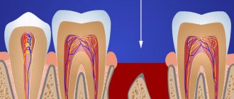
Very often, the patient’s complaints that his gums near the tooth are festering are not associated with inflammation at the apex of the tooth root during periodontitis, but with the formation of periodontal pockets in local or generalized forms of periodontitis. With periodontitis, there is destruction of the attachment of the gingival margin to the necks of the teeth, destruction of the alveolar bone around the teeth, as well as periodontal fibers, with the help of which the tooth is attached to the bone tissue.
All this leads to the formation of so-called periodontal pockets between the gum and the surface of the roots of the teeth (Fig. 14). They create good conditions for the proliferation of pathogenic bacteria and the development of chronic inflammation. When one of the periodontal pockets becomes too deep, this can lead to disruption of the discharge of inflammatory serous-purulent exudate through the lumen of the pocket. As a result, an abscess forms in the projection of the periodontal pocket on the gum, which dentists call the term “periodontal abscess” (Fig. 15-16).
You can immediately suspect that gum swelling is associated with periodontitis, and not with periodontitis, if the tooth is completely intact (i.e., externally healthy and does not have a filling, crown or caries), if it has mobility, and mobility was present in this tooth and before the gums become suppurated, and also if, with slight gentle pressure on the abscess, pus comes out from under the gums (as in Fig. 16).
The differences between local and generalized periodontitis are that they are caused by completely different reasons and, accordingly, the treatment will also be different. With local periodontitis, the inflammatory process occurs only in the area of 1-2 teeth - due to exposure to a traumatic factor. For example, a pocket may appear as a result of trauma to the gum margin by the overhanging edge of a filling or crown. The cause may also be the premature closure of several teeth, which leads to chewing overload and destruction of the bone tissue around them.
But with chronic generalized periodontitis, gum suppuration occurs for other reasons. You can immediately suspect this form of periodontitis if you have symptoms of bleeding and pain when brushing your teeth, swelling, redness or bluishness of the gum margins in the area of most teeth. The cause of generalized periodontitis is soft microbial plaque and hard tartar, which accumulate on the teeth as a result of insufficient oral hygiene.
Bacteria in plaque and tartar produce toxins and various pathogens that trigger an inflammatory reaction in the gums. With prolonged inflammation, first the dental-gingival attachment is destroyed, and then the periodontal fibers and bone tissue around the teeth are destroyed. With this form of periodontitis, pockets are found in almost all teeth, and not just in 1-2 (as with local periodontitis). When the outflow of inflammatory serous-purulent exudate in one of the pockets is disrupted, a periodontal abscess is formed in the gums.
Treatment of local periodontitis –
Based on the examination, the identified amount of tooth mobility, probing the depth of the periodontal pocket and analysis of the x-ray, the doctor will determine the possibility of saving the tooth and the algorithm for further treatment.
If the tooth can be saved, then the first thing to do in case of local periodontitis is to eliminate the impact of the traumatic factor. This means that you need to remove the overhanging edge of the filling or crown, and selectively grind the contacts of the chewing surface of the causative tooth and its antagonists. Next, under anesthesia, the periodontal abscess is opened to allow the outflow of pus and to rinse the periodontal pocket with antiseptics. If pus comes out of the pocket without an incision, but only little by little, then after anesthesia you still need to widen the mouth of the pocket with a stroker. Next, the doctor prescribes systemic antibiotic therapy, anti-inflammatory drugs, and antiseptic rinses.
Opening of periodontal abscess –
Next, the issue of the need to fill the root canals in the causative tooth is resolved. This must be done if the depth of the periodontal pocket reaches 2/3 or more of the length of the root of this tooth. Removing the nerve from the tooth and filling the canals is required here because infection from a deep pocket very easily penetrates through the bloodstream into the neurovascular bundle (tooth pulp), as a result of which the pulp itself becomes a source of infection. But all this is just initial basic treatment!
The main treatment consists of open curettage of the periodontal pocket. This operation allows you to remove inflammatory granulation tissue (which forms at the site of destroyed bone tissue) from under the gums, as well as fill the periodontal pocket cleared of granulations with bone material, which allows you to partially restore the level of bone tissue around the tooth.
Progress of open curettage operation –
During open curettage, the gums are first moved away from the teeth and bone tissue to create good access to the periodontal pocket. Then the granulations are removed from the pocket, the root surface is polished and the pocket is filled with material based on artificial or bovine bone tissue. Next, the flaps of the gum mucosa are placed in place and the gum is sutured. Figure 19 shows that the bone level differs between radiographs taken before and 4 months after surgery (an increase in bone level of approximately 2.5 mm).
Moreover, if an abscess on the gum occurs near a moving tooth, then in addition to all of the above treatment, splinting of the moving tooth may be required. For this purpose, fiberglass and filling material are used, with the help of which the movable tooth is fixed to the adjacent stable teeth. Detailed information on curettage and splinting in the articles:
→ How the open curettage operation is performed, → Teeth splinting technique
Treatment of generalized periodontitis –
With generalized periodontitis, periodontal pockets occur not in 1-2, but in almost all teeth. Typically, this form of periodontitis has a sluggish chronic course. Common complaints from patients include bleeding gums and inflammation of the gingival margin. In severe cases, tooth mobility, changes in the position and inclination of teeth, and suppuration from periodontal pockets occur.
Against the background of decreased immunity, an exacerbation of chronic inflammation may occur, and then abscess formation (i.e., the formation of purulent abscesses) may occur in the area of one or more periodontal pockets. Treatment of the generalized form of periodontitis is very complex, and we have devoted a separate article to this topic, which you can read at the link above. We hope that our article on the topic: What to do if your gums are swollen turned out to be useful to you!
Sources:
1. Higher prof. The author’s education in therapeutic and surgical dentistry, 2. Based on personal experience as a periodontist and dental surgeon, 3. National Library of Medicine (USA), 4. “Outpatient surgical dentistry” (Bezrukov V.), 5. “Therapeutic dentistry: Textbook" (Borovsky E.).
Dangerous symptoms
If a lump appears after tooth extraction, you should inform your doctor about it. Particularly dangerous symptoms are:
- discharge of pus;
- severe swelling;
- acquisition of bluish tint by fabrics;
- pain on palpation;
- absence of a protective blood clot.
Trying to cure a disorder without medical help is not only unwise, but also dangerous. By engaging in amateur activities and experimenting with folk recipes, a person risks creating conditions for the further spread of the inflammatory process. Then it is possible that the extraction will be followed by a second operation. This time it will be aimed at removing tissues affected by the purulent process.
Diagnostic methods
To understand why a lump on the gum formed, it is necessary to conduct a thorough examination. The main method for diagnosing seals on the gums is a visual examination by a doctor with further palpation. Additional methods are x-ray and computed tomography.
Visual inspection and palpation
During the first visit, the doctor conducts a thorough examination and palpation of the affected area of the gum. By palpating the seal, a specialist can determine what structure the ball has formed (soft or hard) and the degree of its filling with liquid. The choice of further diagnostic methods will depend on the result of the examination.
X-ray and CT
The radiography method allows you to consider:
- filling of the lump (pus or blood);
- structure;
- depth of penetration of the seal into the gum tissue.
Computed tomography is used for differential diagnosis of growth and to exclude the possibility of a tumor and the development of an oncological process.
How to treat a lump after tooth extraction
The technique of dealing with a dental disorder depends on the factors that caused it. First, the doctor studies the dominant symptoms and carries out various diagnostic measures. Based on the data obtained, he makes a decision on an effective treatment technique. She may be:
- conservative;
- surgical.
In the first case, we are talking about cleansing tissues from purulent masses and carrying out drainage measures. The patient is prescribed medications of different pharmacological groups. These can be antibiotics, anti-inflammatory, antiseptics, agents that suppress the activity of pathogenic flora, regeneration stimulants, pain relievers.
Medicines can be taken orally. They are also applied to the affected area. The rinses have proven themselves to be excellent. With their help, it is possible to speed up the evacuation of the contents of a purulent lesion.
Surgical therapy is indicated if there is a dense sac on the gum containing dead tissue. Then the surgeon makes a small incision and installs a drainage tube into the formed cavity. The exudate leaves the pathological focus along it. When the lump subsides and all the pus is removed, the doctor stitches it up. To avoid complications, the patient is prescribed antibacterial agents.
Indications and contraindications
The growth is treated surgically. Indications for using this option for removing pathology are:
- the bone growth grows quickly;
- pronounced cosmetic changes, progression of pathology;
- physical discomfort, the growth becomes too large;
- before prosthetics.
There are a number of the following contraindications to surgical intervention:
- pathologies of the endocrine system;
- adrenal dysfunction;
- diabetes of any form;
- bleeding disorders.
Treatment
Before treatment, the doctor conducts an examination and prescribes a number of diagnostic measures. It is necessary to perform fluoroscopy, determine the level of blood clotting, and determine whether there are contraindications to surgery. If there are no contraindications, the doctor removes the growth:
- anesthesia is performed (for uncomplicated pathology, the operation is performed under local anesthesia);
- the oral cavity is treated with antiseptic drugs;
- an incision is made on the gum, then the mucosal tissue is dissected to open access to the bone;
- the growth is removed using a laser beam or chisel (depending on the state of the pathology);
- the bone tissue is smoothed to an even texture, at which time cooled water is supplied to the surface;
- The tissues are sewn together and a bandage is applied.
The duration of the procedure depends on the complexity of the treatment. For mild cases, the operation takes about forty minutes; for complex pathologies, removal may take an hour and a half.
During the recovery period, the patient must follow the doctor's recommendations. For the first day, the intake of food and solid foods is limited, smoking and drinking alcohol are excluded. It is necessary to avoid physical activity, which can provoke bleeding and cause swelling.
What happens if you don’t treat a lump after a tooth extraction?
There are two options here:
- the neoplasm will resolve on its own within three to five days;
- the inflammatory process will progress and the situation will worsen.
Should we hope for a favorable outcome without medical help? Of course it is possible. Moreover, this is what happens in most cases. However, the risk of refusing to receive medical advice is always large and unfounded. There is no need to expect that the lump will resolve on its own. Having received medical advice, a person avoids many troubles.
If the doctor determines that the tumor does not pose a threat to health and is a normal variant, he will simply tell you to wait. If the diagnosis shows the presence of a dental disease, it will be treated. In both cases, the patient will benefit - he will maintain the health of his smile and avoid complications dangerous to the body.
Take care of yourself and don’t let dental problems take their course. Otherwise, very soon you will need prosthetics or implantation.