Pus in a postoperative wound indicates the development of infectious complications. In orthopedics and traumatology, they are difficult to treat and lead to disability of patients. Paraprosthetic infections cause an increase in hospital stay and require costs to combat them.
Discharges from the TBS.
Infections after endoprosthetics can be superficial or deep, acute or chronic, and develop in the early or late postoperative period. The inflammatory process can affect only the soft tissues of the lower limb or spread to the operated joint.
If after endoprosthetics you have pus in your scar, your temperature has risen and you have pain in your leg, go to the doctor immediately. He will examine you, order the necessary tests and find out how serious your condition is. You will have to go to the hospital and undergo a course of treatment.
Relevance of the problem
According to various data, the incidence of early paraprosthetic infection after primary replacement of large joints is 0.3-0.5%, after revision - 9%. Inflammatory processes are detected during the first three weeks after surgery.
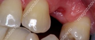
An example of a “quiet” seam.
If we talk about the incidence of late infectious complications, they most often occur in the first two years after endoprosthetics (1.63% of patients). Less commonly (in 0.59% of those operated on), deep paraprosthetic infections develop in the next 8 years after surgery.
The frequency of infectious complications has remained unchanged for several decades. However, the total number of arthroplasties has noticeably increased, and the total number of complications has also increased. Therefore, their prevention, early diagnosis and treatment are becoming increasingly important.
Fact! As scientific studies have shown, the risk of developing infectious complications depends on the type of endoprosthesis. It turned out that in total, implantation of domestic models leads to inflammation more often (3-10% of cases) than installation of imported ones (0.3-4.8%).
What is a surgical site infection?
An SSI is an acute or chronic inflammation that develops at the site of an incision or in an area that has been iatrogenic during surgery. In 67% of cases, the infection affects only the surgical incision area, and in 33% it spreads to the implanted joint.
Factors for the development of SSI:
- the duration of the operation is more than 3 hours;
- technical difficulties during surgery;
- intraoperative blood loss more than 1 liter;
- instability of the installed endoprosthesis;
- the use of additional synthetic and biological materials during the operation;
- the presence of severe chronic diseases.
Inflammatory processes that do not spread to the operated joint can be overcome without revision arthroplasty. If the infection affects bone tissue, components of the endoprosthesis, remnants of the joint capsule or other parts of the knee or hip joint, it will be extremely difficult to treat. In this case, the patient will most likely require repeat arthroplasty.
There are a number of factors that aggravate the patient’s condition, slow down recovery and worsen the prognosis: decreased immunity, previous surgeries, frequent treatment with antibiotics. The resistance of the detected microflora to antibacterial agents, poor blood circulation in the joint area, and massive purulent lesions also complicate therapy.
Knee replacement in the Czech Republic: guarantees, prices, rehabilitation, reviews and statistics.
Find out more
Minimally invasive endoprosthetics in the Czech Republic: doctors, rehabilitation, terms and prices.
Find out more
How do dentists treat pus in gums?
Typically therapy includes the following steps:
- Survey. The doctor finds out when the purulent neoplasm appeared, how quickly it grows in size, and how it manifests itself.
- Examination of the oral cavity. The doctor studies the location of the abscess, its color, shape. If necessary, gives a referral for an x-ray of the affected side of the jaw. X-rays are needed to understand the condition of the internal structures.
- Removing food debris, stone and soft plaque from teeth and gums. Sanitation of the oral cavity before treatment of an abscess.
- Opening of a purulent neoplasm. Performed under local anesthesia. First, the patient is given an injection of anesthetic, and then the affected area is carefully dissected. As a result, purulent masses come out.
- Installation of drainage. To prevent exudate from beginning to accumulate in the gums again, a small drainage tube is installed into the incision. You need to walk with her for several days.
A mandatory stage of treatment is anti-inflammatory therapy. The patient is prescribed antibiotics and mouth rinses to prevent the recurrence of a purulent lesion.
It happens that pus is released when pressing on the tooth. In this case, there is no “bump” on the surface of the gum. In this case, the above scheme will be ineffective. The doctor removes the filling (if it was installed), cleans the dental canals and washes out all purulent accumulations. Places a special medicine and closes it with a temporary filling.
On the appointed day, the patient must come for a follow-up appointment. Then the doctor will assess whether the inflammation was removed. If yes, then he will replace the temporary filling with a permanent one; if not, he will re-place the anti-inflammatory composition in the canals and re-install the temporary filling material.
Types of paraprosthetic infection
In orthopedics and traumatology, several classifications of SSI are used. Systematization and assignment of infection to a specific type helps doctors assess the severity of the patient’s condition. The Coventry-Fitzgerald-Tsukayama classification is the most common.
Table 1. Types of deep paraprosthetic infection according to Coventry-Fitzgerald-Tsukayama.
| Type | Development time | Treatment tactics | |
| I | Acute postoperative | 1st month | Revision of the postoperative wound, removal of necrotic tissue, and, if necessary, replacement of some parts of the endoprosthesis while maintaining its main components. |
| II | Late chronic | From 1 month to 1 year | Mandatory revision endoprosthetics. |
| III | Acute hematogenous | After 1 year | It is entirely justified to try to preserve the installed prosthesis. |
| IV | Positive intraoperative cultures | Asymptomatic bacterial colonization of the implant surface | Conservative treatment consisting of parenteral antibiotic therapy for 6 weeks. |
In the classification created by the Novosibirsk Research Institute of Traumatology and Orthopedics, SSIs are divided into early acute, late acute and chronic. The first develop within three months after endoprosthetics, the second - at 3-12 months, the third - after 1 year. Infectious complications can occur in a latent, fistulous, phlegmon-like or atypical form.
According to the prevalence, infections are epifascial (superficial) and subfascial (deep). May be accompanied by total, femoral or tibial instability.
Superficial and deep infections
Occurs in the first month after endoprosthetics. Characterized by the development of inflammation in the soft tissues of the lower limb. The hip or knee joint itself remains intact, that is, it is not involved in the pathological process. The cause of the complication is most often the introduction of pathogenic microorganisms into the wound during surgery or in the postoperative period.
Superficial SSI:
- necrosis of the skin;
- ligature fistulas;
- divergence of wound edges;
- subcutaneous hematoma.
Deep infections:
- necrosis of paraprosthetic tissues;
- deep fistulas;
- infected subfascial hematoma.
Fact! Mild tenderness, local swelling, redness and hyperthermia of the skin in the scar area usually indicate a superficial infection, which can be treated. The appearance of fever, spontaneous dehiscence of sutures and severe pain in the leg suggest inflammation of deep tissues. In this case, the prognosis is less favorable.
Content:
- Why does pus come out of the gums?
- Signs indicating a problem
- Causes of pus discharge from the gums
- How do dentists treat pus in gums?
- Additional procedures to help get rid of purulent gum disease
- Recovery after treatment
- Is it possible to cure an abscess at home?
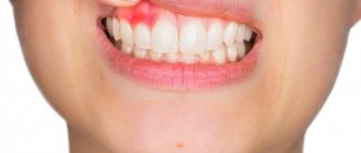
The presence of purulent exudate in the gum tissue is a dangerous symptom that should never be ignored. If it appears, you should immediately consult your dentist. Otherwise, the inflammatory process will progress, which will lead to damage to neighboring structures and premature tooth loss.
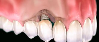
Prosthetic joint infections
In pathology, inflammation spreads to the cavities and membranes of the operated joint, remnants of the synovial membrane, bones at the site of fixation of the endoprosthesis and adjacent soft tissues. The cause of the complication is the colonization of joint surfaces by pathogenic microflora. Bacteria can come from the external environment or be introduced hematogenously.
This is what an infection looks like on an x-ray.
Prosthetic joint infections are the most serious complication among all SSIs. They do not respond to conservative therapy, so they have to be treated surgically. Doctors replace the endoprosthesis, but sometimes they still manage to save it.
There are three methods for treating infections of a prosthetic joint: wound revision without removing the implant, one-stage revision and two-stage endoprosthetics. The choice of technique depends on the patient’s condition, the time of manifestation of the infection, the stability of the prosthetic components and the nature of the pathogenic microflora.
Why does pus come out of the gums?
Suppuration is the result of infection penetrating into the deep tissues of the oral cavity. This is a kind of protective reaction of the body aimed at cleansing the affected area from a dangerous viral, bacterial or infectious agent.
As soon as pathogenic microflora begins to spread inside the gums, blood circulation there increases. Due to this, immune cells are faster delivered to the dangerous focus. As a result, the tissues become red, swollen, and painful to the touch.
Pus in the gums is dead microbes and dead immune cells. As its volume increases, an abscess forms.
X-ray studies
X-ray fistulography plays an important role in the differential diagnosis of fistulous forms of infection. With its help, you can determine the size, shape and location of fistulas, identify purulent leaks and their connection with foci of bone destruction. This makes it possible to distinguish superficial from deep SSIs.
Photo: X-ray fistulography, fistula in the lower third of the thigh.
X-rays are most often used to diagnose prosthetic joint infection. The method does not give 100% correct results, but it allows one to suspect pathology. The presence of a paraprosthetic infection is indicated by the sudden appearance of a periosteal reaction and osteolysis. If these signs appear suddenly, soon after a successful operation, there is reason to suspect something is wrong.
Curious! MRI, ultrasound and radioisotope scanning are rarely used for diagnostic purposes due to their low information content. For example, an installed endoprosthesis interferes with magnetic resonance imaging, which makes the image blurry and unclear.
Lab tests
Taking tests helps identify acute and chronic inflammatory processes in the body. An increase in indicators is not a reliable sign of SSI. To make a diagnosis, it is necessary to take into account the presence of certain clinical symptoms, radiographic data and other research methods.
Clinically significant laboratory parameters:
- Leukocyte count. It is important in the diagnosis of acute paraprosthetic infection. A clear sign of inflammation is an increase in the total number of leukocytes and neutrophils, a shift in the leukocyte formula to the left.
- ESR. It is a non-specific indicator. A normal erythrocyte sedimentation rate indicates the absence of inflammatory processes, an increased rate indicates their presence.
- C-reactive protein. CRP is an acute phase protein and a highly sensitive marker of SSI in people who have undergone arthroplasty. When diagnosing paraprosthetic infections, you need to pay attention to this indicator.
Knee replacement in the Czech Republic: guarantees, prices, rehabilitation, reviews and statistics.
Find out more
Minimally invasive endoprosthetics in the Czech Republic: doctors, rehabilitation, terms and prices.
Find out more
Microbiological studies
Bacterioscopic and bacteriological studies make it possible to identify and identify the causative agent of infection, as well as determine its sensitivity to antibiotics. Quantitative studies make it possible to determine the number of microbial bodies in purulent discharge.
The following materials can be used for research:
- discharge from a wound;
- fabric samples;
- fluid from the joint cavity;
- prosthetic material.
In case of implant-associated infection, it is almost impossible to detect bacteria in biological fluids and tissues. Pathogenic microorganisms are found on the surfaces of endoprostheses themselves. They cover the implants in the form of an adhesive film.
Fact! In addition to bacteriological examination, PCR (polymerase chain reaction) can be used for diagnosis. The method has high sensitivity but low specificity. Because of this, it often gives false positive results.
Treatment
Before deciding how to deal with the infection, doctors carefully examine the patient. Only after establishing a diagnosis and determining the sensitivity of pathogenic microflora to antibiotics do they make a final decision.
Table 2. Treatment methods for paraprosthetic infections:
| Method | Indications | results |
| Wound sanitation while preserving the endoprosthesis | It is carried out in cases where an SSI occurs in the first 3 months after surgery. It is possible to save the endoprosthesis only in the absence of purulent leaks and severe concomitant diseases. In this case, the implant must be stable, and the microflora must be highly sensitive to antibiotics. | It is the least traumatic treatment method. According to various sources, the effectiveness of surgical debridement is 18-83%. |
| Revision (repeated) endoprosthetics | One-stage or two-stage implant replacement is performed in cases where it is not possible to save the joint. A similar situation is observed with instability of endoprosthesis components, late development of infection, low sensitivity of microflora to antibiotics, and the presence of severe somatic diseases. | Allows you to completely cope with the problem in 73-94% of cases. Unfortunately, during treatment the patient has to completely change the installed endoprosthesis. |
| Arthrodesis with transosseous osteosynthesis | Deep recurrent paraprosthetic infection, microflora insensitive to antibiotics, the presence of severe concomitant pathology. | In 85% of cases, it eliminates the inflammatory process and restores the support ability of the lower limb. |
| Disarticulation in the hip joint | Chronic recurrent inflammation that threatens the patient's life, or complete loss of function of the lower limb. | A man permanently loses his leg. It is cut off at the level of the hip joint. |
Is it possible to cure an abscess at home?
Doctors do not allow you to open a flux tumor on your own. This may lead to the addition of a secondary infection. If you are unable to visit a dental clinic in the coming days, you can alleviate the uncomfortable symptoms with the help of:
- Therapeutic and prophylactic rinses. When performing them, you need to use water at room temperature, salt and soda, and pharmaceutical antiseptics.
- Apply cold compresses to the sore cheek.
Under no circumstances should you heat the inflamed area. This will lead to activation of purulent-necrotic processes and the spread of infection, which is very dangerous.
Implant-sparing tactics
Its main goal is to eliminate the infectious process while preserving the endoprosthesis. The patient undergoes surgical treatment of the wound, during which pus and necrotic tissue are removed. If the joint itself is involved in the pathological process, arthroscopic debridement is performed. The patient is prescribed massive antibacterial therapy.
Curious! Scientific studies have proven the effectiveness of non-surgical treatment of early deep infections. As it turned out, a combination of antibiotics and enzyme preparations helps eliminate inflammation in 5-7 days.
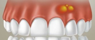
Signs indicating a problem
A purulent neoplasm is difficult to miss. A person complains about:
- discomfort in the affected area, which intensifies with palpation, pressure, chewing food, and talking;
- unpleasant taste in the mouth (caused by periodic discharge of blood and necrotic masses from the abscess);
- tissue swelling;
- swelling of the cheek (indicates that the inflammation has gone very far and emergency dental care is required).
If the patient continues to ignore the symptoms, his health will worsen. His body temperature will rise, headaches and weakness will appear.
A violent inflammatory process will disrupt the dentogingival apparatus. Then the unit will become mobile and can fall out at any moment.