|
|
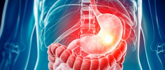
Have you often had toothache? Probably every second person has encountered this problem at least once in their life. At such moments, he has only one desire - to get rid of her as quickly as possible. Various means are used, ranging from medications to traditional methods. After a few minutes, the pain goes away and the person feels much better. Often it is at this stage that his life-saving actions stop. Many people forget that pain is the first sign of the development of a pathological process in the oral cavity. Moreover, if it is accompanied by an increase in body temperature and swelling of the gums. All of these symptoms indicate a dental disease called periostitis or otherwise flux.
Causes of occurrence.
The periosteum is a thin vascular tissue that consists of many nerve endings that completely cover the jaw bone. It securely connects the teeth together with the muscles and ligaments in the oral cavity. If the gum is damaged and an infection gets into it, inflammatory processes begin in the periosteum, the mucous membrane swells, and the person experiences pain. Many patients consider gumboil to be a temporary problem and try to get rid of it using folk remedies. Such an attitude and untimely professional treatment can cause an abscess in the periosteum area. Also, pathology occurs due to difficulty in the eruption of “eights”, i.e. wisdom teeth or as a result of a growing cyst. In addition to these factors, there are several other causes of periostitis:
- Complications of caries.
- Poor oral hygiene.
- Poor quality treatment and improper canal filling.
- Mechanical injuries of the jaw.
- Diseases (sinusitis, sore throat), due to which the infection penetrates the gum tissue through the circulatory system.
- Mistakes during tooth extraction.
Biomechanics of injury
The main difference between running and walking is the absence of a double support phase (when both legs are on the ground at the same time), as a result of which the body weight falls on one limb when landing, which leads to 1) a sharp increase in the impact load on the leg; 2) the need to increase the activity of the foot stabilizer muscles. That is, muscles that provide not so much power (like the gastrocnemius and soleus muscles), but rather the correct position of the landing and pushing off foot, adaptation to uneven surfaces.
As a result of increased load, the stabilizer muscles become clogged and shortened, just like any muscle after training. And here the second mechanism that causes injury is activated - the distribution of the shock load.
When we land on our foot, the muscles, ligaments and bones absorb and dissipate the shock. If the muscle is elastic and in normal tone, then most of the blow is absorbed by the lengthening of its abdomen (central part), and the tendons and places of attachment to the bone take on the rest of the blow. If the muscles are shortened and not elastic, much more shock load falls on the attachment site.
periosteum (a dense layer of connective tissue that covers the entire bone). When receiving a blow, the muscles seem to pull the periosteum, which leads to its microtrauma and inflammation.
Structural disorders of the foot can also increase the shock load on the lower leg muscles, such as an excessively high arch of the foot (Figure 3), which reduces its shock-absorbing ability, and flat-valgus feet, which often lead to hyperpronation (excessive roll of the ankle joint and the foot inwards when landing ) and, consequently, additional load on the tibialis posterior muscle. {banner_st-d-2}
Causes of tire splint:
1) Weak and clogged foot stabilizer muscles 2) High impact load.
High impact loads may be caused by:
overweight; structural features of the foot - hyperpronation, high rigid arch of the foot; shoes with low cushioning; hard surface for running; hard contact with the surface (excessively high tension of the feet upon contact); acceleration or running down an inclined surface; running on uneven surfaces.
Moreover, you can clog up your muscles one day, for example, when training in the park, and damage the periosteum during the next workout when running on your usual hard surface, simply by shortening the muscles.
Signs of inflammation.
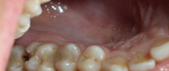
As soon as an infection gets into the gum, an inflammatory process begins in it, it swells and increases in volume. After some time, pathogenic bacteria spread to nearby soft tissues, as a result of which the cheeks and lip begin to swell. There may also be an increase in body temperature, a deterioration in general condition, and the occurrence of pain in the ears and temples. All these signs indicate a problem that requires immediate correction. There are several stages of periostitis:
Acute serous form.
Symptoms appear 1-3 days after the gums become infected. There is redness and swelling of the mucous membrane, mostly imperceptible changes occur.
Acute purulent.
All previous manifestations of the disease remain, and in addition to them, the patient begins to be bothered by acute pain, due to which the movement of the jaw is limited. Pus accumulates in periodontal tissues, which can open on its own, i.e. rupture and leak out. When biting a damaged tooth, unpleasant sensations appear, and it also becomes mobile.
Acute diffuse.
Loss of appetite, deterioration in health, lethargy. If the abscess has not opened, it provokes the formation of severe edema at the site of damage to the periosteum. Flux in the acute diffuse stage is accompanied by throbbing, unbearable pain that can radiate (give) to the head: temples, ears, neck.
Chronic form.
Sluggish course of the disease with periodic exacerbations. The patient has slight facial asymmetry and inflammation of the lymph nodes. On the x-ray you can see thickening of the bone tissue, due to sharply changed dimensions of the periosteum.
Diagnostic measures
The doctor conducts an examination. It is not difficult to understand that a bone is damaged. This is indicated by external signs:
- redness of the gums;
- pain when pressing;
- the presence of a cushion containing pus;
- mobility of the incisor, canine, molar (noted if the disease is advanced).
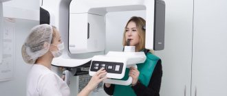
To determine the stage of the disease and the depth of damage to bone formations, the doctor performs an X-ray or radiovisiography of the jaw.
Diagnostics.
Treatment of inflammation of the periosteum of the tooth is carried out in several stages; first you need to undergo a comprehensive examination by a dentist. It includes:
- Initial examination. The doctor will assess the condition of your oral cavity, interview you and give you a referral for tests and x-rays.
- Having received all the necessary data, the specialist must determine what kind of disease the patient has. Because signs of periostitis are similar in symptoms to such pathologies as: acute periodontitis, phlegmon and osteomyelitis. Also, at this stage it is important to establish the exact form of the flux in order to draw up a suitable treatment plan.
Treatment.
Depending on the severity of the disease, the method of therapy is selected. If the patient has acute serous periostitis, then it will be sufficient to depulpate the tooth, clean its canals and treat the opened wound with antiseptic agents. In the case of an acute purulent form, in addition to basic dental procedures, physiotherapy may be prescribed. But most often, combined techniques are used to treat inflammation of the periosteum of the tooth; let’s look at them in more detail:
Surgical method.
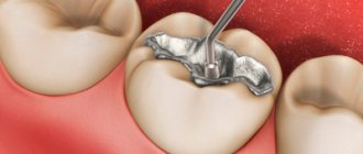
It is used when pus forms in the area of damaged gums and needs to be eliminated. The dentist opens the abscess, removes infected soft tissue and remaining suppuration. Then the oral cavity is sanitized, the tooth canals are filled with medicine and filled with temporary composite materials. After about 2-5 days, a permanent filling is installed and an x-ray examination is performed to exclude the possibility of re-formation of inflammation.
Physiotherapy.
Most often prescribed for chronic flux and in conjunction with other treatments. With the help of physiotherapy, you can influence neoplasms in the bone area. For this purpose: laser, electrophoresis, darsonvalization, UV radiation, etc. are used.
Drug treatment.
It is appropriate only at the initial stages of inflammation formation. Its essence lies in the administration of medications to eliminate swelling, stop (interrupt) the disease and for prevention purposes.
Traditional methods.
Naturally, such a solution to the problem is allowed only as an auxiliary method and exclusively with the permission of the doctor. If you notice symptoms of inflammation of the periosteum of the tooth, in order to relieve pain and reduce swelling of the mucous membrane, it is recommended to use a salt or soda solution to rinse the mouth.
Important! In order not to worsen your condition and not to provoke the spread of inflammation to other areas, you should not apply hot compresses to the affected tooth, do not abuse traditional medicine, and it is strictly forbidden to try to open the suppuration on your own.
What structures can become inflamed in the lower leg?
Pain in the lower leg and ankle occurs suddenly after an injury - bruise, sprain, fracture, dislocation. It can also be caused by a chronic illness. The following structures can become inflamed in the lower leg:
- Ligaments. They may become inflamed or stretched. When a tendon or muscle is sprained, severe tension occurs. Severe trauma also causes muscle or tendon rupture.
- Joints. Arthritis or arthrosis is a common pathological condition that affects the joints and is accompanied by pain. Ankle pain also often accompanies the development of rheumatoid arthritis.
- Tendons. The most common inflammatory tendon pathology is tendonitis. Also, with constant friction of the tendons and a deficiency of interarticular fluid, bursitis develops. This is accompanied by irritation and discomfort.
- Bones. After an injury, the integrity of the bone is often compromised. This is a fracture or crack. In this case, it is very important to visit a doctor as soon as possible. Only through timely and correct treatment will the bone heal and there will be no consequences of the injury.
- Tarsal tunnel syndrome. It develops when the tarsal tunnel is not pinched. This is a nerve located along the entire length of the lower leg.
- Arch. There are tendons in the leg that work synchronously. This is how the arch is formed. When the tendons are connected correctly, a regular and symmetrical arch is formed. If they are connected asymmetrically, an arch of a small size is formed or it is completely absent. With this condition, discomfort or pain occurs.
- Connective tissue. Connective tissue elements are located on the bottom of the foot. When they become inflamed, it is called plantar fasciitis. This happens when wearing incorrectly selected shoes, too narrow and uncomfortable, or with poor posture.
Preventive measures.
As you can already understand, periostitis is a serious and dangerous dental disease. To prevent its occurrence, you must adhere to several rules:
- Maintain good oral hygiene.
- Have a comprehensive examination with your dentist at least once a year, and don’t forget about x-rays. It will help diagnose inflammation of the periosteum in the early stages.
- Avoid fights, and protect your face from injuries and blows.