Types, functions and number of teeth in adults and children
During normal development, a person erupts 20 temporary and 28-32 permanent teeth.
The range of their number varies depending on the hereditary predisposition - whether the rudiments of the third molars, called “eights” or wisdom teeth, have formed. The units are sequentially located in the jaws and form rows - upper and lower. Each contains 16 teeth: 4 incisors, 2 canines, 4 premolars and 6 molars. In children, the arch consists of 10 units on each jaw.
Teeth anatomy. Anatomy of the teeth of the upper and lower jaw
Dental buds are formed in the fetus already in the first trimester of pregnancy, during the 7th week of development. At the same time, at the site of future alveolar processes, the epithelial tissue thickens and, forming a symmetrical arc, grows into the depths of the mesenchyme. Subsequently, secondary plates are formed under it, located perpendicularly.
In the tooth buds, meanwhile, tooth enamel begins to form from epithelial cells. As the dental plate grows, the enamel organs appear in front and become separated from it. It is then that the components of the future tooth are formed.
With normal dental anatomy in humans, the epithelium is transformed into enamel, and the mesenchymal tissue forms dentin and pulp, and a cement shell appears that protects the tooth root. The rudiments themselves remain in the alveolar processes, awaiting the time of their eruption.
Based on their structural parts, teeth are usually divided into crown, neck and root:
- the crown is the visible part that is located above the gum and is directly involved in grinding food;
- the neck is the part located inside the gum, not covered with enamel, but protected by cement;
- the root is hidden in the alveolus, connecting the teeth with the bone tissue of the jaw, and through the canal of which nerves and blood vessels run into the tooth cavity.
The cavity itself is filled with soft tissue, penetrated by many nerve and vascular endings, and is called pulp.
The main part of the dental tissue consists of dentin, which is located around the pulp and is protected from damage by tooth enamel on the crown and cement in the neck and root area.
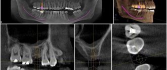
Upper jaw teeth
The crown of the tooth is spade-shaped.
The lateral surfaces gradually converge towards the neck. The vestibular surface is convex and often has the shape of a rectangle. In young people it is wavy, the waves go longitudinally and seem to divide the vestibular surface into three parts, forming three bends along the cutting edge.
With age, the waviness of the vestibular surface of the crown and cutting edge disappears (erases), and it becomes smooth. The crown is wider at the cutting edge and narrower at the neck of the tooth, the medial angle of the cutting edge is straight, the distal one is slightly rounded.
The outer line of the incisor is rounded on the mesial side, and somewhat concave on the distal side. The oral surface is concave and has the shape of a triangle with the apex directed towards the neck of the tooth. In the upper third there is a tubercle.
In young people, the palatine tubercle is divided into several small tubercles. The approximal surface has the shape of a triangle with the apex facing the cutting edge. The line of the neck of the tooth (enamel-cementum border) is curved.
The labial surface is convex only in the upper half (closer to the neck), its half, going to the cutting edge, is flattened. Lateral incisor of the upper jaw The lateral incisors are smaller in size than the central incisors, their shapes vary significantly.
The crown of the tooth is spade-shaped. The lateral surfaces of the crown are almost parallel. The crown of the lateral incisor is smaller than the central one in all dimensions (shorter and narrower by about 1 mm). The medial angle of the lateral incisor is more rounded than that of the central one.
The vestibular surface is convex (and the narrower the crown of the tooth, the more pronounced it is) and has the shape of a triangle with the apex facing the neck of the tooth. With a relatively wide crown, its shape is the same as that of the central incisor, that is, flattened in the lower part of the crown. In the dentition, the neck of the lateral incisor is located somewhat distally compared to the cutting edge.
Maxillary canine
The canine, located distal to the lateral incisor, forms the angle of the dental arch - the transition from cutting teeth to chewing teeth. In the dentition, the crown of the canine is slightly deflected vestibularly and, accordingly, protrudes from the arch of the dentition. The shape of the crown is cone-shaped, its anteroposterior size is larger at the base, and its transverse size is larger at the middle.
When examining the canine crown from above, its mesial-distal curvature is clearly outlined. The vestibular surface is convex and... has a vaguely defined longitudinal ridge, better visible at the cutting edge; the roller divides the labial surface into two unequal parts: the smaller - medial and the larger - distal.
The oral surface is narrower than the vestibular one, slightly convex and, like the vestibular surface, has a longitudinal ridge running from the neck to the cutting tubercle. The roller divides the surface into two parts - medial and distal. There are often indentations on either side of it.
Examples of dental formulas of permanent teeth of some mammals
| View | Latin name | Squad | Family | Dental formula |
| Hedgehog | Erinaceus europaeus Linnaeus, 1758 | Insectivores | Hedgehogs | $ I{3 over 2} C{1 over 1} P{3 over 2} M{3 over 3} $ |
| Kutora | Neomys fodiens Schreber, 1776 | Insectivores | Shrews | $ I{1 over 1} in{4 over 2} M{4 over 3} $ |
| Ushan | Plecotus auritus Linnaeus, 1758 | Chiroptera | Leather | $ I{2 over 3} C{1 over 1} P{1 over 2} PmP{1 over 1} M{3 over 3} $ |
| Wolf | Canis lupus Linnaeus, 1758 | Predatory | Doggystyle | $ I{3 over 3} C{1 over 1} P{4 over 4} M{2 over 3} $ |
| Cat | Felis catus Linnaeus, 1758 | Predatory | Felines | $ I{3 over 3} C{1 over 1} P{3 over 2} M{1 over 1} $ |
| Horse | Equus caballus Linnaeus, 1758 | Odd-toed ungulates | Equine | $ I{3 over 3} C{1 over 1} P{4 over 3} M{3 over 3} $ |
| Boar | Sus scrofa Linnaeus, 1758 | Artiodactyls | Pork | $ I{3 over 3} C{1 over1} P{4 over 4} M{3 over 3} $ |
| Elk | Alces alces Linnaeus, 1758 | Artiodactyls | Deer | $ I{0 over 3} C{0(1) over1} P{3 over 3} M{3 over 3} $ |
| Mouse | Mus musculus Linnaeus, 1758 | Rodents | Mouse | $ I{2 over 2} C{0 over 0} P{0 over 0} M{3 over 3} $ |
| Elephant | Elephas maximus Linnaeus, 1758 | Proboscis | Elephantids | $ I{1 over 0} C{0 over 0} P{3 over 3} M{3 over 3} $ |
| View | Latin name | Squad | Family | Dental formula |
| Koala | Phascolarctos cinereusGoldfuss, 1817 | Two-incisor marsupials | Koalas | |
| Hedgehog | Erinaceus europaeusLinnaeus, 1758 | Insectivores | Hedgehogs | |
| Kutora | Neomys fodiensSchreber, 1776 | Insectivores | Shrews | |
| Ushan | Plecotus auritusLinnaeus, 1758 | Chiroptera | Leather | |
| White hare | Lepus timidus Linnaeus, 1758 | Lagomorpha | Zaitsevy | |
| Wolf | Canis lupus Linnaeus, 1758 | Predatory | Doggystyle | |
| Cat | Felis catus Linnaeus, 1758 | Predatory | Felines | |
| Horse | Equus caballus Linnaeus, 1758 | Odd-toed ungulates | Equine | |
| Boar | Sus scrofa Linnaeus, 1758 | Artiodactyls | Pork | |
| Elk | Alces alces Linnaeus, 1758 | Artiodactyls | Deer | |
| Cattle | Bos taurus taurus Linnaeus, 1758 | Artiodactyls | Bovids | |
| Mouse | Mus musculus Linnaeus, 1758 | Rodents | Mouse | |
| Elephant | Elephas maximus Linnaeus, 1758 | Proboscis | Elephantids |
What formulas are used in pediatric dentistry?
The structure of the dental system record has some features:
- the arrangement of teeth in the arch is strictly sequential;
- the row is divided into two symmetrical parts;
- the formula is compiled in such a way that the units of each half of the jaws are written in it;
- numbering is carried out using letters and numbers;
- the dental formula is read from the doctor’s perspective (from left to right, clockwise, where your right side will correspond to the upper left segment).
We invite you to familiarize yourself with Painkillers for children with toothache
The entry in the universal formula is based on dividing teeth according to group affiliation and assigning them a letter equivalent:
- incisors – I;
- fangs – C;
- premolars – P;
- molars – M.
| M3 | M2 | M1 | P2 | P1 | C | I2 | I1 | I1 | I2 | C | P1 | P2 | M1 | M2 | M3 |
| M3 | M2 | M1 | P2 | P1 | C | I2 | I1 | I1 | I2 | C | P1 | P2 | M1 | M2 | M3 |
| 1 | 2 | 3 | 4 | 5 | 6 | 7 | 8 | 9 | 10 | 11 | 12 | 13 | 14 | 15 | 16 |
| 32 | 31 | 30 | 29 | 28 | 27 | 26 | 25 | 24 | 23 | 22 | 21 | 20 | 19 | 18 | 17 |
This method was created in 1876 and is used in modern dentistry. Its other name is square-digital. The structure of the record is based on the numbering of dental units from 1 to 8, depending on their group affiliation and arrangement order, where:
- 1 – central incisor;
- 2 – lateral;
- 3 – fang;
- 4 – first premolar;
- 5 – second;
- 6 – first molar;
- 7 – second;
- 8 – third.
| 8 | 7 | 6 | 5 | 4 | 3 | 2 | 1 | 1 | 2 | 3 | 4 | 5 | 6 | 7 | 8 | |
| 8 | 7 | 6 | 5 | 4 | 3 | 2 | 1 | 1 | 2 | 3 | 4 | 5 | 6 | 7 | 8 | |
Children's teeth are indicated in Roman numerals, in segments from the center:
- central incisor – I;
- lateral – II;
- canine – III;
- first primary molar – IV;
- the second is V.
| V | IV | III | II | I | I | II | III | IV | V |
| V | IV | III | II | I | I | II | III | IV | V |
This technique was adopted in 1971 by the International Dental Association. Currently it is the most popular in use by doctors. The essence of the two-digit calculation is to assign a two-digit number to the tooth. In this case, the rows are divided in half, forming segments. There are four in total:
- top right side;
- top left;
- lower left;
- lower right.
To determine the position of a molar in the oral cavity, it is assigned the value of the corresponding segment. An Arabic numeral indicating the location is placed in front of its number.
| 18 | 17 | 16 | 15 | 14 | 13 | 12 | 11 | 21 | 22 | 23 | 24 | 25 | 26 | 27 | 28 |
| 48 | 47 | 46 | 45 | 44 | 43 | 42 | 41 | 31 | 32 | 33 | 34 | 35 | 36 | 37 | 38 |
We suggest you read: How often should you change dentures?
When filling out a patient's outpatient chart, the doctor will begin to assign a double value to each tooth. The signs will be located through a dot. For example, the lower left first premolar will acquire code 3.4, where the number 3 indicates the segment and 4 the account number.
The method is also used in children's practice. Decoding of quadrants is based on values from 5 to 8:
- top right side – 5;
- top left – 6;
- lower left – 7;
- lower right – 8.
When designating milk units, Arabic numerals are used:
- central incisor – 1;
- lateral – 2;
- fang – 3;
- first molar – 4;
- second – 5.
| 55 | 54 | 53 | 52 | 51 | 61 | 62 | 63 | 64 | 65 |
| 85 | 84 | 83 | 82 | 81 | 71 | 72 | 73 | 74 | 75 |
Haderup system
The technique is based on the Zsigmondy-Palmer method. The teeth of the same name in the upper and lower jaws are designated by the same Arabic numerals from 1 to 8. As in the two-digit Vilor system, a symbol is assigned to indicate the quadrant in which the tooth is located.
| 8 | 7 | 6 | 5 | 4 | 3 | 2 | 1 | 1 | 2 | 3 | 4 | 5 | 6 | 7 | 8 | |
| 8- | 7- | 6- | 5- | 4- | 3- | 2- | 1- | -1 | -2 | -3 | -4 | -5 | -6 | -7 | -8 |
| 05 | 04 | 03 | 02 | 01 | 01 | 02 | 03 | 04 | 05 |
| 05- | 04- | 03- | 02- | 01- | -01 | -02 | -03 | -04 | -05 |
The eruption of baby teeth has a strict sequence and specific timing. Each time period corresponds to a certain number of units. Parents often worry whether their children are okay? How many baby teeth should they have at a certain age? Starting from the eruption of the first temporary units, it is necessary to check their number with the dental formula corresponding to the given period.
In pediatric practice, dentists resort to various schemes for designating primary occlusion. The doctor works with the one who, in his opinion, fully meets his requirements.
We invite you to familiarize yourself with the effect of anesthesia on the body during dental treatment
An easy way to find out the age of a puppy or adult dog by its teeth
If you adopted a dog from a shelter, picked up an adult animal on the street, or purchased an older puppy without documentation, in order to organize proper care, you need to know how old the dog is. You can determine the age of your four-legged friends using various methods. The most reliable is an analysis of the condition of the animal’s teeth.
Dog dental formula
Healthy dogs that do not have chronic pathologies or congenital defects have only 42 teeth (20 on the upper jaw and 22 on the lower jaw), which differ in appearance and purpose.
Important! Some dogs may have more teeth (polyodontia) or fewer (oligodontia).
Teeth are divided into premolars, molars, canines, and incisors.
- The first six teeth on the upper and lower jaw are called incisors. The two front teeth are hooks, to the right and left of them are the middle incisors, then the outer ones (edges).
- Behind the incisors are the fangs (two on each jaw). These are the longest, sharpest teeth a dog has.
- Two groups of molars - molars (4 on the upper, 6 on the lower jaw), premolars (four on each side) are located immediately behind the canines.
- To determine the number of teeth in dogs, veterinarians use a dental formula. Each tooth has a letter designation. K – canine, R – incisors, P – premolars, M – molars.
The dental formula of adult dogs is as follows:
- Upper jaw: P—6, K—2, P—8, M—4;
- Lower jaw: P—6, K—2, P—8, M—6.
How to determine the age of a puppy by its teeth
Puppies are born not only blind, but also completely toothless. The first milk teeth (canines, hooks) erupt in the third or fourth week of life. The canines appear first, then the middle incisors and molars.
A full set of milk teeth (28 pieces) is noted in puppies at 1.5-2 months of age, of course, if the animals do not have congenital pathologies and grow and develop normally. At this age, puppies' teeth are snow-white or cream-colored, small, but very sharp. Located as close to each other as possible.
Important! If all of the baby's teeth have not erupted before two months, this indicates a developmental disorder and health problems.
- At 2.5-4 months, the front teeth and incisors change.
- From 3 to 5 months, premolars (middle incisors) change.
- Before six months, the puppy's edges should change; after six months, new permanent fangs should appear.
Dogs, like humans, change their teeth once in a lifetime. In puppies, the change of milk teeth occurs from 2 to 6-7 months, up to a maximum of 8 months. At this time, puppies become restless and try everything by heart. Possible loss of appetite and increased temperature.
By 4.5-5 months, adult incisors have erupted, first premolars and molars appear; by six months, canines, second and fourth premolars, and second molars appear. By 7 months - third molars.
With normal development, all 42 molars in a dog should appear by the age of one year.
Determining the age of adult dogs
In young, adult dogs, to determine the approximate age, pay attention to the degree of wear on the dental cusps.
On the molars and incisors there are specific bulges that resemble a trefoil in appearance. As the dog gets older, they will gradually disappear.
Determination of age by abrasion of dental cusps:
- In one-and-a-half-year-old and two-year-old dogs, the tubercles on the toes and on the lower jaw are worn out. Snow-white enamel.
- At 3.5-4 years old, the dog's tubercles on the upper toes wear off. Tooth enamel takes on a matte tint. The tops of the teeth are a little worn.
- At 4.5-5 years old, the dog should have completely erased the tubercles from all its incisors. Yellowish enamel. The ends of the fangs become dull.
- In six-year-old dogs, the incisors take on a more concave shape. A slight deformation of the bite is noted.
- At the age of 8-9 years, the dental crowns are worn out, the ends of the fangs are blunt, and their length decreases. The color of the teeth is yellowish.
- In adult 10-11 year old dogs, the bite is disturbed, and the process of loss of molars begins. The teeth on the lower jaw fall out first. The development of dental diseases (periodontal disease, caries, tartar) is possible.
- In dogs at the age of 11-12 years, the teeth become loose, yellow, and fall out on the upper and lower jaws. Plaque appears, tartar forms, and crowns wear off.
- After 15 years – complete loss of teeth and fangs.
Source: https://zen.yandex.ru/media/vashipitomcy/prostoi-sposob-uznat-vozrast-scenka-ili-vzrosloi-sobaki-po-zubam-5e3bce0ecf37790f172e67ff
Literature
- N. A. Bobrinsky, B. A. Kuznetsov, A. P. Kuzyakin. Key to mammals of the USSR. State Publishing house "Soviet Science", M., 1944.
- N. A. Bobrinsky, B. A. Kuznetsov, A. P. Kuzyakin. Key to mammals of the USSR. State Publishing house "Soviet Science", M., 1944.
This page uses content from the Wikipedia section in Russian. The original article is located at: Dental formula. A list of the original authors of the article can be found in the edit history. This article, like the article posted on Wikipedia, is available under CC-BY-SA.