Gastroesophageal reflux disease (GERD) is one of the most common diseases of the digestive system [1]. Its frequency in Western populations varies from 10 to 20% with a permanent upward trend [2]. According to the definition proposed by the Russian Gastroenterological Association, GERD is defined as a chronic relapsing disease characterized by the development of inflammatory changes in the mucous membrane of the distal esophagus and/or characteristic clinical symptoms associated with repeated reflux of gastric and/or duodenal contents into the esophagus [3]. It is important to note that GERD is often accompanied by extraesophageal pathology, including lesions of the red border of the lips and tongue [4, 5].
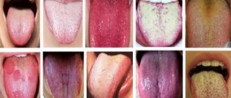
Pathological changes in the tongue with GERD are usually nonspecific and are manifested by the appearance of white plaque, swelling, varying degrees of epithelial desquamation, atrophy or hypertrophy of the papillae [4, 6, 7]. The appearance of plaque is mainly promoted by disturbances in the normal process of keratinization and desquamation of the epithelium, due to neurotrophic disorders, the nature and consistency of food, etc. [1, 2]. It is believed that the basis of plaque is made up of enlarged, keratinized filiform papillae with impaired rejection of keratinocytes, food debris, microorganisms, desquamated epithelial cells and fibrin. The processes of keratinization and desquamation of the epithelium of the mucous membrane of the tongue are influenced by various endogenous and exogenous factors, and their violations are considered not only as a consequence of various diseases, especially of the digestive organs, but also as a physiological adaptation of the body to environmental conditions [8-11].
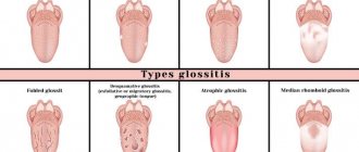
The lamina propria and the stratified squamous epithelium of the tongue form five types of papillae: filiform, cone-shaped, mushroom-shaped, grooved and leaf-shaped. Of these, the most numerous, especially on the dorsal surface of the tongue, are the filiform papillae ( papillae filiformes
), which do not contain taste buds, function as “organs” of touch and help keep food on the tongue. Filiform papillae 0.6–2.5 mm long and 0.1–0.6 mm thick represent a central bell-shaped body (“primary papilla”), crowned with filiform outgrowths of the lamina propria of the mucous membrane, covered with keratinizing epithelium (“secondary papillae” or "hair-like growths") As a rule, there are 3-8 secondary papillae on the primary papilla [12-15]. It is known that normally the dorsal surface of the tongue is home to more than 160 saprophytic and opportunistic species of bacteria. Any change in the properties of their habitat, in particular the morphology of filiform papillae, for example in diseases of the digestive organs, entails qualitative and quantitative changes in the composition of this microflora [14, 15].
The purpose of the study was to identify changes in the filiform papillae and localization of the microflora of the dorsal surface of the tongue in patients with GERD.
Causes of inflammation of the papillae on the tongue
If you have an inflammatory process in your mouth, contact your dentist immediately. Family dentistry in St. Petersburg is a clinic where every patient will be provided with assistance. The doctor will determine the causes of the disease and prescribe treatment. Problems may arise:
- When the patient’s diet includes excessive amounts of sour, sweet, spicy foods;
- If chronic stress develops in the body;
- When the patient received a tongue injury, burn, cuts, punctures;
- If the organ is affected by an allergic reaction;
- For some diseases of internal organs;
The surface of the muscular organ changes significantly under aggressive influence. The provoking factor is xerostomia, disease of the salivary glands, Sjogren's syndrome, diabetes mellitus. With glossitis, the sensitivity of the papillae increases. During the examination, the doctor must exclude HPV, leukoplakia, aphthous stomatitis, syphilis, and oral fibroma. Children are examined for scarlet fever.
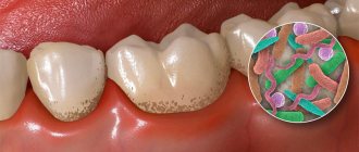
Sometimes the disease can be triggered by acid reflux, gastritis, throat infections, systemic inflammation of the gingival tissues when the pathogenic microorganism develops rapidly. Taking some medications can cause pain. High concentrations of alkali or acid have a negative effect on the condition of the muscular organ. Injuries on the tongue can occur from a hard brush, dentures, or tartar.
Symptoms
When the papillae on the tongue become inflamed, tissue sensitivity increases significantly and taste changes. Patients complain of unpleasant discomfort in the mouth. The taste spectrum is distorted and becomes unnatural. In addition, a number of other symptoms appear.
- Noticeable tissue swelling.
- Marked increase in size of the papillae.
- Tingling and itching.
- Burning, mild soreness.
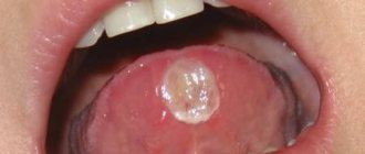
If you do not consult a dentist in time, complications may develop: lingual eruptive papillitis. This disease occurs over a long period of time with an increase in body temperature. Most often the disease affects children. An alarming symptom is the appearance on the surface of the vocal organ of bubbles (pustules) filled with a transparent liquid. When the first signs of illness appear, you should immediately visit the dentist. Timely treatment of the disease minimizes complications and speeds up recovery.
Material and methods
Biopsies of the dorsal surface of the tongue were studied in 7 (3 men and 4 women) patients with a non-erosive form of GERD and, as a comparison group, in 6 (3 men and 3 women) people without pathology of the upper gastrointestinal tract. Informed consent from the patients for the study was obtained. The average age of patients with GERD was 35 years (interquartile range 23; 42), disease duration was 4.8±2.7 years. The diagnosis of GERD was based on diagnostic criteria recommended by the Russian Gastroenterological Association: a set of clinical data, 24-hour pH-metry, esophagoduodenoscopy and a specialized GerdQ questionnaire [3]. At the time of biopsy, patients were not receiving therapy.
Biopsy specimens were studied using scanning electron microscopy (SEM) and fluorescence confocal microscopy (FCM) in the laboratory of molecular biology and genetics (Dr.Med.Sci., Prof. S.A. Gusev, DSc.Med.Sci., Prof. G. I. Lukina) Federal State Budgetary Institution Federal Scientific and Clinical Center for Physico-Chemical Medicine of the Federal Medical and Biological Agency of Russia. To perform SEM, biopsies measuring 0.5×1.0 mm were fixed in a mixture of 2% formaldehyde solution and 1.4% glutaraldehyde solution (pH 7.4) on the 1st day at a temperature of 40 °C, washed in 0.1 M phosphate buffer (pH 7.4) and distilled water for 2 hours at each stage. After dehydration in ethyl alcohol of increasing concentration, the samples were dried using the liquid substitution method by transitioning the critical point using a Hitachi HCP-2 apparatus (Hitachi, Japan) and coated with a 10 nm thick layer of gold by sputtering in a JEE-420 vacuum unit (Joel) , Japan). The study of filiform papillae in the obtained preparations was carried out using a scanning electron microscope S-570 (Hitachi, Japan).
For FCM, unfixed biopsy specimens were immediately stained with fluorescent dyes - synthetic fluorophores carboxyfluorescein (fluorescence emission in the green-yellow region of the absorption spectrum, λ 520 nm) and tetramethylcarboxyrhodamine (fluorescence emission in the red region of the absorption spectrum, λ 576 nm) (Synthol, Russia ). These dyes freely penetrate cell membranes and stain the nuclei of epithelial cells, as well as bacterial chromatin.
Localization
If the papilla on your tongue is inflamed, visit the dentist. Only he will be able to correctly determine the causes of the disease, find out what caused the damage to the mucous membrane of the mouth and the body of the tongue. The area where the disease is localized plays an important role in making the correct diagnosis. Pathology can cover the entire organ, part of it, or the sides. Based on this factor and the clinical picture, we can assume what exactly caused the inflammation.
In all languages
When the mucous layer of an organ is affected over its entire surface, the most common cause is a thermal or chemical burn. One can assume the infectious nature of the pathology. If the damage is severe, there may be no taste at all. Patients complain of severe burning.
On the root
Most often, the papillae on the tongue at the base become inflamed due to the action of allergens. The affected root of the muscular organ causes difficulties such as the perception of bitterness, because the inflammatory process has affected the circumvallate papillae. They are the ones responsible for this function. The patient's salivation increases, and the tissues in the oral cavity swell. This condition can also be caused by problems with the digestive system, too high or low acidity of gastric juice. It is on the back of the speech organ that bacterial or fungal glossitis is localized.
On the tip
The disease can affect the tip. This often occurs due to mechanical injuries or damage. The edge of the tongue is the first to come into contact with excessively hot food or aggressive drinks, which burns this area. Soft tissues often rub against sharp chips of crowns and are subject to accidental biting.
Side
The delicate surface on the sides often suffers from various anomalies. Leaf-shaped, conical, filamentous, mushroom-shaped structures are exposed by the teeth. This leads to an increase in the papillae, their hyperemia due to mechanical damage from fangs and molars. Also provoking factors are painful microorganisms and burns of chemical origin.
Clinical picture
The onset of pathology may be indicated by the appearance of redness and swelling in the affected area. If timely treatment is not started, inflammation spreads to healthy tissue areas, which leads to bleeding, discomfort, and itching.
The gum begins to cover more of the length of the tooth than usual. In the absence of qualified dental care, a change in the shape of the gingival papilla is often observed, and in advanced situations, the gum covers almost half of the element of the dentition.
The listed signs are accompanied by pain, bleeding (especially during and after eating), and a decrease in the quality of chewing function.
Among other characteristic features it is worth highlighting:
- Formation of purulent foci in the space between the teeth.
- The appearance of an unpleasant odor.
- An increase in the volume of soft tissue around the tooth.
- Difficulty eating.
- Uncharacteristic shade of the mucous membrane at the site of the lesion.
- Increased sensitivity.
- Uncharacteristic tissue structure.
In advanced cases, the inflammatory process is complemented by various diseases of the oral cavity, which lead to the destruction of both soft and hard tissues.
Diagnostics
Basic diagnostic methods in dentistry allow a thorough examination to be made in order to make the correct diagnosis. During the initial examination, the dentist determines the size of the pathological zones, appearance, color, and shape of the organ. The doctor finds out whether there is swelling of the tongue, ascertains plaque and its localization, abrasions, punctures, eczema, ulcers. The health of gum tissue and teeth is important.
In order to determine the type of pathogen and the form of the disease, a number of tests are prescribed:
- Histological smear;
- Sensitivity is determined using a special test;
- A general blood test is informative;
Systemic pathologists are also excluded: HIV, syphilis, hepatitis, AIDS. If necessary, the patient is referred for consultation to a gastroenterologist, dermatologist, endocrinologist, otolaryngologist, or immunologist.
Treatment
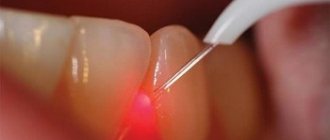
The diagnostic result allows you to prescribe treatment. Therapeutic dentistry has new methods and technologies that will return the patient to health.
- The patient is advised to adjust his diet and reduce spicy and sour foods in his diet. Dishes should not be hot.
- Fillings and dentures that injure the mucous membranes need to be replaced.
- You should have your oral cavity professionally sanitized.
- Remove soft and hard deposits on crowns.
- Cure caries, gum inflammation.
The therapeutic course may include taking antibiotics, anti-inflammatory drugs, and a course of treatment for diseases of internal organs. Regular visits to the dentist are a way to prevent oral problems and complications of existing diseases.
An injured, inflamed muscle organ requires tissue regeneration. For this purpose, drugs containing carotene are prescribed: Aekol, Retinol, Chlorophyllipt, Lugol. Ointment applications are made from Solcoseryl and Cholisal. For pain, anesthetics are used: Trimecaine solution, Anastezin emulsion. Patients are recommended medications to stimulate the body's defenses and vitamins.
If the disease is infectious, a scraping is first done. A pathogenic microorganism is identified in the laboratory. Then therapy is carried out according to the obtained strains. Candidiasis is eliminated with fluconazole. Acyclovir is prescribed against herpes. Metrogil-denta treats protozoal infections.
Prevention of oral diseases
Inflammation of the papillae is a signal of a malfunction in the body. Simple and affordable preventive measures will help prevent illness and maintain oral health:
- using properly selected brushes, pastes, rinses,
- limiting the intake of rough, sour, spicy, hot foods,
- strengthening the immune system and treating chronic ailments that lead to red pimples,
- getting rid of bad habits.
An important point of prevention is regular visits to the dentist. Preventing and promptly treating oral problems will help keep your breath fresh and healthy.
Sources:
- https://FB.ru/article/274996/vkusovyie-sosochki-na-yazyike-opisanie-vidyi-prichinyi-vospaleniya-i-sposobyi-lecheniya
- https://anZub.ru/novosti/vospalenie-sosochkov-na-yazyke/
- https://dentoland.com/polost-rta/vospalenie-sosochkov-na-yazyke.html
- https://100simptomov.ru/rotovaya-polost/yazyk/uvelichennye-sosochki-na-yazyke
- https://StomaGet.ru/bolezni/yazyka/vospalenie-sosochkov-na-yazyke
- https://MedBoli.ru/zabolevaniya/uvelichenie-i-vospalenie-sosochkov-na-yazyke
- https://diseases.medelement.com/disease/%D0%B3%D0%B8%D0%BF%D0%B5%D1%80%D1%82%D1%80%D0%BE%D1%84%D0 %B8%D1%8F-%D1%81%D0%BE%D1%81%D0%BE%D1%87%D0%BA%D0%BE%D0%B2-%D1%8F%D0%B7%D1 %8B%D0%BA%D0%B0-k14-3/4577
- https://www.gomeo-patiya.ru/simptomy/vospalenie-sosochkov-na-yazyke-240.html
- https://spacream.ru/stomatologiya/vospalilis-i-uvelichilis-sosochki-na-konchike-ili-korne-yazyka-prichiny-gipertrofii-i-sposoby-lecheniya
Inflammation of the papillae on a child's tongue
If the child’s body is weakened and he complains of discomfort in the mouth and pain in the muscle organ, parents should immediately show him to the pediatrician and pediatric dentist. The doctor will determine whether his papillae are red and whether there is an inflammatory process in the mouth. Once the diagnosis is made, therapy is prescribed. As a preventive measure, parents should ensure that the baby regularly brushes his teeth and rinses his mouth thoroughly. Children's immunity is not strong enough, so the body cannot cope with the disease on its own. The situation is complicated by the fact that children are curious; they put most objects in their mouths. To avoid serious complications, take your child to the dentist regularly.