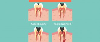
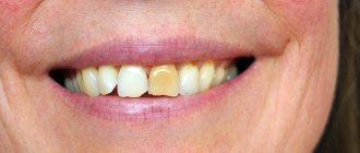
If caries appears on a tooth and it is not detected and treated in time, it can take on a more threatening form called pulpitis, which may be followed by periodontitis. Once the infection reaches the pulp, doctors can use a variety of treatment methods, both biological and surgical, and amputation or pulp extirpation is considered especially effective.
Dental pulp amputation is a gentle treatment method in which neurovascular tissue is partially removed. It is used primarily in the treatment of children's teeth. The maximum effect is achieved with slight damage to the dental tissue or in cases where the roots of the tooth are significantly curved. For adult patients, extirpation is mainly used, in which the dental cavities are completely cleansed.
After the difference in the structure of the various sections of the pulp - root and coronal - was established, the very possibility of amputation arose. If the neurovascular bundle is damaged only in its coronal region, slightly or superficially, it can be removed. This procedure is of particular importance for children whose tooth roots continue to develop, and for their proper development it is necessary to preserve the possibility of their nutrition.
Pulp amputation can be performed by two methods - vital and non-vital. Let's take a closer look at both of these methods.
Vital method of pulp amputation
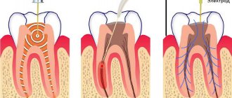
With this method of pulp removal, called pulpotomy, tissue damaged by caries is mechanically removed using local anesthesia. This technique is very popular. Using a bur, all damaged tissues are removed, after which the open cavity is treated with medications. The pulp is amputated before the root canals begin, in which it is kept alive. Its surface layers are fixed. The cavity is sealed with a temporary filling installed for six months. If after this time the patient does not complain of pain, percussion and palpation are painless, a permanent filling is placed in place of the temporary filling.
In children, the vital method of pulp amputation is most often used. This is due to the fact that permanent teeth, in the process of developing their roots, have winding canals in the form of ribbons, with many branches, and milk teeth need to preserve the ability of their root parts to resorption and at the same time the rudiments of future permanent teeth should not be affected.
Whether amputation can be performed using the vital method depends on how viable the part of the pulp located in the roots is, as well as on the presence of inflammation in the tissues surrounding the tooth. Deep amputation of the vital type differs from the ordinary one in that not only the coronal area of the pulp is removed, but also that located in the canals - at different levels of depth.
Modern approaches to the treatment of pulpitis in primary teeth in children
L.P. Kiselnikova, Doctor of Medical Sciences, Professor, Head. Department of Pediatric Therapeutic Dentistry, Moscow State Medical University
O.S.Kovylina, Ph.D., Ass. Department of Pediatric Therapeutic Dentistry, Moscow State Medical University
E.A. Savinova, Ph.D., Ass. Department of Pediatric Therapeutic Dentistry, Moscow State Medical University
T.P.Plyukhina, Ph.D., Ass. Department of Pediatric Therapeutic Dentistry, Moscow State Medical University
S.V. Goncharova, dentist. MSHI Group of Companies
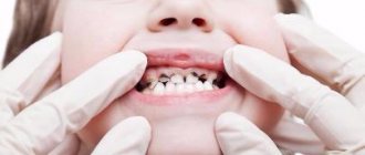
The main problem in pediatric dentistry is still caries and its complications. The presence of carious teeth as a source of chronic infection contributes to allergization of the body, decreased immunity and supports diseases of other organs and systems.
Early removal of primary teeth leads to malocclusion in the child and disruption of the sequence of teeth eruption. Lack of complete chewing of food leads to problems in the functioning of the gastrointestinal tract. Speech therapy and aesthetic problems also arise, which can lead to adverse psychological consequences.
In the therapeutic practice of a pediatric dentist, the most common form of complication of caries in primary teeth is pulpitis. Living, intact dental pulp is necessary for its normal function. The therapeutic and preventive focus of pediatric therapeutic dentistry necessitates tooth-preserving treatment of pulpitis and, if possible, preservation of the entire pulp or part of it.
Dental pulp is a richly vascularized and innervated specialized loose fibrous connective tissue that fills the pulp chamber of the crown and root canal (coronal and root pulp). In the crown, the pulp forms outgrowths corresponding to the tubercles of the chewing surface - the pulp horns. The pulp performs a number of important functions:
- plastic - participates in the formation of dentin due to the activity of odontoblasts located in it;
- trophic - ensures trophism of dentin due to the vessels located in it;
- sensory - due to the presence of a small number of nerve endings;
- protective and reparative - through the production of tertiary dentin, the development of humoral and cellular reactions, inflammation (V.L. Bykov, 1998).
The pulp of primary teeth functions for a relatively short time. It goes through three periods, which correlate with the stages of development of the primary tooth.
- Stage of tooth formation (the root of the tooth is not formed) - the period of pulp growth corresponds to the development of the crown and root.
- The stage of a formed tooth, or relative physiological rest (the root has completed its development) is the period of pulp maturation, which covers the time from the completion of root development to the beginning of its resorption.
- The stage of root resorption (loosening) is a period of regression that lasts from the beginning of root resorption until tooth loss.
During the period of regression, involutive processes occur in the pulp, disruption of its functions and gradual resorption along with resorption of the tooth root.
The development of inflammation in the dental pulp is due to the anatomical and histological features of the structure of temporary teeth:
- thin enamel-dentin layers and a large pulp chamber;
- the pulp horns of primary teeth come close to the occlusal surface and are located in close proximity to the contact surfaces;
- the pulp of temporary teeth is well supplied with blood, pre-collagen fibers predominate in it, there are many cellular elements, the connective tissue stroma of the pulp is less pronounced;
- lack of a clear boundary between the coronal and root pulp in single-rooted teeth;
- wide orifices of the root canals, a wide apical foramen and a wide periodontal fissure, the presence in 50% of cases of additional communication with tissues during the growth period in the area of the root furcation.
The indicated anatomical and histological features of the structure of temporary teeth determine the following features of the course of pulpitis:
- the predominance of chronic forms in the absence of complaints from the child;
- rapid transition of inflammation to the entire pulp and periodontal tissue;
- the presence (in 50-57% of cases) of destructive changes in the periodontal tissues.
Acute forms of pulpitis are much less common than chronic forms. Mostly, pulpitis in children is detected during a preventive examination or a routine visit to the pediatric dentist.
A complete history taking, which must be carried out taking into account the presence of concomitant chronic diseases in the child and objective examination data, helps the pediatric dentist make the correct diagnosis and choose the most rational method of treatment.
For clinical diagnosis of pulpitis of primary teeth in children, survey, examination, percussion, palpation, temperature tests, and x-ray diagnostics are used. The diagnostic value of the listed methods is different and depends on the age of the child, his individual psychological characteristics, as well as on behavior in the dental office. Diagnosis of pulpitis in primary teeth is based on objective examination data and data received from parents.
Particular attention should be paid to the external examination of the patient and the condition of the regional lymph nodes. With acute diffuse pulpitis and exacerbation of chronic gangrenous pulpitis, there may be a change in the configuration of the face due to swelling of the soft tissues.
Palpation along the transitional fold in the area of the causative tooth in chronic pulpitis does not cause pain without exacerbation (if the child behaves adequately).
Percussion does not always help to identify the causative tooth, since we are dealing with a small patient and the child may indicate painful percussion of all teeth (even healthy ones). In acute diffuse pulpitis or exacerbation of its chronic forms, there may be pain due to the accumulation of exudate in the tooth cavity or periodontal reaction.
Probing the walls and bottom of a carious cavity in young children is not recommended due to the occurrence of pain and the child’s inadequate response to the examination, which casts doubt on further reliable research and successful treatment.
After adequate anesthesia, it is necessary to remove the softened dentin with an excavator and remove the overhanging edges of the enamel. It is also important to determine whether there is a connection between the carious cavity and the tooth cavity, because we need to take into account the appearance of the pulp: it may be pink, brightly hyperemic, bleeding on probing, or dirty gray in color and not bleeding. Usually, the dentist discovers the connection between the carious cavity and the tooth cavity in the places where the pulp horns are closest to each other. In chronic forms of pulpitis, the pulp is covered with pigmented, softened dentin and the message can only be detected after its removal.
Pediatric dentists usually do not conduct temperature tests and electroodontodiagnostics (EDD) in primary teeth, because temperature tests cause an inadequate reaction in a small patient, and EDI will obviously not be indicative due to the child’s age.
When collecting anamnesis, it is necessary to take into account the psycho-emotional state of the small patient, which will allow the pediatric dentist to choose the optimal treatment method.
Clinical manifestations of various forms of pulpitis in primary teeth in children are often erased, so for diagnosis it is necessary to carry out additional examination methods. The most informative method in pediatric practice is the x-ray method. In case of pulpitis of primary teeth, an X-ray examination is mandatory, since there is often a discrepancy between the clinical picture of pulp inflammation and the condition of the periodontal tissues. An X-ray image allows you to find out the stage of formation of the root of a temporary tooth, assess the depth of the carious cavity and determine the presence of communication with the pulp chamber, the relationship of the roots of a temporary tooth with the germ of a permanent tooth, and determine the presence of changes in adjacent structures and bone tissue.
In the fibrous form of chronic pulpitis of primary teeth, changes at the bifurcation on the radiograph are detected in 57% of cases (T.F. Vinogradova, 1988), and in the gangrenous form - up to 81% (A.A. Kolesov, V.V. Zhilina, 1991) . The method is painless, which is important in pediatric practice, takes little time and allows you to choose the most appropriate tactics for treating pulpitis.
Types of radiographs: intraoral contact and extraoral (contact in the lateral projection, contact in the oblique projection, panoramic and orthopantomograms).
Figure 1. – X-ray of the jaws in the second oblique projection.
A contact intraoral radiograph allows you to objectively assess the condition of the teeth in the temporary occlusion only in the lower jaw, since in the upper jaw the permanent tooth germ is superimposed on the roots of the temporary one. But with multiple caries, multiple exposures are required, which significantly increases the tissue dose and the effective equivalent dose. Another problem is the need to place the film in the oral cavity, which causes a negative reaction in unprepared children.
An extraoral contact radiograph in a lateral projection provides an image of the chewing group of teeth in the lower jaw. The tissue and effective equivalent dose are significantly lower than with contact intraoral radiographs. This is due to an increase in the distance from the X-ray tube to the object under study and the use of intensifying screens (Yu.I. Vorobyov, V.T. Truten, 1988). With this method of research in the temporary dentition, it is possible to give an objective assessment of the X-ray picture in the area of 2-3 teeth. This method does not require placing the film in the oral cavity, which facilitates contact with the child.
The method of extraoral contact radiography in an oblique projection allows you to obtain a reliable image of not only the lower dentition, but also the upper one. To study primary molars, the method of extraoral contact radiography in the second oblique projection is used. The image of the lateral sections of the jaws is obtained in full size without significant projection distortions. The method does not require manipulation of the film in the oral cavity, which facilitates contact with the child. One image provides an image of both jaws, which reduces the number of studies and reduces radiation exposure. The tissue and effective equivalent dose is 28-30 times lower than when examining the same number of teeth using intraoral contact radiography. Moreover, it becomes possible to take pictures in identical projections, which allows you to objectively monitor the dynamics of the pathological process. The method is performed using the 5D-2 dental X-ray machine, widely used in dental practice, and requires special, expensive equipment.
A panoramic radiograph provides an enlarged image of the complete dentition of the upper or lower jaw. Orthopantomography (panoramic zonography) allows you to obtain the most complete image of the entire dental system while minimizing radiation exposure to the child’s body. However, in children during the period of temporary occlusion, a panoramic photograph of the upper jaw and an orthopaptomogram of the upper teeth do not provide a detailed picture due to the overlap of the projection of the rudiments of permanent teeth on the roots of temporary ones.
Radiovisiography as a method for studying the condition of teeth has been used relatively recently. It does not require a darkroom; the computer program provides automatic differentiated dose setting for each tooth, which improves the quality of images and significantly reduces the radiation dose to the patient. One of the disadvantages of this method is the need for intraoral placement of the sensor, which is often negatively perceived by young children.
One of the modern methods for assessing changes in bone tissue is osteodensitometry. Carrying out dynamic osteodensitometry to study the density of bone tissue makes it possible to determine the degree of destruction of bone beams in various forms of pita bullets and helps the doctor choose the optimal method of treatment (L.P. Kiselnikona, M.A. Chibisova, 2003).
In Fig. Figure 2 shows an x-ray of an intact second primary molar. Dynamic osteodensitometry data at the root bifurcation within 192 cu. e. optical density.
In Fig. 3, 4, respectively, show X-ray images of chronic fibrous pulpitis of temporary molars with changes in bone tissue at the bifurcation of the roots, a decrease in osteodensitometry values from 100 to 20 cu. e. optical density.
Figure 2. – Drawing of bone tissue without changes.
Figure 3. – Changes at the bifurcation of the roots of a temporary tooth with pulpitis.
Figure 4. – Pronounced changes in bone tissue at the bifurcation of the roots of a temporary tooth with pulpitis.
Treatment of pulp inflammation in primary teeth in children
The main tasks in the treatment of pulpitis:
- eliminate the inflammatory process in the pulp and eliminate pain;
- prevent the spread of the infectious process to the periodontium;
- restore tooth function.
- in some cases, preserve and restore pulp function.
The choice of treatment method for pulpitis in children depends on the form of pulpitis, the group of the tooth, the degree of formation of the roots, the topography of the carious cavity, the degree of tooth destruction, the multiplicity and activity of the carious process.
Methods for preserving the viability of the entire pulp in temporary teeth, due to anatomical and physiological characteristics, are used extremely rarely, since they give a high percentage of complications after treatment (V.S. Ivanov, 2003).
The method of amputation (pulpotomy and deep pulpotomy) in single-rooted temporary teeth with unformed roots is in most cases impractical. It is possible that part of the infected pulp may be preserved and the infection may spread to deeper tissues, which leads to tooth extraction.
As a rule, in clinical practice, pulpitis of single-rooted teeth with unformed roots is a consequence of early carious lesions under the age of 1 year. The reactivity of the body in such children (as well as the resistance of hard tissues) is reduced, and there is no hope for the success of vital treatment methods.
There are several methods for treating pulpitis that can be widely used in the clinic for temporary teeth: pulpotomy (vital removal of coronal pulp), the method of dental amputation followed by mummification of the root pulp, pulpectomy (extirpation method).
Based on many years of clinical practice and taking into account the introduction of modern baking methods, we recommend the following algorithm for the treatment of pulpitis of primary teeth.
Table 1
| Treatment methods for pulpitis | |||
| Maintaining the viability of all or part of the pulp | Removal of pulp with loss of its viability | ||
| Complete | Partial | Partial | Complete |
| Biological method | Vital amputation | Amputation | Complete |
| 1. Indirect coating 2. Direct coating | 1. Amputation of the coronal pulp 2. High amputation | Devital amputation | 1. Vital extirpation 2. Devital extirpation |
Method of pulpotomy (vital amputation).
Indications: temporary molars, regardless of the stage of root formation, but without signs of resorption, without pronounced changes in periodontal tissues (initial densitometry values at the bifurcation are in the range of 192-128 optical density units).
The method is carried out using drugs:
- 20% formocresol solution (35% tricresol, 19% formaldehyde in 15% aqueous and glycerin solution);
- Pulpevit No. 3 - formocresol (VladMiVa);
- Krezatin, Palpak;
- glutaraldehyde (GA), Endo-ji No. 3 (VladMiVa), containing glutaraldehyde, has an antiseptic effect;
- Endo-Zhi No. 4 (VladMiVa), contains aluminum chloride, has a hemostatic effect;
- Ferrous sulfate (15.5-55%) - ViscoStat, Astringedent (Ultradent).
The pulpotomy method using a 20% solution of formocresol or Endo-Zhi liquid No. 3 (GA) is carried out as follows. After adequate anesthesia, necrotomy is performed, the pulp chamber is opened, the overhanging edges of the pulp chamber roof are removed, and the coronal pulp is amputated. After this, special attention should be paid to stopping bleeding from the mouths of the root canals. It should be remembered that remnants of coronal pulp in the pulp chamber can cause bleeding, which can only be stopped by removing them completely. If, with a correctly performed pulpotomy, the bleeding cannot be stopped, this may indicate inflammation of the root pulp. In the latter case, this treatment method is unacceptable - pulpectomy is performed.
After stopping the bleeding, a tampon moistened with a 20% solution of formocresol or Endo-Zhi liquid No. 3 (GA) is applied to the mouths of the root canals for 5 minutes. After the specified time, the tampon is removed - a so-called “scab” is formed at the site of contact of the drug with the root pulp (the pulp becomes dark brown in color). Then zinc-oxide-eugenol cement Eodent (VladMiVa) is applied to the orifices of the root canals. Next, preparations are made for a (permanent) restoration made of glass ionomer cement or compomer, and a standard crown is fixed.
It is worth dwelling on the experience of using ferrous sulfate 20% - ViscoStat (Ultradent) during pulpotomy. This drug does not belong to the group of resorcinol-formalin compounds. Consequently, when carrying out the technique, even short-term contact of drugs containing resorcinol-formalin with the child’s body is excluded. In immature molars, coronal pulp amputation and hemostasis are performed. The ostial pulp is treated with ViscoStat - iron sulfate 20% (Ultradent), which has hemostatic and antiseptic properties.
ViscoStat gel is applied for 1-3 minutes, then removed with a cotton ball, and the tooth cavity is dried. Then Eodent zinc oxide eugenol cement (VladMiVa) is applied to the canal mouths.
It is also possible to perform pulpotomy on vital molars using the drugs Pulpotek (RP) and Pulpodent (VladMiVa). After amputation of the coronal pulp and hemostasis under anesthesia, the above drugs are applied to the canal mouths in the form of a paste. Then an insulating glass ionomer lining is applied and the crown of the tooth is restored with a composite or a standard metal crown.
Method of devital amputation followed by mummification of the root pulp
Indications: pulpitis of temporary multi-rooted teeth, regardless of the stage of root formation, without signs of physiological resorption with moderate changes in periodontal tissues (initial densitometry value at the bifurcation is in the range of 128-64 cu optical density).
Treatment is carried out in three visits. On the first visit, after anesthesia and partial treatment of the carious cavity, a devitalizing paste is applied to the exposed pulp horn. To devitalize the pulp, preparations based on paraformaldehyde or trioxymethylene are used. During the second visit, the final treatment of the carious cavity is carried out, the overhanging edges of the roof of the pulp chamber are removed, the coronal pulp is amputated, and a tampon with mummifying liquid is applied to the mouths of the root canals. For this purpose, formalin-containing preparations are used, which have antiseptic and drying properties. On the third visit, the temporary bandage and tampon with mummifying liquid are removed. A resorcinol-formalin-based paste is applied to the mouths of the root canals.
Restoration of the tooth crown is carried out using various filling materials: glass ionomer cements or composites. If the crown is significantly damaged, restoration of the anatomical shape of the tooth is carried out using standard metal crowns.
Pulpectomy (vital or devital pulp extirpation) is carried out in formed temporary single-rooted teeth or molars in case of acute diffuse and chronic gangrenous pulpitis, in the presence of a weakening of the pattern of bone beams and fiberization of the cortical plate of the tooth socket on a radiograph and osteodensitometry indicators of less than 64 y, e. optical density
After trephination of the roof of the pulp chamber, it is necessary to completely remove it using a bur; the canals are processed with hand instruments to a depth of 1-2 mm less than the radiographic length of the chamber. For medicinal treatment of infected root canals, it is advisable to use Katalyugem or Alkasept (Nord-Ost) - drugs based on quaternary ammonium compounds that have a wide range of antiseptic effects and pronounced hemostatic properties (N.B. Potapova, 2005). The material for filling the canals of temporary teeth should be absorbed during resorption and not interfere with the eruption of a permanent tooth. To fill the root canals of temporary teeth, pastes with eugenol and iodoform are used: zinc oxide eugenol paste, Vitapex (Neo Dental Chemica Prod), Metapsx (META). Then X-ray control of the filling and permanent filling with glass ionomer cement or composite are carried out.
Thus, at present, there are various methods for treating pulpitis in primary teeth, which, with differentiated diagnosis, allow in the maximum possible number of cases not only to preserve the tooth in the dentition until its physiological change, but also in some cases to ensure the viability of the pulp as a physiological barrier.
published 12/12/2011 11:43 updated 09/26/2014 – Dentistry
Devital method of pulp amputation
Devital amputation involves preliminary killing of the pulp in its coronal part using special pastes. After which it acquires a fibrous structure that can be easily removed. In this case, the pulp located in the roots remains intact. After removal of the coronal part, the root pulp is mummified under the influence of drugs that impregnate it, dehydrates and becomes a sterile strand. There are a large number of antiseptic pastes based on various anti-inflammatory active substances that have certain advantages and disadvantages.
The devital pulp amputation technique is used for various types of pulpitis - acute and chronic. Or if the patient is afraid of syringes and injections or other phobias associated with dental treatment.
According to a number of experts, the technique of devital amputation for pulp in permanent teeth has low effectiveness, since in some cases it leads to inflammation of periodontal tissue. Such treatment is advisable only during the growth of tooth roots, and if they are already fully formed, their canals are cleaned by pulp extirpation.
Acute pulpitis
The acute form of the disease causes severe paroxysmal pain, which becomes even more intense at night. Even without an external irritant, attacks of pain increase or decrease, but they usually occur when eating cold or hot food or drinks.
Due to the irradiation of the pain impulse along the nerves, sometimes the patient cannot say exactly which tooth hurts. If the pain becomes more and more intense, this may indicate the development of a purulent process. Its symptom is throbbing and almost constant pain, sometimes shooting and almost never subside.
A clear sign of pulpitis is a prolonged subsidence of pain after removal of the irritant. This takes about 15 minutes. This is its difference from caries, in which the pain disappears immediately if the irritant is removed.
Devital pulpotomy
The devital pulpotomy method is used in the case of elderly patients with already overgrown root canals or if the patient suffers from severe somatic pathology. To do this, open the pulp chamber and put a drug into the hole that kills the pulp. The dead pulp is then removed down to the border of the root canal. A product is applied that mummifies the root part, after which a special gasket is placed and the hole in the tooth is filled. In this case, pulpotomy is performed as a barrier method to prevent the inflammation process from spreading to periodontal tissue.
Surgical treatment
Surgical treatment involves removing the pulp in several stages. Depulpation is performed in the root and coronal zones in advanced cases. Sometimes it is impossible to cure a tooth without this procedure.
If the tooth is single-rooted, then it will have to be removed in two or even three visits to the dentist. And if this is a multi-channel case, then even more activities are needed. Therefore, the number of visits to the doctor is determined by the complexity of the lesion and the dynamics of treatment.
If a canal has been filled, the filling cannot be placed in one visit, since the material inside the canals must first harden.
Chronic pulpitis
In the chronic course of pulpitis, there are no obvious symptoms. Sometimes a slight aching pain may occur, especially after cold or hot drinks or meals. Exacerbations, which are similar in their symptoms to acute pulpitis, are also possible.
When pus accumulates, flux appears. If a person is not provided with proper medical care, this can lead to phlegmon and related complications:
- damage to the jaw bones and facial nerve;
- abscess;
- apical periodontitis;
- sepsis;
- chronic hyperplastic pulpitis (occurs in childhood and adolescence).
Pulpotomy process
The method of performing vital pulpotomy is that the process is divided into two stages and is performed in two visits to the dentist with a break of 6 months. The first stage of vital pulpotomy:
- After anesthetizing the desired area, a layer of enamel and affected dentin is removed using a drill.
- The pulp area is then opened and the infected portion is removed, usually at the level of the root canal orifice.
- The bleeding is stopped and a drug is applied, which serves as a therapeutic dressing for the remaining part of the pulp.
- Lastly, a temporary filling is placed.
After this procedure, the dentist closely monitors the operated tooth for six months. Monitors for any negative manifestations that indicate that the pulpotomy was ineffective. If there is no pain or changes in the jawbone, the pulpotomy is considered successful and a permanent filling can be installed.
Vital pulpotomy is considered an effective procedure, but it should be performed in the initial stages of pulpitis. In order not to risk starting the inflammatory process to an irreversible level, when this treatment procedure will no longer help preserve the pulp of a child’s tooth, it is necessary to regularly visit the dentist with the child. The specialist will be able to notice the problem in time and begin treatment immediately.
How is the operation performed?
Most often, dentists perform vital pulp extirpation. It includes the following steps:
- professional cleaning of the affected tooth,
- pain relief using local anesthetics,
- isolation of the working area from salivary secretions,
- incision in the arch of the dental cavity,
- removal of the neurovascular bundle,
- restoration of the correct shape of the root canal,
- antiseptic treatment of the tooth cavity,
- installation and grinding of the filling.